S H O R T R E P O R T
Open Access
Human cardiac stem cells inhibit
lymphocyte proliferation through paracrine
mechanisms that correlate with indoleamine
2,3-dioxygenase induction and activity
Maria J Sebastião
1,2†, Ramón Menta
3†, Margarida Serra
1,2, Itziar Palacios
3, Paula M Alves
1,2, Belén Sanchez
3,
Olga DelaRosa
4, Wilfried Dalemans
5, Eleuterio Lombardo
4*and Patrícia Gomes-Alves
1,2*Abstract
Transplantation of allogeneic human cardiac/stem progenitor cells (hCSCs) is currently being tested in several phase
I/II clinical trials as a novel and promising therapy for restoration of myocardial tissue function in acute myocardial
infarction (AMI) patients. Previous findings demonstrate that these cells have an immune suppressive profile
interacting with different populations from the immune system, resulting in overall attenuation of myocardial
inflammation. However, transplanted hCSCs are still recognized and cleared from the injured site, impairing
long retention times in the tissue that could translate into a higher clinical benefit.
In this work, through modeling allogeneic hCSC/T lymphocyte interaction in vitro by direct contact, transwell inserts,
and hCSC conditioned medium, our results demonstrate that hCSCs exert an immune-suppressive effect on T
lymphocyte proliferation not only through the previously described cell contact-dependent programmed cell
death-1 (PD1)/programmed death ligand-1 (PDL-1) axis but also through a paracrine mechanism associated
with indoleamine 2,3-dioxygenase (IDO) enzyme-mediated tryptophan metabolism. Such findings constitute a
step forward in better understanding the mechanisms of action of transplanted hCSCs in allogeneic settings.
Keywords: Cardiac stem/progenitor cells, Allogeneic stem cell therapy, Tryptophan metabolism,
Immunosuppression, T lymphocytes
Introduction
Human cardiac/stem progenitor cell (hCSC)
transplant-ation is becoming a promising therapy for acute
myocar-dial infarction (AMI), one of the most prevalent causes of
death worldwide [
1
]. CSCs are considered by several
au-thors as the preferred candidate cell source for AMI
pa-tients, mainly due to their function in the heart,
well-documented paracrine regenerative properties [
2
,
3
],
and the success of transplantation studies in myocardial
infarction animal models [
4
,
5
].
Such success in preclinical stages has led to a rapid
translation to the clinic, and several phase I and II
clinical trials using hCSCs as an autologous therapy
have emerged (e.g., SCIPIO and CADUCEUS trials).
Autologous transplantation, although carrying lower
immunogenicity risks, holds several limitations. The
quality of the cells might be compromised by patient
age and comorbidities [
3
,
6
,
7
], as well as logistic,
eco-nomic, and time-constraints issues. To overcome such
limitations, in the last years the field has been moving
towards allogeneic CSC sources (e.g., the ALLSTAR
and CAREMI trials).
Clinical trials have demonstrated physiological
improve-ments and increases in viable tissue and heart functional
outcome [
8
]. However, an obstacle still preventing CSCs
from meeting their full clinical potential and to provide
evident clinical benefit over a standard-of-care is their
* Correspondence:[email protected];[email protected]
†Maria J Sebastião and Ramón Menta contributed equally to this work. 4TiGenix SAU, Tres Cantos, Madrid, Spain
1Animal Cell Technology Unit, iBET, Instituto de Biologia Experimental e
Tecnológica, Oeiras, Portugal
Full list of author information is available at the end of the article
© The Author(s). 2018 Open Access This article is distributed under the terms of the Creative Commons Attribution 4.0 International License (http://creativecommons.org/licenses/by/4.0/), which permits unrestricted use, distribution, and reproduction in any medium, provided you give appropriate credit to the original author(s) and the source, provide a link to the Creative Commons license, and indicate if changes were made. The Creative Commons Public Domain Dedication waiver (http://creativecommons.org/publicdomain/zero/1.0/) applies to the data made available in this article, unless otherwise stated.
limited retention and engraftment in the heart [
9
,
10
], a
problem aggravated in the allogeneic setting [
11
,
12
].
An immune response is triggered upon AMI that,
although essential for proper tissue remodeling and
stabilization, carries unwanted inflammatory-mediated
damage [
13
] and, in the case of cell therapy approaches,
might be also involved in the elimination of injected cells.
Effective activation of T cells, one of the main mediators
of inflammatory damage upon AMI, requires
simultan-eous engagement of the T-cell receptor (TCR) and CD28
receptor. TCR binds to antigens present in major
histo-compatibility complex (MHC; human leukocyte antigens
(HLA) in humans) and CD28 receptor binds to B7
(CD80/CD86) costimulatory molecules. hCSCs express
HLA class I molecules that attract killer cytotoxic T cells,
but very low levels of HLA class II molecules that
stimu-late antibody-producing B cells, and do not express the
costimulatory molecules CD80/CD86 [
14
], therefore
pre-senting a weak immunogenic profile. Besides depicting the
immune phenotype of hCSCs, several studies have shed
some light on how hCSCs interact with monocytes [
15
],
natural killer cells [
16
], and T lymphocytes [
14
,
17
].
Simi-lar to other studies with mesenchymal stem cells (MSCs)
[
18
–
21
], all the referred studies with hCSCs suggest that
the immunologic behavior of cells might be linked to
their therapeutic effects rather than eliciting
deleteri-ous immune reactions, with an immunomodulatory
effect resulting in attenuation of inflammation.
Fur-thermore, programmed death ligand 1
(PDL-1)-me-diated cell-cell interaction has been identified as one
of the main mechanisms for the immunomodulatory
properties of hCSCs, promoting stimulation of regulatory
T cells and subsequent inhibition of T lymphocyte
activa-tion and proliferaactiva-tion [
14
].
Besides direct cell-cell interactions, paracrine
immu-nomodulatory effects based on extracellular vesicles have
been described for hCSCs [
17
]. Tryptophan (Trp)
me-tabolism through the enzymatic activity of indoleamine
2,3-dioxygenase (IDO) has been described as a key
im-munosuppressive mechanism for human adipose-derived
mesenchymal stem cells (hASCs) [
22
–
24
], a cell type
already used in several allogeneic cell transplantation
studies [
25
].
With the aim of better understanding the
immuno-modulatory mechanisms of hCSCs in an allogeneic
set-ting, we further investigated the capacity of hCSCs to
inhibit the proliferation of T lymphocytes in vitro. Taken
together, our results add to knowledge on the
tolero-genic immune behavior of hCSCs, showing that
hCSC-mediated immune modulation is not dependent
exclu-sively on the PDL-1/ programmed cell death-1 (PD-1)
pathway axis, but also via Trp degradation by IDO
en-zyme action. Such findings open new avenues for
de-signing novel hCSC allogeneic transplantation therapies
for AMI patients, including strategies to promote a
higher hCSC engraftment and longer residence time in
the tissue.
Results
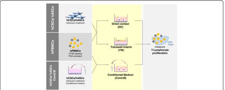
The immunomodulatory experimental design is
illus-trated in Fig.
1
. Briefly, stimulated human peripheral
blood mononuclear cells (hPBMCs) were cultured in the
presence of mitomycin C-treated hCSCs or hASCs.
Cul-tures were performed in culture well plates in: 1) direct
hPBMCs
CFSE labeling PHA activation
hCSCs/hASCs
mitomycin treatment Direct contact
(DC) Transwell inserts (TW) hCSCs/hASCs mitomycin treatment Conditioned medium Conditioned Medium (Cond.M) hPBMCs hCSCs/ hA SCs Cond.M hCSCs/ hA SCs measure T-Lymphocyte proliferation CD3+ 7AAD
-Fig. 1 Schematic representation of immunomodulatory assay experiments. Carboxyfluorescein succinimidyl ester (CFSE)-labeled and phytohemagglutinin (PHA)-activated human peripheral blood mononuclear cells (hPBMCs) were cultured with mitomycin-treated human cardiac/stem progenitor cells (hCSCs)/ human adipose-derived mesenchymal stem cell (hASCs) either in direct contact (DC), in a transwell support (TW), or in contact with mitomycin-C-treated hCSC/hASC conditioned medium (Cond.M.). Viable T lymphocyte proliferation was accessed by CFSE labeling of the CD3+7AAD–hPBMC population
contact (DC); 2) using transwell (TW) inserts to allow
exchange of soluble factors but separation of both cell
types; and 3) using hCSC conditioned medium. hASCs
were used as a positive control for T cell proliferation
in-hibition via IDO. See Additional file
1
for detailed
mate-rials and methods.
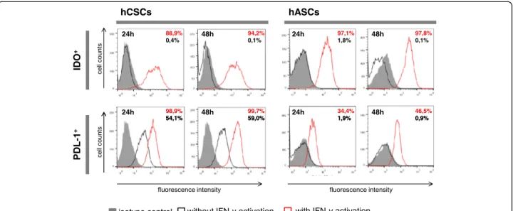
IDO expression is induced in hCSCs in response to
interferon (IFN)-
γ
We first characterized the hCSC expression of the
immune-relevant molecules PDL-1 and IDO with and
without IFN-γ stimulation. While hASCs only express
PDL-1 when activated, hCSCs express PDL-1
constitu-tively, and activation with IFN-γ further upregulates its
expression (Fig.
2
). These results agree with data
previ-ously reported by Lauden et al. [
14
]. Neither hCSCs
nor hASCs displayed any constitutive expression of
IDO but both cell types displayed significant expression
upon stimulation with IFN-γ (Fig.
2
).
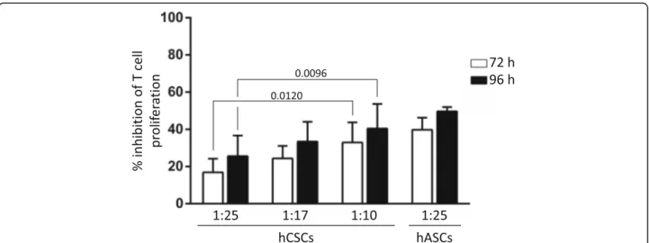
hCSCs impair activated T lymphocyte proliferation
Following the characterization of a favorable
immune-suppressive phenotype of hCSCs, we then examined
whether these cells were capable of inhibiting T
lympho-cyte proliferation in an allogeneic setting. Stimulated
hPBMCs were cultured in direct contact with hCSCs at
different ratios.
As previously reported [
14
], hCSCs exert an immune
suppressive role by inhibiting T lymphocyte proliferation.
hCSCs have a significant suppressive effect in T
lympho-cyte proliferation in a dose-dependent manner, although
to a lesser extent compared with hASCs (Fig.
3
). Although
no significant difference was seen between the time
points, there is a tendency for higher proliferation
in-hibition at 96 h versus 72 h of incubation (Fig.
3
) in all
ratios analyzed, suggesting that this effect may also be
time dependent.
hCSCs immunomodulatory capacity can occur in the
absence of cell-cell contact
To evaluate the importance of IDO enzyme and Trp
metabolism in the immunosuppressive capacity of
hCSCs, we carried out T lymphocyte proliferation assays
in which hCSCs were not in direct contact (DC) with
hPBMCs and therefore cannot exert their
immunomod-ulatory activity through the PDL-1/PD1 axis.
We carried out hCSC-hPBMC coculture under
trans-well conditions (TW), allowing paracrine interaction
be-tween the cell types. At 72 h of incubation, although
slightly lower when compared with DC, hCSCs do exert
a significant suppressive effect on T lymphocyte
prolifer-ation under TW conditions. Moreover, such a difference
between TW and DC conditions was lost after 96 h of
incubation (Fig.
4a
).
Trp metabolism was also assessed by measuring Trp
and kynurenine (Kyn; a Trp metabolite described as
cytotoxic for T lymphocytes [
26
]) concentrations in the
conditioned medium. As shown in Fig.
4b
, Trp is fully
depleted at 72 h under the DC condition, and in the
TW setting it is also significantly diminished when
compared with stimulated hPBMCs alone. Furthermore,
Fig. 2 Human cardiac/stem progenitor cells (hCSCs) display a favorable immune-suppressive phenotype. Representative expression of the immune relevant molecules indoleamine 2,3-dioxygenase (IDO) and programmed death ligand 1 (PDL-1) after 24 and 48 h in untreated (black line histograms) and interferon (IFN)-γ-activated (red line histograms) cells against isotype controls (gray-filled histograms). The percentages (%) of positive cells are indicated. Human adipose-derived mesenchymal stem cells (hASCs) were used as a positive control for IDO expression. hCSC results are shown for donor hCPC8. Other donors presented similar results
Fig. 3 Human cardiac/stem progenitor cells (hCSCs) inhibit T lymphocyte proliferation in a time- and hCSC concentration-dependent manner. CFSE-labeled hPBMCs were stimulated with PHA and cultured alone or in direct contact with hCSCs (ratios 1:10, 1:17, and 1:25 hCSCs:hPBMCs). After 72 h (white bars) and 96 h (black bars), proliferation of the viable population of CD3-viable T lymphocytes (CD3+/7AAD–) was assayed by loss of CFSE staining. Percentage of inhibition of proliferation was determined using FSC Express software against proliferation of activated hPBMCs alone. Human adipose-derived mesenchymal stem cells (hASCs) were used as a positive control for T cell proliferation inhibition (ratio
1:25 hASCs:hPBMCs). Adjustedp values are shown
A
B
C
D
Fig. 4 Human cardiac/stem progenitor cells (hCSCs) inhibit T lymphocyte proliferation via a paracrine mechanism. a CFSE-labeled hPBMCs were stimulated with PHA and cultured alone, in direct contact (DC), or in a transwell setting (TW) with hCSCs (ratio 1:10 hCSCs:hPBMCs). b Concentrations of tryptophan (Trp) and kynurenine (Kyn) were determined by HPLC in the supernatants. c CFSE-labeled hPBMCs were
stimulated with PHA and cultured alone or in conditioned medium (Cond.M.) from hCSCs cultures activated or not with interferon (IFN)-γ.
Conditioned media were generated for 24 h (white bars), 36 h (grey bars), and 48h (black bars). d Concentrations of Trp and Kyn were
determined by HPLC in the conditioned media. Proliferation of the viable population of CD3 T lymphocytes (CD3+/7AAD–) was assayed
by loss of CFSE staining after 72 h (white bars) and 96 h (black bars) for TW and DC experiments (a) and after 96 h for Cond.M. experiments (c). Percentage of cells per generation and percentage of inhibition of proliferation was determined using FSC Express software against proliferation of activated hPBMCs alone. Human adipose-derived mesenchymal stem cells (hASCs) were used as a positive control for T cell proliferation inhibition (ratio 1:25 hASCs:hPBMCs). Adjustedp values are shown
the accumulation of Kyn occurred in both experimental
setups (Fig.
4b
).
Besides the TW experiments, hCSC conditioned
medium was generated for 24 h, 36 h, and 48 h using
control and IFN-γ-stimulated hCSCs. Similar to the
hASC control, hCSC-derived conditioned medium
sig-nificantly inhibited T lymphocyte proliferation, with a
significant increase in IFN-γ-stimulated cells (51.79 ±
11.67 % versus 15.49 ± 8.10% with 36-h conditioned
medium; 100 ± 0.00 % versus 19.01 ± 7.22% with 48-h
conditioned medium; Fig.
4c
). Moreover, conditioned
medium from longer IFN-γ-stimulated hCSC cultures
prompted higher inhibition of T lymphocyte
prolifera-tion (Fig.
4c
). Such findings are also in accordance
with the Trp and Kyn measurements, where Trp is
gradually depleted and Kyn gradually accumulates in
the supernatant of hCSC cultures (Fig.
4d
).
Discussion
Allogeneic hCSC-based therapies continue to be explored
as an alternative for AMI patients. However, hCSC
regen-erative medicine approaches have yet to show an evident
and robust clinical benefit over standard-of-care.
Al-though described as having a positive immunomodulatory
role rather than eliciting further inflammation [
14
–
17
],
one of the main challenges to be addressed in hCSC
transplantation-based therapies is the rapid elimination of
the injected cells. A better knowledge of the
immuno-logical properties of hCSCs is therefore paramount in
de-veloping strategies to increase the retention time of the
cells in the myocardium that would consequently increase
their regenerative benefits.
Similar to what is described for MSCs [
18
–
21
], hCSCs
have been described as having an immune-suppressive
pro-file. Several studies show that hCSCs in allogeneic settings
have an anti-inflammatory profile by modulation of natural
killer cell cytokine secretion and cytotoxicity [
16
], by
modu-lation of monocytes, macrophages, and dendritic cells [
15
],
and by activation of T regulatory lymphocytes with
subse-quent inhibition of T lymphocyte proliferation via PDL-1/
PD1 direct cell communication [
14
], as well as via
extracel-lular vesicle-mediated paracrine communication [
17
].
In this work, we assayed the immunomodulatory
capacity of hCSCs in an inflammatory setting by IFN-
γ
activation and explored the hypothesis of paracrine
IDO-mediated T lymphocyte proliferation inhibition.
IFN-
γ is highly expressed under inflammatory settings
(such as after AMI). This proinflammatory pleiotropic
cytokine is produced primarily by the host T lymphocytes
in response to antigen recognition and has also been
shown to induce the expression of immune-relevant
mole-cules in several stem cell populations, including neural
stem cells [
27
] and MSCs [
24
,
28
]. IFN-
γ was shown to
cause hCSCs to upregulate the expression of both class I
and II HLA molecules [
14
], which indicates that their
administration into an inflammatory myocardium
envir-onment probably increases their recognition by host T
lymphocytes. On the other hand, IFN-γ supplementation
has also previously been shown to upregulate hCSC
ex-pression of PDL-1, resulting in a stronger hCSC
immune-suppressive profile [
14
].
IFN-
γ activation has also been correlated with increased
IDO expression in human MSCs, including hASCs, which
in turn has been shown to be a key enzyme involved in
the immunomodulatory capacity of these cells [
22
,
29
].
IDO suppresses T lymphocyte proliferation and promotes
T lymphocyte death through degradation of Trp, an
es-sential amino acid required for cell proliferation and
sub-sequent accumulation of cytotoxic Trp metabolites
(including Kyn) [
26
,
30
]. IFN-
γ also causes the activation
of tryptophanyl-transfer RNA synthetase (WRS; an
ami-noacyl synthetase that incorporates Trp into proteins) in
IDO-expressing cells, which has been postulated to be a
compensatory mechanism allowing IDO-expressing cells
to better cope with Trp depletion [
31
].
In our study, we show that IFN-
γ activation is correlated
with an increase in hCSC PDL-1 and IDO expression.
Al-though with an overall weaker immune-suppressive
pro-file when compared with hASCs, we also showed that
hCSCs are able to inhibit T lymphocyte proliferation in a
time-and hCSC cell concentration-dependent manner
when in direct coculture.
Moreover, we showed Trp depletion and Kyn
accumu-lation in activated hCSC conditioned medium.
Concor-dantly, stimulated hCSC conditioned medium showed a
superior antiproliferative effect when compared with
un-stimulated hCSC conditioned medium, suggesting a
rele-vant role of IDO-mediated Trp metabolism in the
immunomodulatory paracrine effect of hCSCs.
We also showed no significant differences between DC
and TW in terms of T lymphocyte proliferation,
suggest-ing that paracrine-mediated effects are a central
mech-anism of action for hCSCs, as previously demonstrated
with Sca1
+hCSCs [
17
].
These findings provide evidence that, although playing
a role in the process, PDL-1 mediated T-regulatory cell
modulation is not the exclusive nor the central
mechan-ism involved in T lymphocyte proliferation inhibition, at
least under our experimental conditions. This finding
further supports the prominent paracrine-based
benefi-cial CSC activities in the host tissue.
In this work, we provide evidence of Trp metabolism
as a novel mechanism involved in the hCSC-mediated T
lymphocyte proliferation suppression properties. We
also hypothesize that, similar to what is already
de-scribed for hASCs [
22
–
24
], IDO is the main player in
hCSC Trp metabolism in inflammation settings in vitro.
Complementary studies to further test this hypothesis
should include analysis of WRS expression, Trp
supple-mentation, and IDO inhibition experiments to further
validate IDO Trp metabolism as a main player in hCSC
immunomodulatory properties in the host tissue.
Additional file
Additional file 1:Detailed material and methods: a full description of material and methods including cell isolation and culture, immunomodulatory assays, and statistical analysis. (DOCX 26 kb)
Abbreviations
AMI:Acute myocardial infarction; CSC: Cardiac/stem progenitor cell; DC: Direct contact; hASC: Human adipose-derived mesenchymal stem cell; hCSC: Human cardiac/stem progenitor cell; HLA: Human leukocyte antigen; hPBMC: Human peripheral blood mononuclear cell; IDO: Indoleamine 2,3-dioxygenase; IFN: Interferon; Kyn: Kynurenine; MHC: Major histocompatibility complex; MSC: Mesenchymal stem cell; PD1: Programmed cell death-1; PDL-1: Programmed death ligand 1; TCR: T-cell receptor; Trp: Tryptophan; TW: Transwell; WRS: Tryptophanyl-transfer RNA synthetase
Funding
This work was supported by the project NETDIAMOND (SAICTPAC/0047/2015) financially supported by FEEI– Lisboa2020 and FCT/POCI-01-0145-FEDER-016385; and iNOVA4Health - UID/Multi/04462/2013, financially supported by FCT/Ministério da Educação e Ciência, and co-funded by FEDER under the PT2020 Partnership Agreement. MJS is a recipient of the FCT fellowship SFRH/ BD/52339/2013.
Availability of data and materials
Data sharing is not applicable to this article as no datasets were generated or analyzed during the current study.
Authors’ contributions
Conceptualization: RM, IP, OD, WD, and EL; Methodology and investigation: MJS and RM; Writing the original draft: MJS, PGA, and MS; Review and editing: MJS, RM, IP, PMA, and EL; Resources: BS, IP, and EL; Funding acquisition: PGA and PMA. All authors read and approved the final manuscript.
Ethics approval and consent to participate
All experiments were performed in accordance with the local institutional guidelines and regulations and the approval of the local ethic committee, and with informed consent of all cell donors. Human cardiac stem cells were obtained from human cardiac biopsies after signed informed consent in accordance with the Declaration of Helsinki. The ethical committees of the Hospital 12 de Octobre and the Fundación Jiménez Dias (Madrid, Spain) have approved the project. hPBMCs were isolated from blood donors after signing an informed consent following the human ethics committee from the Centro de transfusion de la Comunidad de Madrid.
Consent for publication Not applicable. Competing interests
RM, EL, IP, BS, OD, and WD are employees of TiGenix Group. The remaining authors declare that they have no competing interests.
Publisher
’s Note
Springer Nature remains neutral with regard to jurisdictional claims in published maps and institutional affiliations.
Author details
1Animal Cell Technology Unit, iBET, Instituto de Biologia Experimental e
Tecnológica, Oeiras, Portugal.2ITQB-NOVA, Instituto de Tecnologia Química e Biológica António Xavier, Universidade Nova de Lisboa, Oeiras, Portugal.
3Coretherapix, S.L.U. (TiGenix Group), Tres Cantos, Spain.4TiGenix SAU, Tres
Cantos, Madrid, Spain.5TiGenix NV, Leuven, Belgium.
Received: 9 August 2018 Revised: 4 September 2018 Accepted: 17 September 2018
References
1. Benjamin EJ, Blaha MJ, Chiuve SE, Cushman M, Das SR, Deo R, et al. Heart disease and stroke statistics—2017 update: a report from the American Heart Association. Circulation. 2017;135:e146–603.
2. Chimenti I, Smith RR, Li T-S, Gerstenblith G, Messina E, Giacomello A, et al. Relative roles of direct regeneration versus paracrine effects of human cardiosphere-derived cells transplanted into infarcted mice. Circ Res. 2010; 106:971–80.
3. Sharma S, Mishra R, Bigham GE, Wehman B, Khan MM, Xu H, et al. A deep proteome analysis identifies the complete secretome as the functional unit of human cardiac progenitor cells. Circ Res. 2017;120:816–34.
4. Tang X-L, Rokosh G, Sanganalmath SK, Yuan F, Sato H, Mu J, et al. Intracoronary administration of cardiac progenitor cells alleviates left ventricular dysfunction in rats with a 30-day-old infarction. Circulation. 2010; 121:293–305.
5. Crisostomo V, Baez-Diaz C, Maestre J, Garcia-Lindo M, Sun F, Casado JG, et al. Delayed administration of allogeneic cardiac stem cell therapy for acute myocardial infarction could ameliorate adverse remodeling: experimental study in swine. J Transl Med. 2015;13:156.
6. Dimmeler S, Leri A. Aging and disease as modifiers of efficacy of cell therapy. Circ Res. 2008;102:1319–30.
7. Wu Q, Zhan J, Pu S, Qin L, Li Y, Zhou Z. Influence of aging on the activity of mice Sca-1+CD31- cardiac stem cells. Oncotarget. 2016;8:29–41.
8. Cahill TJ, Choudhury RP, Riley PR. Heart regeneration and repair after myocardial infarction: translational opportunities for novel therapeutics. Nat Rev Drug Discov. 2017;16:699–717.
9. Mathur A, Fern Andez-Avilés F, Dimmeler S, Hauskeller C, Janssens S, Menasche P, et al. The consensus of the Task Force of the European Society of Cardiology concerning the clinical investigation of the use of autologous adult stem cells for the treatment of acute myocardial infarction and heart failure: update 2016. Eur Heart J. 2017;0:1–6.
10. Hong KU, Bolli R. Cardiac stem cell therapy for cardiac repair. Curr Treat Options Cardiovasc Med. 2014;16:324.
11. Huang X-P, Sun Z, Miyagi Y, McDonald Kinkaid H, Zhang L, Weisel RD, et al. Differentiation of allogeneic mesenchymal stem cells induces
immunogenicity and limits their long-term benefits for myocardial repair. Circulation. 2010;122:2419–29.
12. Malliaras K, Li T-S, Luthringer D, Terrovitis J, Cheng K, Chakravarty T, et al. Safety and efficacy of allogeneic cell therapy in infarcted rats transplanted with mismatched cardiosphere-derived cells. Circulation. 2012;125:100–12.
13. Epelman S, Liu PP, Mann DL. Role of innate and adaptive immune mechanisms in cardiac injury and repair. Nat Rev Immunol. 2015;15:117–29. 14. Lauden L, Boukouaci W, Borlado LR, López IP, Sepúlveda P, Tamouza R, et al.
Allogenicity of human cardiac stem/progenitor cells orchestrated by programmed death ligand 1. Circ Res. 2013;112:451–64.
15. Dam N, Hocine HR, Palacios I, DelaRosa O, Menta R, Charron D, et al. Human cardiac-derived stem/progenitor cells fine-tune monocyte-derived descendants activities toward cardiac repair. Front Immunol. 2017;8:1413. 16. Boukouaci W, Lauden L, Siewiera J, Dam N, Hocine H-R, Khaznadar Z, et al.
Natural killer cell crosstalk with allogeneic human cardiac-derived stem/ progenitor cells controls persistence. Cardiovasc Res. 2014;104:290–302. 17. van den Akker F, Vrijsen KR, Deddens JC, Buikema JW, Mokry M, van Laake
LW, et al. Suppression of T cells by mesenchymal and cardiac progenitor cells is partly mediated via extracellular vesicles. Heliyon. 2018;4:e00642 Elsevier Ltd.
18. Zhou C, Yang B, Tian Y, Jiao H, Zheng W, Wang J, et al. Immunomodulatory effect of human umbilical cord Wharton’s jelly-derived mesenchymal stem cells on lymphocytes. Cell Immunol. 2011;272:33–8.
19. Giuliani M, Fleury M, Vernochet A, Ketroussi F, Clay D, Azzarone B, et al. Long-lasting inhibitory effects of fetal liver mesenchymal stem cells on T-lymphocyte proliferation. PLoS One. 2011; 6(5):e19988.
20. Rasmusson I, Ringdén O, Sundberg B, Le Blanc K. Mesenchymal stem cells inhibit lymphocyte proliferation by mitogens and alloantigens by different mechanisms. Exp Cell Res. 2005;305:33–41.
21. Rasmusson I, Ringdén O, Sundberg B, Le Blanc K. Mesenchymal stem cells inhibit the formation of cytotoxic T lymphocytes, but not activated
cytotoxic T lymphocytes or natural killer cells. Transplantation. 2003;76: 1208–13.
22. Mancheño-Corvo P, Menta R, del Río B, Franquesa M, Ramírez C, Hoogduijn MJ, et al. T lymphocyte prestimulation impairs in a time-dependent manner the capacity of adipose mesenchymal stem cells to inhibit proliferation: role of interferonγ, poly i:c, and tryptophan metabolism in restoring adipose mesenchymal stem cell inhibitory effect. Stem Cells Dev. 2015;24:2158–70. 23. Menta R, Mancheño-Corvo P, Del Río B, Ramírez C, DelaRosa O, Dalemans W, et al. Tryptophan concentration is the main mediator of the capacity of adipose mesenchymal stromal cells to inhibit T-lymphocyte proliferation in vitro. Cytotherapy. 2014;16:1679–91 Elsevier Inc.
24. DelaRosa O, Lombardo E, Beraza A, Mancheno-Corvo P, Ramirez C, Menta R, et al. Requirement of IFN-gamma-mediated indoleamine 2,3-dioxygenase expression in the modulation of lymphocyte proliferation by human adipose-derived stem cells. Tissue Eng Part A. 2009;15:2795–806. 25. Bajek A, Gurtowska N, Olkowska J, Kazmierski L, Maj M, Drewa T.
Adipose-derived stem cells as a tool in cell-based therapies. Arch Immunol Ther Exp (Warsz). 2016;64:443–54.
26. Terness P, Bauer TM, Röse L, Dufter C, Watzlik A, Simon H, et al. Inhibition of allogeneic T cell proliferation by indoleamine 2,3-dioxygenase-expressing dendritic cells: mediation of suppression by tryptophan metabolites. J Exp Med. 2002;196:447–57.
27. Kulkarni A, Ganesan P, O’Donnell LA. Interferon gamma: influence on neural stem cell function in neurodegenerative and neuroinflammatory disease. Clin Med Insights Pathol. 2016;9:9–19 SAGE Publications.
28. Polchert D, Sobinsky J, Douglas G, Kidd M, Moadsiri A, Reina E, et al. IFN-gamma activation of mesenchymal stem cells for treatment and prevention of graft versus host disease. Eur J Immunol. 2008;38:1745–55.
29. Li Z, Jiang C-M, An S, Cheng Q, Huang Y-F, Wang Y-T, et al. Immunomodulatory properties of dental tissue-derived mesenchymal stem cells. Oral Dis. 2014;20:25–34.
30. Fallarino F, Grohmann U, Vacca C, Orabona C, Spreca A, Fioretti MC, et al. T cell apoptosis by kynurenines. Adv Exp Med Biol. 2003;527:183–90. 31. Mellor AL, Munn D, Chandler P, Keskin D, Johnson T, Marshall B, et al.
Tryptophan catabolism and T cell responses. Adv Exp Med Biol. 2003;527: 27–35.