UNIVERSIDADE DE LISBOA
FACULDADE DE CIÊNCIAS
DEPARTAMENTO DE BIOLOGIA ANIMAL
Morphological and Genetic Variability Analysis in
the Rhipicephalus sanguineus (
Parasitiformes,
Ixodidae)
Portuguese Populations
Mestrado em Biologia Humana e Ambiente
Rúben Miguel Ângelo Rodrigues Simões
Dissertação orientada por:
Doutora Fernanda Rosa - Instituto Superior de Agronomia
Professora Doutora Deodália Dias
-
Faculdade de Ciências da Universidade de Lisboaii
UNIVERSIDADE DE LISBOA
FACULDADE DE CIÊNCIAS
DEPARTAMENTO DE BIOLOGIA ANIMAL
Morphological and Genetic Variability Analysis in
the Rhipicephalus sanguineus (
Parasitiformes,
Ixodidae)
Portuguese Populations
Mestrado em Biologia Humana e Ambiente
Rúben Miguel Ângelo Rodrigues Simões
Dissertação orientada por:
Doutora Fernanda Rosa - Instituto Superior de Agronomia
Professora Doutora Deodália Dias
-
Faculdade de Ciências da Universidade de Lisboaiii
Nota prévia
Devido ao facto do Inglês ser a língua científica universal, a presente dissertação de mestrado encontra-se escrita na língua inglesa.
As partes desta dissertação escritas em Português, nomeadamente os agradecimentos e o sumário não respeitam o novo acordo ortográfico.
As referências bibliográficas foram elaboradas segundo os parâmentros da revista científica
Parasites & Vectors, uma vez que esta é uma das mais relevantes na área da parasitologia
iv
Acknowledgements
As previously stated, all of this thesis will be written in English, however I would like to acknowledge everyone that helps me to concluding this stage of my academic life in Portuguese.
Naturalmente quero agradecer a todos os que me ajudaram a concluir a tese, desde as primeiras entrevistas até à entrega final, aos quais deixo um humilde e sincero obrigado. Queria naturalmente agradecer às minhas orientadoras, Doutora Fernanda Rosa, e Doutora Deodália Dias, com quem estabeleci uma excelente relação profissional e pessoal, tenho muito que agradecer a ambas, pois aprendi muito com elas, sempre me ajudaram e sempre tiveram disponibilidade para me ouvir e aconselhar. Tenho também a agradecer-lhes o voto de confiança e a aposta feita em mim, em particular queria ainda agradecer as moedas dadas pela Professora Deodália para eu pagar os parquímetros, e as sobremesas oferecidas pela Doutora Fernanda.
Quero também agradecer à Maria João que me ensinou muita coisa desde de como fazer o tratamento estatístico dos dados, a trabalhar no laboratório, algo que requereu muita paciência. Simultaneamente, também lhe queria agradecer toda a disponibilidade demonstrada, desde o primeiro dia.
Também tenho imenso a agradecer à Carina Almeida, que me ensinou a fazer o tratamento dos resultados genéticos, a formatar textos, e todo o tipo de dicas e recomendações. A ela devo o meu Verão ter sido muito menos solitário e mais agradável, o que também merece um grande obrigado.
Agradeço a Carolina e ao Leonardo, os dois alunos da Professora Deodalia, que me ajudaram, na parte laboratorial, e dos quais eu apenas tenho excelentes coisas a dizer pois não só ganhei dois colegas mas também dois amigos.
Queria deixar uma palavra de gratidão aos meus pais, tenho muito que lhes agradecer como é óbvio mas vou-me focar apenas na sua contribuição para a minha vida académica, por motivos de economia de espaço, queria então agradecer-lhes o facto de sempre terem suportado os custos da minha educação, acreditarem sempre em mim, respeitarem e suportarem sempre as minhas escolhas.
v Por último, queria agradecer a todos os meus familiares e amigos, em particular, à minha prima Teresa, à minha Tia Alexandrina, que já faleceu, mas que representa uma parte muito importante da minha vida, e aquele que é o meu melhor amigo desde os 5 anos, o Guilherme. Todas estas pessoas foram cruciais para eu concluir esta fase da minha vida académica, mais uma vez muito obrigado a todos e caso me tenha esquecido de alguém, as minhas mais sinceras desculpas.
vi
Abstract
Ticks are arthropods with medical and veterinary importance. In particular R. sanguineus constitutes a risk to public health, being responsible for the transmission of several pathogens, namely Ricketsia conorii, the etiologic agent of Mediterranean spotted fever. This tick is very frequent in Portugal that currently presents one of the highest rates of incidence of tick borne diseases in Europe.
Ticks belonging to genus Rhipicephalus are extremely difficult to identify morphologically, due to the high level intraspecific variability. Ticks from the R. sanguineus group are associated with controversy, once the species identification and distinction are sometimes difficult due to their morphological similarities specialy between R. sanguineus and R.
turanicus, which is a particularly challenging task. Portugal is not indifferent to the taxonomic
issues between this two species, since the results obtained in previous studies differ, and there is much disagreement around their taxonomic classification.
In order to promote more consistent taxonomic reconstructions, morphological studies should be applied together with biological and molecular approaches. It is in this context that this study appears, once it combined a morphological study, in which several quantitative and qualitative variables were considered and studied through a-statistic analysis and simultaneously a rigorous morphological analysis was conducted on several specimens of Portuguese Rhipicephalus sanguineus. A representative sample from each clusters obtained were selected for a genetic study using 12S and 16S molecular marker.
Results revealed the presence of great morphological variability in the Portuguese populations of R. sanguineus and also the existence of some interesting genetic variability. Although not enough to justify the classification as different species. However phylogenetic analysis highlight the grouping in separate tree branches, suggesting the possibility of the beginning of a speciation.
vii
Súmario
As carraças são artrópodes, da classe Arachnida, ectoparasitas obrigatórios e apresentam grande relevância médica e veterinária, devido à sua acção hematófaga e à sua capacidade de transmitir vários patogéneos, nomeadamente vírus, protozoários, helmintes e fungos. São consideradas o segundo vector mais importante na transmissão de agentes causadores de doenças humanas a seguir aos mosquitos, sendo responsáveis por mais de 100000 casos de doença humana em todo o mundo. De igual modo são, os vectores mais importantes em termos de transmissão de patógenios causadores de doença a animais domésticos e silvestres, e consequentemente responsáveis por grandes danos económicos.
Dentro das várias espécies de ixodídeos existentes, o género Rhipicephalus da família Ixodidae é o que tem maior distribuição mundial, sendo simultaneamente um dos mais controversos, pela grande semelhança interespecífica evidenciada pelas espécies que agrupa. As espécies envolvidas, caracterizam-se ainda pela capacidade de parasitar uma grande diversidade de hospedeiros vertebrados e pela sua eficácia como vectores de diversos agentes patogénicos. Uma das questões dentro deste género está relacionada com a distinção de duas espécies nomeadamente R. sanguineus e R. turanicus que, devido a ausência de características morfológicas, permitam a sua distinção óbvia.
Em particular, as carraças da espécie R. sanguineus constituem um risco para a saúde pública, uma vez que são responsáveis pela transmissão de uma grande diversidade de agentes patogénicos causadores de doenças a cães e humanos. As doenças mais graves em cães são a babesiose, causadas por Babesia canis e a erliquiose monocítica, causada por Erlichia canis. No que diz respeito aos humanos a doença mais grave é a febre botonosa ou escaro-nodular, transmitida pela bactéria Rickettsia conorri. Esta ultima é uma doença de declaração obrigatória em Portugal, apresentando uma taxa de incidência de 9,8/105 habitantes, uma das mais elevadas da Europa.
Esta incidência deve-se ao facto de Portugal exibir condições ecológicas como vegetação adequada, grande variedade de hospedeiros e condições climáticas que propiciam a adaptação de carraças e dos agentes patogénicos por elas transmitidas. Acredita-se ainda que as alterações climáticas, que se têm verificado e que se irão intensificar nas próximas décadas, deverão contribuir para o agravamento desta situação, pois o aumento da temperatura média favorece a proliferação destes vectores.
viii Portugal não é indiferente às questões taxonómicas existentes no género Rhipicephalus, em particular à distinção das espécies R. sanguineus e R. turanicus. Estudos conduzidos anteriormente indicavam a existência destas 2 espécies em Portugal mencionando que R.
sanguineus se encontrava associado ao cão e que R. turanicus se encontrava associado a
ruminantes. No entanto, estudos posteriores relevaram que estas duas espécies são morfologicamente idênticas e não distinguíveis do ponto de vista genético, em Portugal. Uma vez que estas duas espécies poderão estar associadas a capacidades patogénicas e vectoriais distintas e considerando, que Portugal possui características eco-ambientais que favorecem a manutenção e a proliferação de carraças e dos agentes patogénicos por elas transmitidas, é relevante em termos de saúde pública, a compreensão desta questão e conseguir caracterizar as populações portuguesas de R. sanguineus sensu lato.
Sendo as espécies do género Rhipicephalus extremamente difíceis de identificar morfologicamente, devido à elevada variabilidade intraespecífica, os estudos morfológicos devem ser acompanhados de estudos moleculares, de modo a promover reconstruções taxonómicas mais consistentes e é neste contexto que este estudo surge. Assim, foi o principal objectivo desta dissertação avaliar e caracterizar morfologicamente através do estudo estatístico de variáveis quantitativas e qualitativas, o que levaram à formação de clusters qualitativos, quantitativos e morfológicos A partir destes clusters foi possível avaliar as diferenças que os caracterizavam e quais as variáveis que mais contribuíam para a sua distinção. Outro objectivo foi inferir se a variabilidade morfológica correspondia também a variabilidade genética. Para esse objetivo, vários espécimes representantes dos clusters formados foram selecionados para um estudo genético recorrendo os marcadores moleculares (12S e 16S).
Os resultados obtidos revelaram a presença de uma grande variabilidade morfológica, formando 8 clusters morfológicos nos machos, e 5 nas fêmeas, os quais apresentam várias diferenças entre si, especialmente em termos das placas espiraculares nos machos e da abertura genital nas fêmeas. Os resultados obtidos neste estudo vieram ainda confirmar que as placas espiraculares nos machos e a abertura genital nas fêmeas são, de facto, as estruturas mais adequadas para diferenciar R. sanguineus de R. turanicus. Uma vez que se verifica que os machos R. turanicus possuem espiráculos mais largos e curtos e os machos de R.
sanguineus, apresentam espiráculos mais finos e longos; as fêmeas de R. sanguineus
ix as fêmeas de R. turanicus apresentam abertura genital em forma de U fechado com os escleritos próximos um do outro, como havia sido previamente descrito na literatura.
Os resultados moleculares revelaram a existência de variabilidade intraespecífica mas não suficientemente elevada para justificar a classificação em 2 espécies distintas. Foi ainda possível concluir que todos os haplotipos obtidos neste estudo se encontram inseridos no grupo R. sanguineus T2, e são genética e filogeneticamente distintos dos outros 3 grupos filogénicos previamente descritos (R. sanguineus T1, R. sanguineus sensu lato and R.
turanicus). Os resultados suportam ainda a hipótese apresentada em estudos anteriores, que
existem diferenças genéticas consideráveis entre a linhagem norte associada a clima tropical, e a linhagem sul associada a clima mais moderado.
É ainda digno de destaque que alguns haplotipos obtidos com o marcador 16S, quando analisados filogenicamente surgem agrupados num ramo isolado, formando um de mini-clade, sugerindo que está a ser observado é muito provavelmente o início de um processo de especiação. No entanto, estes estudos deverão prosseguir no sentido da maior clarificação desta problemática.
Palavras-chave: Rhipicephalus sanguineus, Rhipicephalus turanicus, análise molecular,
x
Index
NOTA PRÉVIA... III ACKNOWLEDGEMENTS ... IV ABSTRACT ... VI SÚMARIO ... VII INDEX ... X LIST OF TABLES ... XII LIST OF FIGURES ... XIV LIST OF ABBREVIATIONS ... XVII
1. INTRODUCTION ... 1
1.1 HISTORIC BACKGROUND OF RHIPICEPHALUS SANGUINEUS ... 1
1.1.1 Historic perspective ... 1
1.1.2 Taxonomy... 2
1.2 BIOLOGY OF RHIPICEPHALUS SANGUINEUS ... 7
1.2.1 Morphological Characterization: Identification and Sexual Dimorphism ... 7
1.2.2 Lifecycle ...11
1.2.3 Habitat ...15
1.2.4 Population Growth and Abundance ...17
1.2.5 Seasonality ...18
1.2.6 Host specificity ...19
1.3 IMPACT ON SOCIETY ...22
1.3.1 Disease vector role ...22
1.3.2 Control...25
1.3.3 Economic Impact ...27
1.4 GENETIC STUDIES ...28
1.4.1 Molecular identification of species ...28
1.4.2 Molecular Markers associated with R. sanguineus ...29
1.4.3 Population genetics ...30
1.5R. SANGUINEUS IN PORTUGAL ...34
2. BACKGROUND AND AIMS ...36
3. MATERIALS AND METHODS ...37
3.1 TICK COLLECTION AND IDENTIFICATION ...37
3.2 MORPHOLOGICAL AND STATISTICAL DATA ANALYSIS ...37
xi
4. RESULTS ...41
4.1 STATISTICAL AND MORPHOLOGIC ANALYSIS - MALES...41
4.1.1 Hierachical cluster analysis ...41
4.1.2 Quantitative clusters analysis ...44
4.1.3 Qualitative Variable clusters analysis ...49
4.1.4 Correspondence analysis ...50
4.1.5 Morphologic Classification...52
4.2 STATISTICAL AND MORPHOLOGIC ANALYSIS - FEMALES...68
4.2.1 Hierachical cluster analysis ...68
4.2.2 Quantitative clusters analysis ...71
4.2.3 Qualitative Variable clusters analysis ...77
4.2.4 Correspondence analysis ...78
4.2.5 Morphologic Classification...80
4.3 GENETIC ANALYSIS ...97
5. DISCUSSION ... 114
6. CONCLUSIONS AND FUTURE PERSPECTIVES... 133
7. REFERENCES ... 135
APPENDICES ... 143
MALES QUALITATIVE VARIABLES CLUSTERS CHARACTERIZATION: ... 143
FEMALES QUALITATIVE VARIABLES CLUSTERS CHARACTERIZATION: ... 146
MATRIX OF ABSOLUTE NUCLEOTIDE DIFFERENCES AND P-DISTANCE 12S ... 156
xii
List of Tables
Table 1 – Primes used in the amplification of 12S and 16S DNA……….…….….40 Table 2 – Last 10 fusion coefficients obtained with the Hierarchical Cluster Analysis……….…...…42
Table 3 – Males descriptive statistics of quantitative variables within the clusters formed by hierarchical cluster analysis……….………..44 Table 4 – Information of each male element of the sample………..…..148 Table 5 – Males descriptive statistics of quantitative variables within the morphologic clusters………..….55 Table 6 – Last 10 fusion coefficients obtained with the Hierarchical Cluster Analysis……..….….69 Table 7 – Females descriptive statistics of quantitative variables within the clusters formed by hierarchical cluster analysis………..……….71 Table 8 – Information of each female element of the sample……….………153 Table 9 – Females descriptive statistics of quantitative variables within the morphologic clusters……….….83 Table 10 – Matrix of absolute nucleotide differences (in bold) and matrix of p-distance in italics, between the five haplotypes presented by the 12S rRNA gene in this study………..……..100 Table 11 – Matrix of absolute nucleotide differences (in bold) and matrix of p-distance in (italics), between the ten haplotypes presented by the 16S rDNA gene in this study………...100 Table 12 – Matrix of absolute nucleotide differences (in bold) and matrix of p-distance in italics, between the haplotypes obtained in this study, and several R. s. and R. tur isolated from different origins presented by the 12S rDNA gene………104 Table 13 – Matrix of absolute nucleotide differences (in bold) and matrix of p-distance in (italics), between the haplotypes obtained in this study, and several R. s. and R. tur isolated from different origins presented by the 16S rDNA gene………105 Table 14 – Information of each element of the sample from which a sequence was isolated, using the 12S marker………....110
xiii Table 15 – Information of each element of the sample from which a sequence was isolated, using the 16S marker……….111 Table 16 – Matrix of absolute nucleotide differences (in bold) and matrix of p-distance in italics, between all the sequences isolated by the 12S rDNA gene in this study..………....156 Table 17 – Matrix of absolute nucleotide differences (in bold) and matrix of p-distance in italics, between all the sequences isolated by the 16S rDNA gene in this study……….…..158
xiv
List of Figures
Figure. 1: Taxonomic Tree of the R. sanguineus group……….………….4
Figure. 2: Differences between the male and female spiracular plate in R. sanguineus……….….7
Figure. 3: Differences between the R. sanguineus male and female………8
Figure. 4: Differences between the female genital apertures R. sanguineus vs R turanicus……….9
Figure. 5: Differences between males’ adanal plates R. turanicus vs R sanguineus……..…….…10
Figure. 6: Differences between the males and females spiracular plates R. turanicus vs R. sanguineus…...……….11
Figure. 7: Life Cycle of R. sanguineus……….…………12
Figure. 8: Oviposition of R. sanguineus……….……….14
Figure. 9: Life stages of R. sanguineus………14
Figure. 10: Habitat of R. sanguineus………..…………16
Figure. 11: Seasonality of R. sanguineus………...………….19
Figure. 12: Tick infection………..…………..20
Figure. 13: Tick infection on humans………..…22
Figure. 14: Hierarchical Cluster Analysis dendrogram obtained with males’ quantitative variables data……….…… 41
Figure. 15: Hierarchical Cluster Analysis dendrogram obtained with males’ qualitative variables data………...………...41
Figure. 16: Quantitative variables………..……….43
Figure. 17: Qualitative variables………..………43
Figure. 18: “Spiracular area tail angle” quantitative variable male’s clusters mean………..……45
Figure. 19: Clusters averages obtained based on all males quantitative variables less the spiracular area tail angle……….….46
Figure. 20: Bivariate graph acquired from correspondence analysis of the quantitative variables with the qualitative variables of males formed clusters……….51
xv
Figure. 22: Morphologic Classification………..……..54
Figure. 23: Differences of morphological types of male spiracular plates identified………....59
Figure. 24: Morphologic distribution within the Quantitative Cluster 1……….…59
Figure. 25: Morphologic distribution within the Quantitative Cluster 2……….………61
Figure. 26: Morphologic distribution within the Quantitative Cluster 3………...……..62
Figure. 27: Morphologic distribution within the Qualitative Cluster 1………..………….64
Figure. 28: Morphologic distribution within the Qualitative Cluster 2………..…….65
Figure. 29: Morphologic distribution within the Qualitative Cluster 3………..………….66
Figure. 30: Hierarchical Cluster Analysis dendrogram obtained with females quantitative variables data………..……….68
Figure. 31: Hierarchical Cluster Analysis dendrogram obtained with females’ qualitative variables data………..……….68
Figure. 32: Quantitative variables………...70
Figure. 33:Qualitative variables………..70
Figure. 34: “Spiracle area angle” quantitative variable female’s clusters means………73
Figure. 35: “Genital pore aberture” quantitative variable female’s clusters means………73
Figure. 36: Clusters means obtained based on all females quantitative variables less the spiracular angle and the genital pore aperture……….……74
Figure. 37: Bivariate graph obtained from correspondence analysis of the females´ qualitative variables with the quantitative variables formed clusters………..……….79
Figure. 38: Regions were the specimens were collected………81
Figure. 39: Morphologic Classification………..………82
Figure. 40: Main distinctive features of females……….……….. 87
Figure. 41: Morphologic distribution within the Quantitative Cluster 1……….……..88
Figure. 42: Morphologic distribution within the Quantitative Cluster 2………...………89
Figure. 43: Morphologic distribution within the Quantitative Cluster 3…….………..91
xvi Figure. 45: Morphologic distribution within the Qualitative Cluster 2…….……….…..94 Figure. 46: Morphologic distribution within the Qualitative Cluster 3………..…….95 Figure. 47: Morphologic Alignment of nucleotide sequences (5`-3`) of the 12S rDNA gene of the ten haplotypes found in the specimens considered in this study……….98 Figure. 48: Morphologic Alignment of nucleotide sequences (5`-3`) of the 16S rDNA gene of the ten haplotypes found in the specimens considered in this study……….99 Figure. 49: Phylogeny of Rhipicephallus spp. Inferred from 12S rDNA……….106 Figure. 50: Phylogeny of Rhipicephallus spp. Inferred from 16S rDNA……….108
xvii
List of Abbreviations
ANOVA - Analysis of Variance BOLD - Barcode of Life Data Systems CYTB - Cytocrome B
COI or COX1- Cytocrome Oxidade I COIII - Cytocrome Oxidade III CA- Correspondence Analysis DNA- Deoxyribonucleid acid HCA - Hierchical Cluster Analysis ISA- Instituto Superior de Agronomia ITS1- Internal Transcribed Spacer 1 ITS2 - Internal Transcribed Spacer 2 LAS - Leica Application System
mtDNA - Mitochondrial Deoxyribonucleid acid MEGA - Molecular Evolutionary Genetics Analysis NJ - Neighbour Joining
PCR - Polymerase chain reaction N. – Number
RNA - Ribonucleic Acid
rDNA - Ribossomal Deoxyribonucleid acid rRNA - Ribossomal Ribonucleic Acid
R. sanguineus - Rhipicephalus sanguineus
R. sanguineus T1 - Rhipicephalus sanguineus Type 1 R. sanguineus T2 - Rhipicephalus sanguineus Type 2 R. sanguineus af- Rhipicephalus sanguineus sensu lato
xviii
R. sanguineus s. l. - Rhipicephalus sanguineus sensu lato R. sanguineus s. s. - Rhipicephalus sanguineus sensu stricto R. sanguineus Int - Rhipicephalus sanguineus Intermediate R. turanicus - R. Turanicus
R. pusillus - R. Pusillus
SEM - Scanning Electron Microscopy Std. Deviation - Standard Deviation Std. Error - Standard Error
HSD – Tukey Honestly Significant Difference USA - United Stades of America
US Dollars - American Dollars WHO -World Health Organization
1
1. Introduction
1.1 Historic background of Rhipicephalus sanguineus
1.1.1 Historic perspective
The historic origin of R. sanguineus can be divided into two segments, its history and the history of the knowledge concerning itself. One of first mentions to ticks was made by Aristoteles in his famous Historia Animalia, where he described certain aspects of the ticks habits and host relations. Later the Roman Pling wrote a mixture of facts regarding the habits of ticks in his book Historia Naturalis. This subject was also treated by Cato in The
Agriculture [1].
Despite the early realization that ticks are ectoparasites in mammals, few knowledge regarding ticks was added until the eighteen century, when Linnaeus developed the nomenclature system and contributed to the current taxonomic scheme of animalia, wich is still being applied in modern times. In 1746, the first tick was described and included in
System Naturalis with the descriptions of 24 species of the genus Accarus. Posteriorly, in
1795, Latrielle divided the genus Accarus into 11 new genera that preceded the current taxonomic classifications. The early 1900’s saw attempts to investigate the anatomy of ticks exemplified in various papers of Bonnet, Samson, Robinson, as well as the biological studies carried on by Bishop in the USA and Loundsby in Africa [1].
One of the most significant discoveries, which lead to further investigations, occurred in 1893 by Smith and Kilbourne, who identify the pathogen responsible for the Texas Fever in humans, Babesia microti, whose transmission was made by a tick, Boophilus annulatus. It was the first moment in which the transmission of a protozoan by an arthropod was confirmed. After that, the field of taxonomy evolved with a large number of papers by Cooley, Hoogstraal, Delpy, Theiler, Posmerantzev and Roberts, the biological field was also the subject of an intense study and in more recent decades, pathological studies has emerged from the identification of ticks as vectors in a great diversity of pathogens worldwide.[1].
2 Regarding the particular history of R. sanguineus, it is believe that this species existed for a very long time in Egypt; this conclusion is supported by the recent finding of a dog mummy infected by ticks, in a tomb surrounding a Roman fortress. This discovery also raises an interesting question on the origin of dogs and their ticks. During the Roman Empire and its colonization, which started about 270 B. C., the Mediterranean witnessed a serious of relevant historic events, namely the intense waves of migration that occurred during and after the fall of the Roman Empire.These migration might have been a crucial factor to the dissemination of dog ticks around the Mediterranean region. Indeed, the Roman Empire expanded for more than four centuries and in maximum of its extension reached all countries surrounding the Mediterranean Sea, as far as Turkey, Lebanon, Iran, Arabia in the East, Germany and Britain in the North of Europe. So, considering that Rhipicephalus is typically an African genus, the most probable hypothesis, explaining the introduction of the R. sanguineus in Europe, is that at a certain point of time, this occurrence took place as result of the migration from people and their dogs from North Africa, during the Roman Empire or soon after his collapse [2].
So, this hypothesis may explain the introduction and dissemination of R. sanguineus in Europe, however R. sanguineus species is considered as the tick with a wider geographical distribution worldwide and currently it is established in North, Central and South America, Europe, Africa, Asia and Oceania [3]. This broad distribution is possible due to another historic occurrence, namely the globalization, which has increased the mobility of pets, in particular that has occurred since the fifteenth century, which allowed this species expansion [4]. In the last few decades the number of pets (particularly dogs) has increased considerably in many countries, which also contributed for the establishment of this tick species in several geographic locations [5].
1.1.2 Taxonomy
Ticks are an ancient lineage with origin in the cretaceous, about 100 million years ago, several data indicate that the two most important families of ticks existent today had differentiate by that time [6].
These parasites are obligatory hematophagous mites are included in the suborder Ixodida (phylum Arthropoda, class Arachnida, subclass Acari, order Parasitiformes), containing 3 families: Argasidae, mainly characterized by the absence of dorsal shield, being designated as
3 soft-bodied ticks; Ixodidae with dorsal shield, being designated as hard-body ticks; and Nuttalliellidae that have in Nuttalliella namaqua its sole representative, is a rare species only known in South Africa, that has intermediate characteristics of the other two families [6, 7].
A total of 896 species of ticks are recognized currently; the Ixodidea family contains 702 species distributed in 14 genera, including Rhipicephalus, originally described by Koch in 1844, this genus comprises 82 species including 5 from the former genus Boophilus; the Argasidae family contains 193 species and the family Nuttalliellidae is monotypic, therefore contains only one species [6].
R. sanguineus belongs to the subfamily Rhipicephalinae in the Metastriate (one of the two
lineages of hard ticks), within the family Ixodidae. However the specific taxonomic classification of R. sanguineus is an ongoing debate [8].
Because of this, the genus Rhipicephalus was divided into eight groups or complex according to their morphological similarities: R. appendiculatus, R. cliffordi-senegalensis, R. evertsi, R.
kochi, R. pravus, R. sanguineus, R. simus and R. tricuspis. The species of the R. sanguineus
complex assume R. sanguineus sensu stricto as the basis of their taxonomic entity [9].
Although taxonomic status of R. sanguineus is very controversial, it can be said that at least 11 species are considered in this complex, namely: R. sanguineus s.s. (Latrielle, 1806) R.
bergeoni, (described by morel and Balis in 1976) R. camicasi (described by Morel, Rodhain
and Mouchet in 1964) R. guilhoni (described by Morel and Vassiliades in 1963), R. leporis (described by Pomerantsev in 1946), R. moucheti (described by Morel in 1964), R. pumilio (described by Schulze in 1935), R. pusillus (described by Gil Collado in 1938), R. schulzei (described by Olenev in 1929), R. sulcatus (described by Neumann in 1908) and R. turanicus (described by Pomerantsev in 1940) [7]. A representative taxonomic tree of R. sanguineus group is present in figure 1.
4
Fig. 1: Taxonomic Tree of the R. sanguineus group: NJ phylogenetic tree of 12S partial sequences (287bp).
Numbers next to the branches represent percentages of replicate trees (out of 1000) in which associated taxa clustered together in the bootstrap test [10].
R. sanguineus species was originally described by Latreille in 1806 as Ixodes sanguineus and
later placed in the genus Rhipicephalus by Koch, in 1884. Posteriorly, in 1911 Newman was the first to critically analyze this group of species and he was responsible for the synonymization of several species. A second attempt to revise this group was performed by Zumpt some years later. Despite that, only in 1940, through the studies of Pomerantsev the reference "sanguineus" was assigned to the genus Rhipicephalus, who provided this name to mention the ticks found on dogs in Mediterranean, because the original specimen described by Latreille was lost and his exiguous description did not provide an appropriate overview of the species [11]. This Pomerantsev pioneer idea was the basis of the contemporary concept of the group R. sanguineus originated by several authors, such as Hoogstral, Feldman, Morel and Filipova [12].
Despite that, R. sanguineus species is surrounded by very little consensus. Controversy begins after the attribution of an African origin by some authors in the opposition to others that
5 proclaim its Mediterranean origin. The fact that the genus Rhipicephalus is considered typically African causes the first theory to be more acceptable [8].
Simultaneously, it was not believed that the R. sanguineus ticks, distributed worldwide, represented a single species, these hypothesis found support in a study of the genital aperture of specimens in R. sanguineus showed that several species could be discriminated [13] . At the moment, it has been proposed the existence of two strains of R. sanguineus, one predominantly associated with the dog, mostly endophilic, and a “wild race” that parasitized wild carnivores [14].
It was then suggested that R. bergeoni should be removed from the R. sanguineus group, because it shares more affinities with R. appendiculatus. However the main taxonomic issue within this group is the morphological variation in the species R. sanguineus and R. turanicus [15]. Several authors consider both R. sanguineus and R. turanicus to be valid species and proposed several morphological features that allow the separation of both species [15–17].
Nevertheless even considering these morphological features, the separation of both species is a very difficult task; such difficulties arise because the species within the group do not have sufficient discriminating features between the different morphological characteristics which is related to a great intraspecific variability [18].
More recent studies use morphological and molecular evidence to understand the morphology of ticks and several phylogenetic studies were performed with members of the subfamily Rhipicephalinae. These studies brought new knowledge that lead to significant alterat ions to the traditional phylogenies, based only in morphological characters.
In that context, several studies were performed, namely with the molecular marker 16S, which demonstrated some significant genetic differences alongside the morphologic ones, between
R. sanguineus and R. turanicus [19].
Later, using the molecular markers COI and 12S, it was found that genus Rhipicephalus was paraphyletic with respect of the species of the genus Boophilus [20]. This result associated with the findings of other studies using the molecular marker 16S [21] and also ITS2, COI and 12S [22] contributed to synonymize the genus Boophilus within the genus Rhipicephalus.
6 However this inclusion within the genus Rhipicephalus is still not accepted by many authors, despite the molecular evidences, due to several morphological and physiological differences within both genus, namely the Boophilus displays oval spiracular plates, does not present festoons and only use one host to complete its life cycle [23].
Despite all these echoes regarding this group, phylogenetic analysis of the R. sanguineus complex, using concatenated amino acid sequences of 13 protein-coding genes by three different computational algorithms (MP, ML and Bayes) provided molecular support that R.
sanguineus represent indeed a species complex [24].
It is also noteworthy that the genus-level taxonomy of the family Argasidae is even more uncertain than the Ixodidae, at the species level, there are two factors responsible for such uncertainty; first the lack of adequate guidelines based on stable morphological features and second the fact that high biodiversity present by that family has been underestimated regarding the taxonomic keys [25].
Currently the main taxonomic issue within the R. sanguineus group is not how to separate R.
sanguineus and R. turanicus, but to recognize the “morphological limits” that define each
species and to accommodate large numbers of specimens within such a range of variation if necessary, new species should be erected and defined, but always within an adequate framework of morphology, ecology and DNA traits. Local or even regional variations of these ticks are frequent and they are not an excuse for species erection [11].
7
1.2 Biology of Rhipicephalus sanguineus
1.2.1 Morphological Characterization: Identification and Sexual
Dimorphism
The ticks external structure is composed by 3 major regions, the anterior called gnathosoma also kwown as capitulum, the posterior idiossoma, usually called body and the legs. The capitulum is formed by the basis capitulum, whose function is to attach the body to the four segmented palps, chelicerae and hypostome that contains rows of teeth. The idiossoma is divided into two regions, the anterior called podossoma, containing 4 pairs of legs and the genital aperture in females, and the posterior denominated opistossoma bearing the anal aperture, the festoons grooves and the spiracular plate. Finally, the legs are sub-divided into 6 segments namely: trochanter, femur, tibia, tarsus, pre-tarsus and coxae, being this last one responsible for connecting the legs to the body. Tarsus in the first pair of legs contains the Haller’s organ. Little is known about this structure but is believed that it is a sensory organ used for detecting heat and several odors in questing new hosts [6, 26].
The species of ticks R. sanguineus in particular are characterized by being small or medium-sized, red-brown in coloration, have elongated body-shape, indistinct anal opening, usually lack staining ornaments, short palps, presence of eyes and festoons. The base of the dorsal basis capituli presents hexagonal shape, coxae I is deeply cleft, the spiracular plates are located near coxae IV, which in males are shaped like commas and in females are oval shaped, shorter and wider than in males (fig.2) [3, 27, 28].
Fig. 2: Differences between the male and female spiracular plate in R. sanguineus: (a) presents the male
spiracle, this presents comma shape and is thinner and narrower upwards (b) presents the female spiracle this present oval shape and is wider and larger than the male´s.
8 These difference between the shape of the spiracular plates among males and females is not only due to the overall wider body presented by females but also the result of different physiological necessities exhibited by these gender such as, the digestion of larger blood meals, egg production, excretion and other metabolic process. It is also believed that several ecological factors, for instance climate, also have an effect on the spiraculars plates form and size [29].
The first stage of development is the egg, which is characterized by being small, spherical, and dark brown. The hatched larvae are small, measuring on average 0.54 mm by 0.39 mm in length and width, respectively, and have only three legs on each side of their body. The next stage, the nymphs, have four pairs of legs, and they are similar to adults, (particularly to females due to the incomplete scutum they show) except that they have smaller dimensions on average 1.3 mm long by 0.60 mm wide and do not have genital opening, because they are immature stages and do not exhibit porose areas [3, 8].
The adult matches the phase of sexual maturity; in this phase the ticks has four pairs of legs and also sexual dimorphism (fig. 3): Males are flattened dorsal-ventrally and have dimensions in the order of 3 mm long and 1.5 mm wide, they present a complete dorsal shield, adanal plates, accessories shields on thethe ventral face, and also comma-shaped spiracles and a reddish-brown coloration as well as punctuations of variable size distributed in the dorsal region. On the other side, females are larger in size and present incomplete dorsal shield allowing them to ingurgitate more than males. Females have oval spiracles with a shorter tail, and also present porose areas on the dorsal surface of the basis capitulli with connection to nerve endings that have chemical-tactile functions, it appears that after the engorgement, the differences are accentuated, because after this process the females swells up to 11.5 mm long by 7.5 mm wide and the part of their body that is increased in size becomes blue-gray [3, 27]. There is also a difference in terms of the hypostomal teeth; males present 6-7 and females present 8-10 [30].
9
Fig. 3: Differences between the R. sanguineus male and female: Scaning electron micrographs of adults’ R.
sanguineus, dorsal view, illustrating basic features of the genus. (A) Female. (B) Male. It is noteworthy the
presence of incomplete dorsal shield in the female and of porose areas in the basis capituli, something that does not occurs in the male specimen, once it presents a complete dorsal shield and the absence of porose areas [11].
R. sanguineus, from the morphological point of view, is very similar to R. turanicus, despite
that there are several morphological structures, that can be used as a tool to differentiate both species, namely examining the females genital aperture, once R. sanguineus presents a circular anterior edge an wider than deep cup and R. turanicus exhibit a narrower U-shape aperture with higher sclerites (fig. 4) [31].
Fig. 4: Differences between the female genital apertures R. sanguineus (a) vs R turanicus (b): (a) this
structure in R. sanguineus it exhibits a broad U-shape and a wider opening, (b) displays this structure in R. turanicus, it exhibits a V-Shape and a narrower opening
Besides the genital aperture, there are also key differences in terms of the spiracular plates and the adanal plates, namely, the tail of the spiracular plate are thinner in R. sanguineus; less than half of the adjacent festoon, by opposite that isn’t observable in R. turanicus. However this observation, is not as evident in females, also R. sanguineus presents rounder adanal plates termination, and R. turanicus is associated with a sharper termination of the adanal
10 plates. In relation to females the major difference is in fact genital aperture: R. sanguineus displays a broad U-shaped aperture and in turn R. turanicus presents a V-shaped aperture. It was also noted that the cervical grooves in males are longer in R. sanguineus, and also that the termination of the females scutum is more linear in R. sanguineus than in R. turanicus [15].
However, it is believed that the most differentiating morphological traits for this two species, are the adanal plates for males, the genital aperture for female, and the spiracular plates for both genders [17]. Posteriorly, it was noted that the intraspecific morphological variation among ticks of R. sanguineus and R .turanicus in females is translated in differences in the female scutum pattern, genital aperture shape and spiracular plates, and in males it is translated in spiracular plates and in the adanal plates shape (fig. 5) [32]. Still, it is important to note that hibridation between R. sanguineus and R. turanicus is possible and, in that case, adanal plates are no longer on effective separation criteria between these two species [33].
Fig. 5: Differences between males’ adanal plates R. turanicus (a) vs R sanguineus (b): (a) presents this
structure in R. turanicus. It presents a sharp termination; (d) presents this structure in R. sanguineus. It presents a rounder termination, and slightly smaller dimension than what occurs in R. turanicus [32].
Although several authors defend different points of view, the main morphological differences between R. sanguineus and R. turanicus are: in male, the ending of the spiracle tail is inferior or equal to half of the adjacent festoon in R. sanguineus; the ending of the spiracle tail is superior to half of the adjacent festoon, in R. turanicus; in females, R sanguineus exhibits a genital opening in the shape of an open U with sclerites slightly wider than lower and far apart from each other; R. turanicus females show a genital opening in the shape of a close U, with sclerites slightly higher than wider and closer to each other; the ending of the spiracular tail is higher and narrower in R. sanguineus while in R. turanicus these structure are wider and shorter (fig. 6) [34].
11
Fig. 6: Differences between the males and females spiracular plates R. turanicus (b) and (c) vs R
sanguineus (a) and (d): (a) Represent the spiracular plate of R. sanguineus in a female and (d) the same
structure in a male. The spiracular tail is higher and narrower; (b) Spiracular plate in R. turanicus in a female and
(c) the same structure in a male. The spiracle tail is wider and shorter.
Both the immature R. sanguineus and R. turanicus have less morphological variation than adult forms, the fact that immature female forms do not possess genital aperture contributes for that occurrence and simultaneously the other distinctive morphological characteristics are less observable, at these stages [11].
1.2.2 Lifecycle
The R. sanguineus species is a three-phase type tick, each stage of development (excluding the egg) larva, nymph and adult feeds on a different host, which may be the same, in certain circumstances (fig 7) [12, 27]. From the ethological point of view, it is endophilic (adapted to indoor living), however is also able to survive in outdoor environment, according to its survival necessities and the surrounding environments [35].
Ticks spend most of their cycle away from the host [36]. However the successful attachment to a host, is crucial for its survival and perpetuation. When seeking for a host, the R.
sanguineus is a hunter, although it can also adopt the ambush strategy, this behavior pattern
displayed is result of its close relation with the domestic dog through its evolutionary history [35].
12
Fig. 7: Life Cycle of R. sanguineus: The complete life cycle, of a 3 host tick from egg to adult [37].
After a host is found, the attachment process follows. R. sanguineus can attach everywhere on the dog, it was commonly believed that ears, interdigital areas and armpits, where the favored areas for their attachment[38]. However it was later demonstrated that adult ticks prefer to attach to head, neck, ears and also to the back of the dog, making difficult to the dog to remove them. On the other hand, immature stages of this tick life cycle attach to lower areas of the dogs body, such as interdigital areas, legs and belly rump, probably because to their more limited mobility [39].
Once attached to the dog, R. sanguineus uses its chelicerae to pierce the host skin and then insert its hypostomeinto the host epidermis. During attachment, ticks secrete a cement-like substance, which forms a cone on the surface of epidermis, while propping for blood, capillary and small blood vessels are lacerated, creating a feeding pool from which the tick extracts the blood [40].
The ticks saliva is a crucial tool to allow the R. sanguineus successfully attach and collect its blood meal, once the saliva components suppress the host immune and inflammatory response, allowing the tick to remain on the host for an extended period of time [8].
R. sanguineus reaches sexual maturity and mates solely on the host, the female would not
become fully engorged unless mated. During mating, males climbs onto the dorsum of the female and crawls to her ventral surface, and then transfers the spermatophore (a
double-13 walked, sperm bag filled) to the females genital aperture with the help of his mouthparts, which penetrates the genital aperture [36].
While larvae and nymphs need blood for their molting process, both adult males and females need blood for reproductive purposes, especially females that require large amounts of blood to produce eggs. Whereas males try to mate with as many females as possible, performing small feeds, then transfer a bag of sperm to the opposite sex and die, females mate only once [11].
The drop-off from the host occurs during the day-time for larvae, and during night for engorged nymphs and females. This difference is related to the activity of the host but, also suggests that different strategies are adopted by the tick’s different phases of its life cycle [41].
Usually, R. sanguineus, life cycle occurs as it follows: an adult female of the R. sanguineus species feeds for 5 to 21 days, when the engorgement is complete it detaches itself from the host to digest their blood meal and lay her eggs in a sheltered place. Oviposition is preceded by a pre-oviposition period, ranging from 3 to 14 days. The average duration of the oviposition period is 16-18 days. The females of the species R. sanguineus usually lay about 1500 to 4000 eggs (Fig 8) and, after finishing this process, the female dies. The eggs incubation period ranges from 6 to 23 days, after which small larvae hatch from the eggs, staying inactive for 2 weeks. During this period the formation of the external walls of the body takes place and, immediately after this process, the larva starts searching for a host. The larva feeds for a period of 3 to 10 days, before leaving the host to become nymph. The molting period is preceded by a seclusion period, and it may last 5 to 15 days, regulated by molting hormones. The nymph feeds for 3 to 11 days before releasing the host to become an adult, a process that lasts between 9-47 days. The life stages of R. sanguineus are present in figure 9 [8, 11, 27, 37].
14
Fig. 8: Oviposition of R. sanguineus: Several engorged females laying eggs, a key process in proliferation [35].
The feeding and molting periods in R. sanguineus species are directly influenced by biotic factors, such as host availability and abiotic factors, such as light cycles, humidity and temperature [42]. R. sanguineus generally completes two generations per year, but under favorable conditions, the life cycle can be completed in 63-91 days and make up to 4 generations [43].
Fig. 9: Life stages of R. sanguineus: Clockwise from top left; larvae, male, female and nymph [37].
When comparing the life cycle of R. sanguineus with the one presented by R. turanicus it is possible to note several differences namely, R. sanguineus is very tolerant from the ecological point of view and as result it is very flexible to a large spectrum of climate conditions. In some areas it is active all year and it has shorter molting periods, so this species has a life cycle with both moderate reproductive success and moderate inter-stage compensatory growth when compared to R. turanicus. Although widely distributed R. turanicus is ecologically more
15 limited, what results in a shortest period of activity, higher reproductive rates and faster development including a greater compensatory growth and a higher rate of metabolism [12].
1.2.3 Habitat
A tick’s habitat is composed of the variety of living and non-living things in the space in which it lives. Ticks are adapted to two contrasting components of their habitat: the physical environment and their host (fig 10). When ticks are moulting and then questing in the physical habitat they are in danger of drying out and starving. The larvae are most susceptible to predators, such as rodents, birds, reptiles and ants, and also to pathogens, such as fungi. These adverse factors impose some limits to the type of habitats, where a species might be found. However the most important component of the physical habitat of a tick is the climate that is defined by temperature and humidity [36].
R. sanguineus species is the tick with the widest geographical distribution worldwide,
currently is established in North, Central and South America, Europe, Africa, Asia and Oceania [3]. As already mentioned this broad distribution is possible due to a number of factors, including globalization, which has increased the mobility of pets, in particular that has occurred since the fifteenth century, which allowed this species expansion, it is believed from the African continent. These factor combined with the ability of ticks successfuly find and establish on new geographic and climatic conditions, with increases in populations of host species and with the great ability to parasitize a very wide host range, beyond the dog, such as migratory birds that can transport them to other habitats and continents, can justify the extension of the worldwide distribution of R. sanguineus [4].
16
Fig. 10: Habitat of R. sanguineus: R. sanguineus engorged nymphs in a dog kennel, Ivory Coast, West Africa
[11].
Another fact that justifies that phenomenon is that R. sanguineus is very tolerant from the ecological point of view and also very flexible to a large spectrum of climate conditions [12]. In this regard it was demonstrated that R. sanguineus can develop well under different conditions of temperature (20-35ºC) and relative humidity (35-95%) [44].
This tick can survive in very different ecological niches and is particularly well adapted to dry environments. In particular it appears that the species R. sanguineus is quite effective at suppressing the rate of dehydration. Furthermore, stages of their life cycle, except the eggs, all can reset the amount of water by absorption of water vapor from the air and by drinking free water. Its ability to retain water, combined with the use of shelters, like vegetation, in order to obtain protection against adverse environmental conditions, allows these parasites to colonize a great variety of habitats. It also appears that this tick is better suited for situations of drought stress than for situations of excess moisture, which is why the species R. sanguineus prefers relatively dry environments [45].
It is believed that this species was originally primarily a parasite of burrowing carnivorous like the fox. However, after the domestication of the dog, this animal has become its preferred host as a result this parasite adopted its habitat and therefore is perfectly adapted to live in or close to human dwellings. In high infested domiciles this tick can be found crawling on carpets, wall and furniture [46, 47] .
Therefore, this is an endophilic species, being adapted to life inside dwellings, and is frequent in burrows, artificial shelters or vegetation that lies close to dwellings. However, when in
17 moist habitats this tick can adapt and adopt an exophilic behavior conducting ambushes to their hosts from the open vegetation. This capacity of changing strategies according to its survival necessities, and the surrounding environments is another indicator that this species is extremely well adapted to survive, perpetuate and colonize different habitats in diverse geographical locations [11].
1.2.4 Population Growth and Abundance
The prevalence and mean intensity of R. sanguineus infection in dogs can vary widely both geographically and seasonally. Despite that there are others ecological parameters such as dog population intensity, proportion of dogs treated with ectoparasiticides or tick repellents within the hosts populations, for instance, in areas associated with untreated dogs, the frequency of infected dogs reached 80%, By the opposite in areas where those were adequate treated with repellents, the frequency of infection varied from 3% to 40%. It was also registered that the highest values were associated with dogs that lived in houses with high grassy yards [35].
It has been observed that the extent of parasitic infection in dogs varies within the kennels, indicating that the susceptibility of the individual host is likely to be a significant factor in determining the size of the population of the R. sanguineus species, for example it was demonstrated that some dog breed are more susceptible than other, namely the English Cocker Spaniel shows particular susceptibility to be infested by ticks [48].
The resistance of dogs normally, also varies with age, once it was verified that young dogs tend to carry infections with larger number of specimens than older dogs. This fact suggests that there are probably several immune mechanisms involved in limiting feeding and reproductive success of ticks in adult hosts [49]. Besides that, with the exposure, it was noted that ticks that infect naive dogs did not encountered great difficulties to complete its life cycle, in turn, the ones that infested dogs previously infected with ticks produced fewer eggs and weren't able to engorge completely [48].
The dog gender may also affect its resistance to tick infections since it was found that males tend to have a higher prevalence of infection than females, however it is uncertain if this difference is gender related or the consequence of different levels of previous exposure [43]. It was also observed that urban and suburban dogs have a higher prevalence of infection,
18 particularly dogs that are not systematically treated with ectoparasiticides [49]. Simultaneously, it appears that dogs from rural areas have lower prevalence of infection, the main reason for this phenomenon is probably the low density of dogs in rural areas [50].
However, in spite of these limitations associated with age, previous exposure, gender and race of the dog and even the treatment with ectoparasiticides, in the presence of suitable climatic conditions, easy contact between ticks and hosts, reduction of mortality rates occurs with the increase of the reproductive success, allowing the populations of R. sanguineus to growth very rapidly in short periods of time [11].
1.2.5 Seasonality
The weather affects the survival of ticks, especially during the non-parasitic phase of its life cycle, which represents a large part of the lifecycle of this parasite. It also appears that warmer conditions favor its presence and survival, whereas colder conditions hamper their survival. This abiotic factor is extremely relevant to the life cycle of R. sanguineus, once unfavorable temperatures, below 14ºC or higher than 35ºC are responsible for causing severe limitations in the development of this parasite [11].
In regions such as the tropics, seasonality is not as evident and R. sanguineus species shows no distinct seasonal activity. It was also reported that this parasite is less endophilic, in warmer areas, such as the tropics (Fig.11). In turn, in areas where seasonality is evident, such as Southern Europe, these ticks tends to show reduced activity in winter, that will gradually raise as the temperature increases, reaching the peak of its activity in spring and early summer. Adictionally in some areas there may be a resurgence of activity in the autumn [35].
The peak of activities of this species varies accordingly to its geographic location.In the USA, it occurs in July and September; in France, the spring is associated with a peak of immature stages and the summer with adults; in Greece, the peak of activity of the adult stages occurs continuously in the spring and summer [8]; in Portugal, ticks are particularly active in the months of Abril, May, June, July and August [51].
19
Fig. 11: Seasonality of R. sanguineus: R. sanguineus engorged females’ crawling in between rocks [35].
Although there are no thorough studies of the role of diapause as a regulatory mechanism, it is believed that diapauses may regulate the seasonality of ticks. It was demonstrated that the light cycles and temperatures that developing ticks are exposed to can affect their feeding behavior [52].
However, R. sanguineus species has the ability to present an endophilic behavior when associated with pets, what allows this tick to live in protected locations, thus reducing their exposure to climate change, which leads to a reduction of the effects of seasonality in its lifecycle [37].
1.2.6 Host specificity
Dogs are the primary host of R. sanguineus and their presence is a necessary condition for the maintenance of large ticks’ populations (fig. 12). However in certain areas R. sanguineus displays opportunistically host selection, according to its development stage, once immature stages are often found on rodents and other small mammals and adults usually parasite larger animals [53] .
20
Fig. 12: Tick infection: A female adult dog highly parasitized by R. sanguineus [46].
Despite the fact that the dog is in any circumstance the primary host, there are records of infection by R. sanguineus in rabbits, cats, rodents, wild canids, birds and humans [3].
The preference for the host appears to be based on instinct behavior preferences, as a result of the close relation with the dog during the curse of its evolutionary history; however other factors come into play during feeding including modulation of the host immune response. The fact that R. sanguineus can occasionally fed on other host namely humans, which normally do not belong to its natural trophic chain, indicates that this tick is able to adopt different strategies to ensure its survival [35].
Although the parasitism in humans is rare, there has being an increase of registered cases over the lasts years, and it’s an occurrence that is more common than what was usually recognized [46, 47]. The parasitism in humans is frequently a consequence of an explosive growth of ticks’ populations that leads to high level of host exposure [8].
There are a serious of risk factors that increase, the risk of tick parasitism in humans, namely, it was being shown that ticks “attack” humans more frequently when subjected to high temperatures, therefore areas with warmer and longer summer are more dangerous in terms of the risk of the transmission of pathogens [54]. There are also other non-ecological risk factors associated with human parasitism namely, dog ownership, presence of infected dogs indoors
21 and high level of environmental infections [46]. The increasing number of dogs in many countries is most likely to create conditions more susceptible to increase the risk for humans than what used to occur several decades ago [5].
It was demonstrated that R. sanguineus is one of the ticks more capable of pre-feeding on dogs and then move to people, potentially increasing the risk of infection with tick-borne diseases agents [55]. This is one major health concern once tick borne diseases are believed to be responsible for more than 100000 cases of illness in humans’ around the world [56].
It was also showed that human parasitism is frequent, but there is no statistically acceptable difference between genders, or different age groups. However rural farmers are associated with the double of the chances of being parasitized by ticks [57]. Another’s groups that are considered to be at risk are people who daily contact with dogs, namely veterinarian, pet shop workers and dog owners [8]. Dog owners in particular are associated with 5 times more risk [5].
When a dog bring an infected tick home, the direct risk of pathogen transmission to humans, is minimal, once attached to a dog, ticks will hardly ever detach and move to another host. On the other hand, the introduction of infected larvae and nymphs into a house could result in human infections and tick borne pathogen transmission by the next developmental stages. Moreover the introduction of engorged females may cause the establishment of an in-house population of R. sanguineus [58].
22
1.3 Impact on society
1.3.1 Disease vector role
Since ticks are blood sucking arthropods, they may transmit pathogens, including virus, bacteria, protozoa, helminthes and fungi [35, 56]. They are considered to be the second most important vectors of human disease worldwide, after the mosquitoes, being responsible for 100000 cases of illness in humans throughout the world and are the most important vector of disease-causing pathogens in domestic and wild animals [8, 56].
The infection of a tick by an infection agent occurs during feeding on the host. The mode of pathogen transmission in tick population happens by a transstadial transmission, meaning the passage to the next life stage and by a transovarial transmission, when pathogens are pass on to offspring.[59]. The transmission of pathogens from the tick to its host occurs during the blood feeding (fig. 13). During this process ticks inject saliva, where the great majority of infective forms of pathogens are located, into the skin of the host; a salivary component has the ability to make the tick bite initially painless allowing it to go undetected for relatively long periods of time. This mechanism allows this parasite to feed more easily but, simultaneously, increases its effectiveness as a pathogenic vector. It is also verified that a higher dose of this anesthetic component is associated with a higher probability of transmission of pathogens [8].
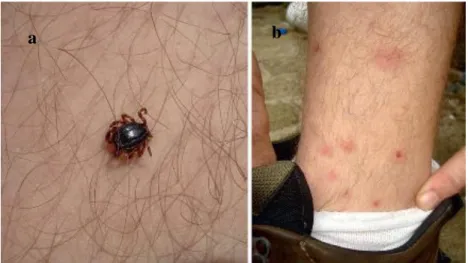
Fig. 13: Tick infection on humans: (a) Hyalomma marginatum male feeding on a man in southern Italy. (b).
Skin reactions 24 hours after being bitten [58]. .
R. sanguineus tick constitutes a possible risk to public health, because it can be responsible
for the transmission of a wide variety of pathogens to dogs and humans. The most troubling
23 canine illnesses transmitted by this tick are babesiosis, caused by Babesia canis and monocytic ehrlichiosis, caused by Ehrlichia canis. The most dangerous pathogens transmitted to humans are Rickettsia conorii, which is responsible for causing Mediterranean spotted fever and Ri. rickettsii, the etiologic agent of Rocky Mountain spotted fever. All of these diseases are associated with morbidity or even death if not treated properly [4, 8, 11, 58].
In severe cases, babesiosis is responsible for causing haemolytic anemia, hypotensive shock, intravascular coagulation, systemic inflammatory response and multiple organ dysfunctions, erythrocyte autoaglutamination and oxidative damage in red blood cells. The susceptibility to this disease varies with the host, its breed and its age, and also the auto-immne status of the dog. In severe cases, Erlichiosis causes immunological destruction of platelets, platelet dysfunction, ocular and central nervous system abnormalities and, in some cases, bone marrow destruction. Certain dogs breeds and younger dogs are more susceptible to this disease [4].
Rocky Mountain spotted fever and Mediterranean spotted fever are characterized for presenting one weak incubation period, frequently asymptomatic, followed by the arise of symptoms such like, headache, myalgia, arthralgia vomits, diarrhea, physical pain, and after one weak, skin spots spots with 0,5 to 2 cm of diameter appear, where the tick has bitten [59]. Severe cases are associated with very high fevers, lethargy, anorexia, icterus, hyperthermia, and trombocyptoria [60].
R. sanguineus species is also associated with the transmission of other pathogenic agents,
such as Anaplasma marginale, A. platys, B. cabalii, B. canis canis, B. canis vogeli, B. gibsoni,
Coxiella burnetii, Dipetalonema dracunculoides, Mycoplasma haemocanis, Rangelia, Salmonella spp, among others [11, 58, 61].
In addition to the entire pathogens previously mentioned, the filaroid Cercopithifilaria grassi is associated with dermal microfilariae, is also transmitted by R. sanguineus. This was demonstrated by the fact that this filaroid appears in every stage of the life cycle of this tick that also presents a high level of toleration to its infections [62].
There is also the suspicion that this parasite can transmit the bacteria Leishmania infantum, responsible for causing visceral leishmaniasis that can affect both dogs and children. This


![Fig. 7: Life Cycle of R. sanguineus: The complete life cycle, of a 3 host tick from egg to adult [37]](https://thumb-eu.123doks.com/thumbv2/123dok_br/15146249.1012379/30.892.286.606.100.392/fig-life-cycle-sanguineus-complete-life-cycle-adult.webp)
![Fig. 8: Oviposition of R. sanguineus: Several engorged females laying eggs, a key process in proliferation [35]](https://thumb-eu.123doks.com/thumbv2/123dok_br/15146249.1012379/32.892.200.695.106.337/fig-oviposition-sanguineus-engorged-females-laying-process-proliferation.webp)
![Fig. 10: Habitat of R. sanguineus: R. sanguineus engorged nymphs in a dog kennel, Ivory Coast, West Africa [11]](https://thumb-eu.123doks.com/thumbv2/123dok_br/15146249.1012379/34.892.259.637.102.388/habitat-sanguineus-sanguineus-engorged-nymphs-kennel-ivory-africa.webp)
![Fig. 11: Seasonality of R. sanguineus: R. sanguineus engorged females’ crawling in between rocks [35]](https://thumb-eu.123doks.com/thumbv2/123dok_br/15146249.1012379/37.892.198.695.106.402/fig-seasonality-sanguineus-sanguineus-engorged-females-crawling-rocks.webp)
![Fig. 12: Tick infection: A female adult dog highly parasitized by R. sanguineus [46]](https://thumb-eu.123doks.com/thumbv2/123dok_br/15146249.1012379/38.892.308.587.105.453/fig-tick-infection-female-adult-highly-parasitized-sanguineus.webp)