ANALYSIS OF THE INFLUENCE OF ANIMAL POSITIONING ON
THE RADIOGRAPHIC STUDY OF THE COXOFEMORAL JOINT
PHD THESIS IN VETERINARY SCIENCES
JOÃO MANUEL CARDOSO MARTINS
Advisor: Professor Doutor Mário Ginja
Co-Advisors: Professor Doutor António Ferreira
Professor Doutor Bruno Colaço
ANALYSIS OF THE INFLUENCE OF ANIMAL POSITIONING ON
THE RADIOGRAPHIC STUDY OF THE COXOFEMORAL JOINT
PHD THESIS IN VETERINARY SCIENCES
JOÃO MANUEL CARDOSO MARTINS
Advisor: Professor Doutor Mário Ginja Co-Advisors: Professor Doutor António Ferreira
Professor Doutor Bruno Colaço
Original thesis presented by João Manuel Cardoso Martins at the
University of Trás-os-Montes and Alto Douro, to obtain the doctor’s
degree on Veterinary Science.
This work is supported by: European Investment Funds by FEDER/COMPETE/POCI– Operacional Competitiveness and Internacionalization Programme, under Project POCI
01- 0145 FEDER-006958 and National Funds by FCT - Portuguese Foundation for Science and Technology, under the project UID/AGR/04033/2013
“Aquilo em que julgamos acreditar, na verdade não tem consequências. A
única coisa que tem consequências é aquilo que fazemos”
Desejo expressar a mais sincera gratidão:
À Universidade de Trás-os-Montes e Alto Douro (UTAD), na pessoa do seu Magnífico Reitor Professor Doutor António Fontainhas Fernandes.
Ao Professor Doutor Mário Manuel Dinis Ginja, orientador da presente tese, o meu sincero agradecimento pela simpatia, amizade, apoio e conhecimento transmitido, sem o qual não teria sido possível realizar a presente tese. Obrigado pela confiança, profissionalismo, sábia orientação, determinação e persistência ao longo deste projeto e por me ter deixado participar em mais um excelente trabalho de investigação.
Ao Professor Doutor António Ferreira, coorientador da presente tese, manifesto o meu sincero agradecimento pela amizade, simpatia, apoio, disponibilidade e conhecimento transmitido ao longo da realização desta tese.
Ao Professor Doutor Bruno Jorge Antunes Colaço, coorientador da presente tese, manifesto o meu sincero agradecimento pela amizade, simpatia, apoio, disponibilidade e conhecimento transmitido ao longo da realização desta tese. Agradeço também a disponibilidade do Laboratório de Anatomia para a conservação e preparação dos cadáveres usados neste trabalho.
À Doutora Maria Sofia Rodrigues Alves Pimenta pela simpatia, ajuda, amizade e contribuição e sugestões dadas ao longo da realização desta tese.
Ao Professor Doutor Jorge Colaço, pela simpatia e inestimável ajuda na análise estatística.
À direção do Hospital Veterinário da Universidade de Trás-os-Montes e Alto Douro, na pessoa da Professora Doutora Maria Isabel Ribeiro Dias, pela disponibilização de meios e infraestruturas para a realização de parte da componente prática do trabalho.
Ao Hospital Veterinário da Universidade de Trás-os-Montes e Alto Douro, à Faculdade de Medicina Veterinária de Lisboa, ao Hospital Veterinário Montenegro, à Clinica Veterinária Alcabideche vet por terem disponibilizado radiografias usadas no desenvolvimento desta tese.
À minha família, à minha mulher Carla, aos meus filhos Beatriz e Pedro pelo apoio, paciência e precioso tempo que me disponibilizaram e sem os quais não seria possível avançar com esta tese.
A displasia da anca (DA) é uma das doenças ortopédicas mais comuns na espécie canina. O controlo desta doença assenta essencialmente na prevenção. Apesar da investigação desenvolvida nas últimas décadas, os resultados obtidos na sua erradicação são ainda insatisfatórios, existindo a necessidade de aprofundar a investigação sobre os atuais e novos métodos de diagnóstico. Na origem da dificuldade em controlar e erradicar a DA está a sua natureza hereditária e poligénica extremamente complexa que impossibilita que na atualidade e num futuro próximo se desenvolvam técnicas eficazes de despiste genético. Na atualidade o despiste e classificação da DA praticado na espécie canina pelas principais organizações mundiais (OFA, FCI, BVA/KC) assentam os seus métodos na análise e classificação de radiografias obtidas na projeção ventrodorsal convencional. As técnicas baseadas na lassidão articular e as novas técnicas de imagem como a tomografia computorizada (TC) e a ressonância magnética (RM), não são ainda usadas de forma rotineira quer devido à necessidade de maior investigação para a sua validação, quer no caso da TC e RM também devido aos seus elevados custos. Apesar das críticas apontadas à projeção ventrodorsal convencional, esta continua a ser a técnica mais utilizada na atualidade para despiste de DA em grande escala. Um dos fatores que é apontado como responsável pela falta de eficácia deste método para reduzir a incidência da DA na espécie canina é o mau posicionamento radiográfico, nomeadamente a rotação pélvica ao longo do eixo corporal longitudinal e a rotação femoral. Baseado nesta problemática estabeleceram-se como objetivos desta tese: criar e validar parâmetros que possam ser usados na identificação de rotação pélvica e femoral na projeção ventrodorsal convencional; analisar de forma objetiva qual o efeito da rotação pélvica e femoral sobre alguns parâmetros usados pelas principais organizações internacionais, nomeadamente o ângulo de Norberg (AN). No desenvolvimento deste trabalho, numa primeira fase foram utilizados cadáveres de raças médias e grandes (> 20Kg), sem atender a critérios de raça ou sexo. Foi criado um suporte especial para estabilização e rotação do terço posterior destes animais. A rotação pélvica ao longo do eixo corporal longitudinal ficou compreendida entre 0 e 6 graus. Para o estudo da rotação femoral, foram obtidas projeções radiográficas com rotação interna e externa tendo esta ficado compreendida entre 6 a 32 graus e 7 a 32 graus, respetivamente.
Os resultados obtidos mostram que os parâmetros diâmetro horizontal do íleo (DHI), largura do forâmen obturador (LFO) são critérios uteis para a identificação de rotação pélvica e o parâmetro índice de deslocamento patelar (IDP) para identificação de rotação femoral. A medição do AN e a distância cabeça femoral- acetábulo (DFA) não são influenciados por reduzidos valores de rotação pélvica. No entanto, a medição dos parâmetros índice de subluxação da cabeça femoral (IS), categorias de subluxação da cabeça femoral (CS) e espessura pélvica (EP) são afetados por baixos valores de rotação pélvica, sendo sobrevalorizados no lado ipsilateral à rotação e subvalorizados no lado contralateral à rotação. A rotação femoral interna beneficia os parâmetros AN, IS e CS ao aumentar a congruência articular entre a cabeça femoral e o acetábulo, por outro lado a rotação femoral externa afeta negativamente o AN, IS e CS ao reduzir a congruência articular entre a cabeça femoral e o acetábulo.
Numa segunda fase deste trabalho foi usada uma base de dados de 68 animais vivos de raças médias e grandes (> 20Kg), radiografados para despiste e classificação de displasia da anca de acordo com as normas da FCI, foram incluídos neste trabalho animais que apresentavam uma radiografia com correto posicionamento e pelo menos uma radiografia com rotação pélvica ou rotação femoral.
A deteção e quantificação da rotação pélvica e femoral foram efetuadas através dos parâmetros DHI e IDP. Secundariamente avaliou-se o efeito da rotação sobre os parâmetros AN, IS e CS.
Este trabalho permitiu a criação e validação dos parâmetros DHI e IDP, que podem ser uteis quando utilizados como forma de controlo e padronização do posicionamento radiográfico na projeção ventrodorsal convencional. Por outro lado, foi possível esclarecer a influência da rotação pélvica e femoral sobre a medição e obtenção de importantes parâmetros utilizados pelas principais organizações mundiais. De acordo com conhecimento dos autores, este é o primeiro trabalho realizado sobre critérios de identificação objetiva e efeitos de rotação pélvica ao longo do eixo corporal longitudinal e rotação femoral na projeção ventrodorsal convencional.
Palavras-chave: Displasia da anca canina, projeção ventrodorsal convencional, rotação
Hip dysplasia (HD) is one of the most common orthopedic diseases in the canine species. This disease control is essentially based on prevention. Despite the research carried out in the last decades, the results obtained in its eradication are still unsatisfactory, and there is a need to deepen research on current and new diagnostic methods. In the source of the difficulty to control and eradicate HD is its extremely complex hereditary and polygenic nature, which probably makes impossible the development of effective genetic screening techniques in the near future. Actually HD screening and classification made in the canine species by the main world organizations (OFA, FCI, BVA / KC) are based on the analysis and classification of radiographs obtained in the standard ventrodorsal projection. Techniques based on joint laxity and new imaging techniques such as computed tomography (CT) and magnetic resonance imaging (MRI) are not routinely used either because of the need for further investigation for their validation, or in the case of CT and MRI also due to their high costs. Despite the critics to the standard ventrodorsal projection, this remains the most widely used technique for large scale HD screening. One of the factors responsible for the lack of effectiveness of this method to reduce the incidence of HD in the canine species is the poor radiographic positioning, namely pelvic rotation along the longitudinal axis of the body and femoral rotation. Based on this problem, the objectives of this thesis were: to create and validate parameters that can be used in the identification of pelvic and femoral rotation in the standard ventrodorsal projection; to analyze objectively the effects of pelvic and femoral rotation on some parameters used by the main international organizations, namely Norberg angle (NA).
In the development of this work, cadavers of medium and large breeds (> 20 kg) were used. There was no gender or breed criteria. A special support was created to stabilize and rotate the pelvis and hind limbs of these animals. The pelvic rotation along the longitudinal body axis ranged from 0 to 6 degrees. For the study of femoral rotation, radiographic projections were obtained with internal and external rotation, the femoral rotation ranged between 6 to 32 degrees and 7 to 32 degrees respectively.
The results show that iliac horizontal diameter (IHD) and maximum obturator foramen width (OFW) are useful for pelvic rotation identification and the patella displacement index (PDI) to identify femoral rotation. The NA and the femoral head-acetabular
distance (FAD) parameters measurement are not affected by slight pelvic rotations.
However the pelvic thickness at the level of the cranial effective acetabular rim (PT),
femoral head subluxation index (SI) and the femoral head subluxation categories (SC)
are affected, being overvalued on the ipsilateral side of rotation and undervalued on the contralateral side of rotation. The internal femoral rotation benefits the NA, SI and SC parameters by increasing joint congruence between the femoral head and the acetabulum. On the other hand, external femoral rotation affects negatively NA, SI, reducing joint congruence between the femoral head and the acetabulum.
In a second phase of this work we used a live animal database of 68 dogs from medium and large breeds (> 20 kg), radiographed for HD screening and classification purposes
according to FCI recommendations. Each animal selected should have at least a normal
radiography and at least another with pelvic or femoral rotation. Detection and quantification of pelvic and femoral rotation were performed using the IHD and PDI parameters. Secondarily the rotation effect on the AN, IS and CS parameters was evaluated.
This work allowed the creation and validation of the IHD and PDI parameters, which may be useful when used for radiographic positioning control and standardization in the standard ventrodorsal projection. On the other hand, it was possible to clarify the effects of pelvic and femoral rotation on important parameters measurement used by the main world organizations. According to the authors' knowledge, this is the first work performed on criteria for objective identification and effects of pelvic rotation along the longitudinal body axis and femoral rotation in the standard ventrodorsal projection.
Keywords: Canine hip dysplasia, standard ventrodorsal projection, pelvic rotation,
ACKNOWLEDGEMENTS xi
RESUMO xiii
ABSTRACT xv
LIST OF FIGURES xxiii
LIST OF TABLES xxvii
LIST OF ABREVIATURES AND SYMBOLS xxxi
CHAPTER 1
1. GENERAL INTRODUCTION 1
1.1 Canine hip dysplasia 3
1.1.1 Hip anatomy 5
1.1.2 Diagnosis and screening 7
1.1.3 Treatment 14
1.1.4 Current challenges in hip dysplasia diagnosis and control 16
1.2 References 18
CHAPTER 2
2. AIMS 29
CHAPTER 3
3. ANALYSIS OF PELVIC ROTATION ON THE STANDARD HIP VENTRODORSAL EXTENDED RADIOGRAPHIC VIEW
33
3.1 Introduction 37
3.2 Material and methods 38 3.2.1 Pelvic radiography 38 3.2.2 Radiographic measurements 40 3.2.3 Statistical analysis 42 3.3 Results 42 3.4 Discussion 49 3.5 Conclusion 52 3.6 References 52
CHAPTER 4
4. EFFECTS OF PELVIS ROTATION ON PROJECTED RADIOGRAPHIC POSITION OF FEMORAL HEAD IN RELATIONSHIP TO ACETABULUM
55
4.1 Introduction 59
4.2 Materials and methods 61
4.3 Results 66
4.4 Discussion 71
4.5 Conclusions 74
4.6 References 75
CHAPTER 5
5. FEMORAL ROTATION AND RELATIONSHIP BETWEEN THE FEMORAL HEAD AND ACETABULUM
79
5.1 Introduction 83 5.2 Materials and methods 84
5.3 Results 90
5.4 Discussion 93
5.5 Conclusions 95
5.6 References 95
CHAPTER 6
6. EVALUATION AND DETECTION OF PELVIC AND FEMORAL MALPOSITIONING IN A LIVE ANIMAL DATABASE
99
6.1 Introduction 103
6.2 Materials and methods 104
6.3 Results 105 6.4 Discussion 108 6.5 Conclusions 111 6.6 References CHAPTER 7 111 7. GENERAL DISCUSSION 115
CHAPTER 8
Figure 1.1 Normal hip joint; 1. Ilion; 2. Round ligament; 3. Femoral head; 4.
Joint capsule; 5. Greater trochanter; 6. Femoral diaphysis.
6
Figure 1.2 Standard ventrodorsal hip extended radiographic view.
12
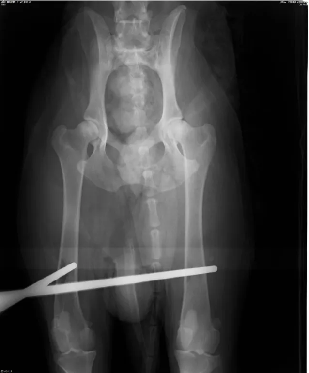
Figure 3.1 Canine cadaver positioned in the special device in dorsal recumbency.
Bilateral pins (b) applied to the femoral diaphysis fixed in a transverse strip of wood (a) for correct positioning of the femurs.
39
Figure 3.2 Ventrodorsal hip extended view with measured parameters: iliac
horizontal diameter (IHD), maximum obturator foramen width (OFW), ischiatic femoral overlap (IFO), pelvic horizontal radius (PHR), femoral head diameter (FHD) and obturator foramen area (ObFA).
41
Figure 3.3 Animal with 6º rotation to right side. 44
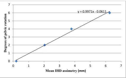
Figure 3.4 Linear regression analysis for calculating pelvic rotation in degrees
based on iliac horizontal diameter (IHD) asymmetry.
48
Figure 3.5 Linear regression analysis for calculating pelvic rotation in degrees
based on obturator foramen width (OFW) asymmetry.
48
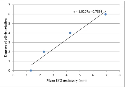
Figure 3.6 Linear regression analysis for calculating pelvic rotation in degrees
based on ischiatic femoral overlap (IFO) asymmetry.
49
Figure 4.1 Illustration outlining the rear views of the special holder device. (A)
Cadaver positioned in the ventrodorsal hip extended view. (B) Modified view with pelvic rotation of 6º to the right side (elevating the left side), making the right hip dependent and the left non-dependent.
61
side. The Norberg angle (NA) was determined as the angle formed by a line joining the centre of the femoral heads and a line joining the centre of the femoral head and the craniolateral aspect of the ipsilateral acetabular rim.
Figure 4.3 Ventrodorsal hip extended view with pelvic rotation of 6º to the right
side. Pelvic thickness (PT) is obtained as the thickness of the pelvis at the level of the cranial effective acetabular rim on dependent (PTd) and on non-dependent (PTnd) side. The femoral head subluxation category was attributed by attending to the position of the femoral head centre (fhc) relative to the dorsal acetabular edge (dae) and taking into account the congruency of the fit between the femoral head and the cranial acetabular edge. Femoral head-acetabular distance (FAD) is determined by drawing a line between both cranial effective acetabular rims (a), and then a second perpendicular line drawn from the lateral aspect of the cranial effective acetabular rim (b). FAD was measured as the minimum horizontal distance between the second perpendicular line and the centre of the femoral head on dependent (FADd) and on non-dependent (FADnd) side.
64
Figure 4.4 Ventrodorsal hip extended view with pelvic rotation of 6º to the right
side. The femoral head subluxation index (SI) was obtained drawing a line (a) between the centre of the femoral head (fhc) and the acetabular centre (ac) and dividing by the radius (r) of the femoral head on dependent (SId) an on non-dependent (SInd) side.
65
Figure 5.1 Modified ventrodorsal hip extended view with internal femoral
rotation (right side 16° and left side 10°).
86
Figure 5.2 Illustration outlining the rear view of the special fixation device. (A)
Cadaver positioned in the normal ventrodorsal hip extended view. (B) Modified view with femoral internal rotation. (C) Modified view with femoral external rotation. (D) Worksheet with the tilt o each pin in normal, internal and external rotation femoral radiographic views.
87
16° resulted in a patella displacement index of 0.27 (0.59 medial index in normal view minus 0.32 medial index in internal rotation view). (C) External femoral rotation of 13° resulted in a patella displacement index of 0.20 (0.41 lateral index in normal view minus 0.21 lateral index in external rotation view). Line drawn from the base of the patella to the apex of the patella (a); horizontal distance between line-a and the lateral femoral cortical (b); horizontal distance between line-a and the medial femoral cortical (c).
Figure 5.4 Scatterplot of lateral (black circles) and medial (grey squares) patella
displacement index and regression line versus femoral rotation in degrees.
91
Figure 5.5 Scatterplot of lateral (black circles) and medial (grey squares) patella
displacement index and regression line versus: (A) Norberg angle variation in degrees; (B) Subluxation index variation; (C) Subluxation category variation
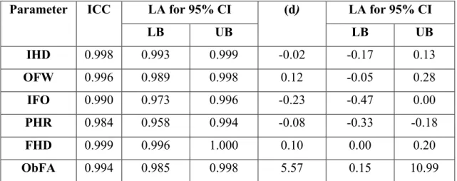
Table 3.1 Evaluation of repeatability for the six parameters using the interclass
correlation coefficient (ICC) and limits of agreement (LA) for the 95% confidence interval (95% CI) for differences in the two measurements.
43
Table 3.2 Evaluation of symmetry for the six parameters using the interclass
correlation coefficient (ICC) and limits of agreement (LA) for the 95% confidence interval (95% CI) for differences in right and left side measurements.
43
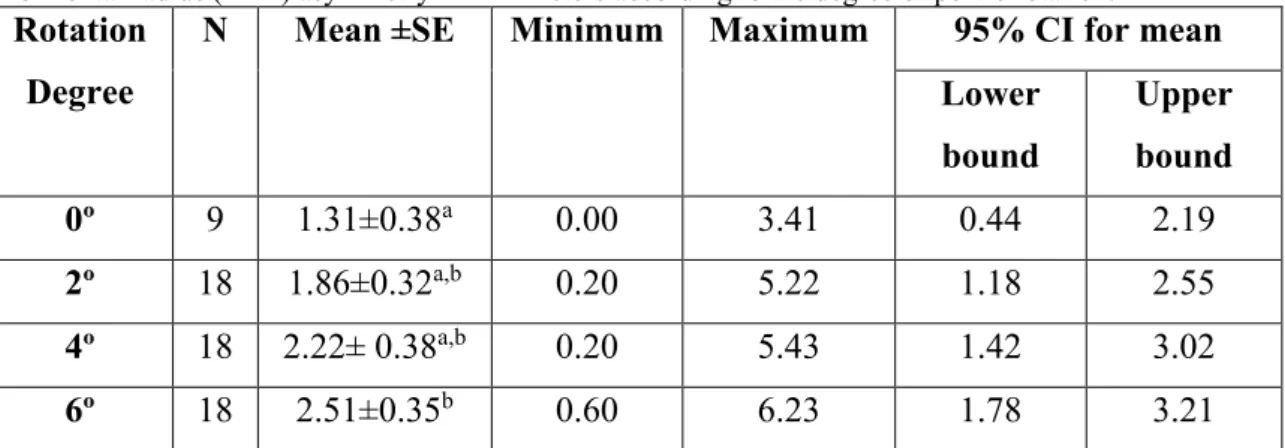
Table 3.3 Descriptive and limits of agreement for 95% confidence interval (95%
CI) for the mean iliac horizontal diameter (IHD) asymmetry in millimeters according to the degree of pelvic rotation.
45
Table 3.4 Descriptive and limits of agreement for 95% confidence interval (95%
CI) for the mean obturator foramen width (OFW) asymmetry in millimeters according to the degree of pelvic rotation.
45
Table 3.5 Descriptive and limits of agreement for 95% confidence interval (95%
CI) for the mean ischiatic femoral overlap (IFO) asymmetry in millimeters according to the degree of pelvic rotation.
45
Table 3.6 Descriptive and limits of agreement for 95% confidence interval (95%
CI) for the mean pelvic horizontal radius (PHR) asymmetry in millimeters according to the degree of pelvic rotation.
46
Table 3.7 Descriptive and limits of agreement for 95% confidence interval (95%
CI) for the mean femoral head diameter (FHD) asymmetry in millimeters according to the degree of pelvic rotation.
46
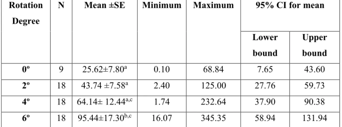
Table 3.8 Descriptive and limits of agreement for 95% confidence interval (95%
CI) for the mean obturator foramen area (ObFA) asymmetry in square millimeters according to the degree of pelvic rotation.
Table 3.9 Pearson correlations for the mean six parameters asymmetry and the
rotation degrees of 0º to 6º.
47
Table 4.1 Evaluation of repeatability for the 4 parameters using the interclass
correlation coefficient and limits of agreement for 95% CI of the mean of the differences in the two measurements.
66
Table 4.2 Descriptive statistical data for the normal Norberg angle in degrees (º),
according to the degree of pelvic rotation on the dependent and non-dependent side.
67
Table 4.3 Descriptive statistical data for normal pelvic thickness in millimetres, according to the degree of pelvic rotation on the dependent and non-dependent side.
68
Table 4.4 Descriptive statistical data for the normal femoral head subluxation
index according to the degree of pelvic rotation on the dependent and non-dependent side.
69
Table 4.5 Descriptive statistical data for the normal femoral head subluxation
category after the direct conversion into a numerical scale with the same value, according to the degree of pelvic rotation on the dependent and non-dependent side.
70
Table 4.6 Descriptive statistical data for the normal femoral head-acetabular
distance in millimetres according to the degree of pelvic rotation on the dependent and non-dependent side.
71
Table 6.1 Internal femoral rotation in degrees (º). 105
Table 6.2 Internal femoral rotation in degrees (º). 106
Table 6.4 Statistical data of the internal femoral rotation. 107
Table 6.5 Statistical data of the external femoral rotation. 107
ANOVA - Variance analysis
BVA/KC - British Veterinary Association / Kennel Club
CAR - Cranial effective acetabular rim
CI - Confidence interval
cm - centimeters
CT -Computed tomography
DAE -Dorsal acetabular edge
DI - Distraction Index
DLS - Dorsolateral Subluxation Score
FAD - Femoral head-acetabular distance
FCI - Fédération Cynologique Internationale
FHC - Femoral head centre
FHD - Femoral head diameter
HD - Hip dysplasia
ICC - Interclass correlation coefficient
IFO - Isquiatic femoral overlap
IHD - Iliac horizontal diameter
JM -João Martins
Mm -millimeters
MMDG -Mário Manuel Dinis Ginja
NA - Norberg angle
OFA - Orthopedic Foundation for Animals
ObFA - Obturator foramen area
OFW - Maximum obturator foramen width
PDI - Patella displacement index
PennHIP - Pennsylvania Hip Improvement Program
PHR - Pelvic horizontal radius
PT - Pelvic thickness at level the cranial effective acetabular rim
SC - Femoral head subluxation categories
SI - Femoral head subluxation index
CHAPTER 1:
GENERAL INTRODUCTION
1. GENERAL INTRODUCTION
1.1 CANINE HIP DYSPLASIA
The original hip joint that is still present in wolfs and in a few canine breeds like the greyhounds, is a tight and well-fitting joint that permits full athletic function throughout the animal’s life without the early development of degenerative joint disease (Dennis, 2012). The unfavorable evolution that hip joint suffered seems to be related to the elimination of natural selection and the introduction of domestication (Dennis, 2012). The domestication process and selection developed in the last decades, introduced the phenotypic selection for other traits, which perpetuated imperfect hips with hip dysplasia (HD) (Galibert et al., 2011; Dennis, 2012; Comhaire, 2014; Weiner et al., 2017).
HD is one of the most common orthopedic diseases in the canine species and can be a common reason for euthanasia (Bonnet et al., 2005; Comhaire and Schoonjans, 2011; Smith et al., 2012). It is characterized by an abnormal development of the hip joint with subluxation of the femoral heads and joint laxity leading to the development of osteoarthritis (Lust et al., 2001; Thrall, 2002; Paster et al., 2005; Macias et al., 2006; Corr, 2007; Simeonova, 2007; Thompson et al., 2007; Wigger et al., 2008; Ciarlini et al., 2009; Schachner and Lopez, 2010; Anderson, 2011; Broeckx et al., 2014; Kimeli et al., 2015). HD in humans presents phenotypic similarity with HD in dogs, with both presenting joint subluxation and development of osteoarthritis (Weinstein, 1987; Jacobsen et al., 2004; Reijman et al., 2005; Zhou et al., 2010; van Bosse et al., 2011; Ginja et al., 2015). It has been demonstrated a direct correlation between passive laxity of the hip joint and the development of osteoarthritis (Fries and Remedios, 1995; Remedios and Fries, 1995; Thrall, 2002; Macias et al., 2006; Flükiger, 2007; Ginja et al., 2009; 2010; Doskarova et al., 2010). The original cause of HD remains unknown, but the main risk factor for HD is considered to be joint laxity (Belkoff et al., 1989; Smith et al., 1995; 2001; Flükiger et al., 1999; Thodhunter et al., 2003; Corr, 2007; Ginja et al., 2008) Although hip laxity appears to be necessary for the development of
HD by causing hip instability and leading to femoral head dislocation (Henricson et al.,
1966; Brass, 1989; Smith et al., 1995; Kate et al., 2006; Gatineau et al., 2012), it seems like it is not sufficient because with similar levels of laxity, canine breeds have different susceptibilities in developing degenerative joint disease (Ginja et al., 2005).
HD may go unnoticed or lead to chronic pain and functional limitation, and is a challenging disease to diagnose and treat effectively (Remedios and Fries, 1995; Ginja
et al., 2005). HD occurs mainly in large and giant breeds but it may also affect small breeds and cats (Alexander, 1992; Plante et al., 1997; Altunatmaz et al., 2003; Anderson, 2011). This disease usually has a bilateral presentation, although unilateral presentation may also occur (Kealy et al., 2000). The incidence in both males and females seems to be the same (Fries and Remedios, 1995; Zorko et al., 2005; Macias et al., 2006; Ginja et al., 2009; Krontveit et al., 2010). HD is not present at birth, being a
disease of animal development contrary to what occurs in humans (Fries and Remedios,
1995; Thrall, 2002; Ginja et al., 2005; Macias et al., 2006; Vieira et al., 2010;Guilliard, 2014).
HD is a complex hereditary polygenic and multifactorial disorder, the phenotypic expression is influenced by genetic and non-genetic factors, being caused by the small additive effect of hundreds of genes, at least one pair of these genes are thought to be recessive (Fries and Remedios, 1995; Swenson et al., 1997; Thrall, 2002; Chase et al., 2005; Ginja et al., 2005; 2009; Zorko et al., 2005; Macias et al., 2006; Vieira et al.,
2010; Kristin and Lewis, 2011; Chalmers et al., 2013). HD heritability ranges between
0.1 to 0.6 (Leighton, 1997; Ginja et al., 2015). The incidence rate of HD vary between breeds and in some breeds can affects more than 50% of the population (Remedios and Fries, 1995; Hara et al., 2002; Ginja et al., 2009; Risler et al., 2009; Dennis, 2012; Smith et al., 2012). Despite the several screening and control programs implemented in some countries for over 50 years we still continue having high incidences due to its complex genetic origin and dogs with normal phenotype can be carriers of some mutations and transmit these genes to their offspring (Zorko et al., 2005; Ginja et al.,
2015; Miranda et al., 2016). Actually, there is no definitive molecular diagnostic test,
being the animals genotype estimated by evaluating the hip phenotype (Ginja et al.,
2015). As a hereditary disease, control involves the selection of individuals
genotypically free from susceptibility to the development of the disease, and their use in selective crosses to maximize unaltered genes in the population (Ginja et al., 2005).
Among the non-genetic or environmental factors that contribute to the phenotypic expression of HD, we found body size, growth rate, nutrition, exercise and muscle mass (Smith et al., 1990; 2006; Alexander, 1992; Plante et al., 1997; Altunatmaz et al., 2003; Corr, 2007; Schachner and Lopez, 2010; Zhou et al., 2010; Gaspar et al., 2016).
1.1.1 HIP ANATOMY
The functional evaluation of the hind limb shows that in normal quadrupedal animals, hind limbs are primarily responsible for the propulsion and support of approximately 30 to 40% of body weight and during locomotion the hip joints are subjected to forces of approximately three times body weight (Prieur, 1980; Weigel and Wasserman, 1992; Breit et al., 2002). The hip joint movement and support are dependent of the anatomic relations between the bone components and the integrity of muscles, tendons and ligaments (Weigel and Wasserman, 1992). The proximal femur and acetabulum create a functional unity that depend each other to allow a synchronized growth, resulting in a normal hip development with adequate transmission force between the articular surfaces (Weigel and Wasserman, 1992; Wenger and Bomar, 2003; Martins et al., 2012). The most critical period in terms of hip joint development are the first 60 days of live because the stress loading can interfere with the acetabular cavity depth and the femoral head and neck conformation (Riser, 1975; Wenger and Bomar, 2003; Ginja et
al., 2015).Any change in the normal development of the proximal femur may result in biomechanical changes, with direct interference in the proper transmission of loads between the cartilaginous surfaces, resulting in joint overload with early destruction of chondrocytes and development of osteoarthritis, as occurs in HD (Martins et al., 2012).
Kinetic studies performed on dogs with unilateral osteoarthritis demonstrate that when the dog is in motion, vertical forces redistribute alternating force between pelvic limbs rather than switching between pelvic limbs to a thoracic limb. The activity patterns of the hip muscles are also different in dogs with HD and osteoarthritis (Bockstahler et al., 2012).
The hip joint (Figure 1.1) is a spheroidal joint of the diarthrosis type composed by the acetabulum, femoral head, regional muscles and ligaments (Barone, 1980; Ficus et al., 1991; Laborda, 1995; Done et al., 1997; Dyce et al., 1997). The acetabulum is a cotiloid cavity, with the crescent-shaped articular surface formed by the union of the ilium, ischium and pubis (Done et al., 1997; Dyce et al., 1997).The femur is a long bone, with a well-defined neck proximally and a regular cylindrical body. In normal animals, the femoral head and neck are inclined relative to the femoral diaphysis at an angle of 130 to 145° and have an anteversion of 12 to 40° (Macias et al., 2006). The
femoral head has a shape of a nearly perfect hemisphere, interrupted by a small central fovea, where the round ligament is inserted (Laborda, 1995; Dyce et al., 1997). In a normal hip the femoral head lies deeply and firmly in the acetabulum (Ficus et al., 1991).
Figure 1.1 Normal hip joint; 1. Ilion; 2. Round ligament; 3. Femoral head; 4. Joint capsule; 5.
Greater trochanter; 6. Femoral diaphysis (Adapted: Ficus et al., 1991).
The acetabular cavity surface is coated by the synovial membrane which is supported externally by a fibrous capsule (Macias et al., 2006). Together, the round ligament and the joint capsule make the primary stabilization of the joint (Macias et al., 2006). Secondary stabilizers include the acetabular labrum, gluteal muscles, adductor and abductor muscles of the hip (Macias et al., 2006) and the negative pressure exerted by the synovial fluid in the joint (Farese et al., 1999). The hip joint does not have peripheral ligaments that limit its movement (Dyce et al., 2001). The articular capsule, the orbicularis ring and the ligaments: iliofemoral (reinforces the cranial part of the capsule), femoral ischium (strengthens the caudal part of the capsule) and round (ligament between the femoral head and the acetabulum) form the capsular and ligamentous structures of this joint (Dyce et al., 1997).
Regional muscles are responsible for the support, stability and locomotion of the dog, being therefore very important for its total development (Riegger-Krugh et al., 2004). The main movements that the hip joint presents are extension and flexion (Evans and de Lahunta, 2004), but can also perform other movements like abduction, adduction, internal and external rotation (Laborda, 1995).
The femoral head and neck blood supply comes potentially from two sources: the first is the intraosseous supply or metaphyseal system that goes to the femoral neck, the second is the epiphyseal system that enters through the joint capsule via cranial and caudal circumflex arteries (Macias et al., 2006).
The acetabular region enervation in humans and animals shows bilateral symmetry. However, the difference between the biped support in humans and quadruped in the canine species generates biomechanical forces in distinct points, promoting different concentration of nerve fibers in the two species. While the first has a greater density of nerve fibers in the antero-medial region, the second one present’s greater density in the craniolateral and dorsal region (Schmaedecke and Ferrigno, 2008).
1.1.2 DIAGNOSIS AND SCREENING
The HD diagnosis is not always easy to perform, and there is no method that can identify all cases. In general diagnosis is based on anamnesis, clinical observation and physical examination, complemented with radiographic examination (Smith, 1997;
Ginja et al., 2005). Clinical signs are highly variable and many dogs may be
asymptomatic or only show mild clinical signs (Anderson, 2011), making that the correlation between radiographic joint changes and HD clinical presentation to be poor (Barr et al., 1987; Heilm-Bjorkman et al., 2003; Schachner and Lopez, 2015). Despite the significant variability in HD progression, in general clinical signs have a bimodal age distribution which includes animals under one year old and adults (Ginja et al., 2005; Corr, 2007; Schachner and Lopez, 2015).
In dogs less than 12 months of age, pain is related to hip laxity and clinical signs are triggered by joint changes such as instability of the hip joint, stretching of the capsule and round ligament, secondary synovitis, acetabular microfractures, and supply disturbances of the proximal femur (Corr, 2007). Most of these dogs have pain when manipulating the hip joint (rotation, extension, flexion) and have positive results in the three main tests for assessing laxity: Ortolani, Bardens and Barlow tests (Chalman and
Butler, 1985; Adams et al., 1998; Puerto et al., 1999; Thrall, 2002; Macias et al., 2006;
Ginja et al., 2010; Gatineau et al., 2012).
In adult dogs, clinical signs are a result of hip joint osteoarthritis, being the pain caused
by subchondral bone exposure and joint inflammation (Macias et al., 2006; Corr, 2007;
Ginja et al., 2005). These animals have weakness of the pelvic limbs with difficulty in
getting up and jumping. In these animals the positive Ortolani signal is rare (Macias et
al., 2006; Ginja et al., 2010).
In the course of HD there is always a variable period of time until radiographic degenerative changes occur. As soon as they are present they generally progress with the aging of the animal. Radiographic changes become more evident in dogs older than
five months old (Macias et al., 2006; D`Amico et al., 2011). In dogs severely affected
with HD, radiographic changes at the hip joint can be detected as early as 30 to 60 days of age (Macias et al., 2006).
Screening programs for HD have traditionally involved radiographic assessment of the hip joints of young adult dogs, making radiography the gold standard to assess and quantify changes associated to HD (Szabo et al., 2007; Chalmers et al., 2013; Ginja et
al., 2015; Schachner and Lopez, 2015).
Currently the radiographic diagnosis of HD is based on two main key features, the detection of signs of degenerative joint disease and the detection of hip joint laxity (Ginja et al., 2005; Smith et al., 2012; Schachner and Lopez, 2015). The primary goal for each screening program is to exclude genetically affected individuals from the breeding pool (Verhoeven et al., 2012).
Various schemes for scoring severity of radiographic changes associated with HD have been used. The Fédération Cynologique Internationale (FCI), the British Veterinary Association / Kennel Club (BVA / KC), the Orthopedic Foundation for Animals (OFA), the Pennsylvania Hip Improvement Program (PennHIP) and the Dorsolateral Subluxation Score (DLS) are the most widespread and investigated screening approaches for canine HD (Smith et al., 1990; Corley, 1992; Farese et al., 1999; Lust et
al., 2001; Comhaire and Schoonjans, 2011; Gatineau et al., 2012; Verhoeven et al.,
2012; Schachner and Lopez, 2015).
To evaluate the hip joint through radiography, the standard method introduced in the 1960s, uses the ventrodorsal hip extended view (VDHE) (Whittington et al., 1961; Leighton, 1997; Swenson et al., 1997).
Depending on the classification system used, the radiographs in VDHE are made between 12 and 24 months of age (Genevois et al., 2008; Ginja et al., 2008). The reliability of the standard radiographic method varies from 85% to 95% at 24 months (Ginja et al., 2005). This view is used by the three main international HD scoring organizations (FCI, BVA / KC and OFA). Despite these main organizations share common goals and make their classifications based on the VDHE view interpretation, each organization has its own specific methods and grading schemes (Genevois et al., 2007, 2008; Comhaire et al., 2009; Skurková et al., 2010; Chalmers et al., 2013).
The FCI was founded in 1911 and her scheme is currently being applied by 84 members, most of them from Europe, Russia, South America and Asia (Skurková et al., 2010; Verhoeven et al., 2012). Dogs should be at least 12 months old, except in the giant breed which is 18 months (Verhoeven et al., 2012; Schachner and Lopez, 2015). Radiographs are evaluated and classified by a specialized veterinarian approved by the national club in which the dog is registered (Verhoeven et al., 2012). The classification system combines the subjective evaluation with the measurement of the Norberg angle (NA) in VDHE view radiographs (Verhoeven et al., 2012). The FCI classifies the animals into five different grades (A to E), in order to classify the severity of the disease. Animals classified as A and B are considered non-dysplastic and can be used in breeding programs. Grades C, D and E are considered dysplastic (Verhoeven et al.,
2007; 2012; Skurková et al., 2010).
The BVA / KC scheme is used since 1965 in the United Kingdom, Ireland, Australia and New Zealand (Flückiger et al., 2007; Verhoeven et al., 2012). It uses the VDHE view in animals with a minimum age of 1 year (Dennis, 2012; Schachner and Lopez, 2015). The BVA/KC system scores hip joints based on the severity of the changes of nine specific radiographic criteria (Dennis, 2012). These are the presence of subluxation (2 criteria), the shape and depth of the acetabulum (5 criteria) and also the shape and signs of degenerative joint disease of the femoral head and neck (2 criteria). Each criterion is rated from 0 (ideal) to 6 (worst). The final classification of the hip is given as the sum between 0 and 53 for each joint and as the sum of both hips from 0 to 106 (Macias et al., 2006; Flückiger et al., 2007; Ginja et al., 2010).
OFA scheme is applied exclusively in the USA and Canada since 1966 (Rendano and Ryan, 1985; Henry, 1992; Chalmers et al., 2013). It is also based on the VDHE view, but dogs must be at least 24 months old (Skurková et al., 2010; Verhoeven et al., 2012).
The phenotypic assessment of the hips divides the dogs into two groups of three degrees: the first group of dogs with normal hip divides into excellent, good and reasonable, the second group of dogs with dysplastic hip is divided into mild, moderate and severe. Hip joints that cannot be inserted in the previous groups are classified as undetermined type, and reassessment is recommended after six months (Henry, 1992; Essman and Sherman, 2006; Macias et al., 2006; Flückiger et al., 2007; Ginja et al., 2010; Verhoeven et al., 2012; Schachner and Lopez, 2015).
It has been pointed out that VDHE view does not demonstrate coxofemoral laxity reliably because of the nonphysiological tensioning of the pelvic muscles and twisting of the joint capsule (Smith et al., 1990). Based on the important role that joint laxity plays in the development of degenerative joint disease in dogs with HD, several alternative methods of radiographic diagnosis have been developed (Kealy et al., 2000; Smith et al., 2001; Todhunter et al., 2003; Ginja et al., 2006; 2010; Macias et al., 2006).
The PennHIP method, developed in 1983 at the University of Pennsylvania was
introduced commercially in 1994 and seems to be the most advantageous (Smith et al.,
1990; Ginja et al., 2005; Macias et al., 2006; Verhoeven et al., 2012; Schachner and Lopez, 2015). The method goal is to detect HD early (approximately 16 weeks of age) so that preventive treatment can be instituted as early as possible or select the animals to create a stock of breeding dogs (Smith et al., 1990; Gibbs, 1997; Ginja et al., 2005; Corley, 2006; Verhoeven et al., 2012; Miranda et al., 2016). The PennHIP method quantifies joint lassitude by measuring the Distraction Index (ID) (Kate et al., 2006; Guilliard, 2014). In this method, the animal is sedated or anesthetized and placed on the x-ray table in dorsal decubitus and three radiographs are performed (Ginja et al., 2005). Animals with normal hip joints present a joint laxity with an ID less than or equal to 0.45. Animals with dysplastic coxofemoral joints have a joint lassitude greater of 0.45 (Ginja et al., 2008). Another method based on joint lassitude is the dorsolateral subluxation method, patented in 1999 (Farese et al., 1998). This method is considered to achieve a more functional joint lassitude, while the PennHIP method results in a passive joint lassitude. This method involves the placing of the anesthetized animal in the ventral decubitus, imitating the physiological positioning of the hip joint. The degrees of dorsolateral subluxation do not change after eight months of age. Dogs> 55% are unlikely to develop HD later, but dogs with <45% are more likely to develop HD (Schachner and Lopez, 2015). This method is not used in large-scale screening
programs. The relationship between the degree of dorsolateral subluxation and the development of degenerative articular disease throughout the dog's life needs further investigation (Verhoeven et al., 2012). There are no reports regarding the evolution of prevalence of canine HD using the PennHIP and the DLS systems (Verhoeven et al., 2012).
To perform the VDHE view, animals should receive a deep sedation or general anesthesia to ensure adequate muscle relaxation and the radiograph must be identified in an immovable way and have adequate definition (Ginja et al., 2005; Flückiger et al., 2007). In this radiographic view the animal is placed in dorsal decubitus with hind limbs extended parallel, avoiding the pelvis rotation and the stifles internally rotated with the
patella being centered inside the trochlear groove (Figure 1.2) (Kealy et al., 2000; Dyce
Figure 1.2 Standard ventrodorsal hip extended radiographic view.
Relationships between the dorsal edge of the acetabulum and the femoral head are of
great importance when the integrity and normality of hip joint is evaluated (Dyce et al.,
2001; Macias et al., 2006). The first recognized change is joint laxity, with loss of congruence between the joint surfaces of the acetabulum and the femoral head (Fries
and Remedios, 1995; Thrall, 2002; Macias et al., 2006; Ginja et al., 2009, 2010).
In the VDHE view analysis it should be observed the presence of joint laxity (percentage of femoral head covered with the limits of the dorsal acetabular edge), the
degree of subluxation, increase of medial joint space, NA and signs of osteoarthritis. In osteoarthritis there are changes such as modification in the dorsal acetabular edge shape, bone neoformation in the acetabular fossa, cranial and caudal acetabular borders,
femoral head and neck, and femoral head and neck remodeling (Macias et al., 2006).
Normal hip joints have a deep acetabulum, a round, uniform femoral head and the femoral head fits perfectly within the acetabulum, and at least half of the femoral head should be on the medial side of the dorsal acetabular margin. The cranial third of the joint space is uniform and regular and does not have an increase in width, the femoral
neck is smooth and thin and there is no evidence of joint changes (Figure 1.2) (Kealy et
al., 2000).
In dogs with HD, the radiographic changes include alterations such as acetabular flattening, the femoral head does not fit adequately in the acetabulum, and there may be subluxation or even dislocation of the femoral head. A common sequel of HD is the development of secondary degenerative joint disease, leading to the appearance of bone production around the acetabulum and over the femoral head and neck (Kealy et al., 2000). The first recognized change that is constant in HD is joint laxity. The lack of acetabular filling, head remodeling and thickening of the femoral neck progress to become radiographically evident in dogs from five months of age. With the evolution of the lesions, there is a flattening of the acetabulum and the femoral head loses its spherical shape. Eventually a gradual thickening of the joint capsule leads to increased joint stability (Macias et al., 2006). An early sign of joint instability and degenerative joint disease is the formation of solitary entesophytes in the caudal aspect of the femoral neck called the Morgan line or caudolateral curvilinear osteophytosis, and represents
bone neoformation on the femoral neck (Morgan, 1987; Kealy et al., 2000; Mayhew et
al., 2002; Macias et al., 2006; Risler et al., 2009; Miranda et al., 2016). The Morgan line is sometimes evident in animals where joint lassitude is camouflaged in the extended ventrodorsal radiographic projection and should therefore be taken into account as an early sign of degenerative disease of the hip joint (Fries and Remedios, 1995; Thrall, 2002; Ginja et al., 2005).
1.1.3 TREATMENT
HD control is always preferable to any treatment option and there is still some
controversy surrounding. The ideal method to be used in the treatment of HD has not yet
been established and therapeutic recommendations are therefore often based on personal preference, dogma or comfort level with a particular procedure (Altunatmaz et al., 2003; Berg and Budsberg, 2014). Generally, this is divided into medical and surgical treatment (Plante et al., 1997; Kirby and Lewis, 2012; Berg and Budsberg, 2014). The discovery of medical or surgical treatment that prevents or stops degenerative joint disease is very unlikely (Ginja et al., 2005). In addition, HD is generally diagnosed with the appearance of symptoms, when osteoarthritis is already in an advanced state making the use of any conservative or surgical treatment that limits the development of the disease or its severity practically useless (Ginja et al., 2010). The goals of treatment are to alleviate pain, maintain or improve limb function and when possible reduce the
progression of osteoarthritis (Macias et al., 2006; Ginja et al., 2010; Anderson, 2011).
In young dogs the clinical signs severity will generally decrease when hip stability increases due to the development of periarticular fibrosis and increased muscle mass. In adult dogs with hip dysplasia and secondary osteoarthritis, the goal is to control the
clinical signs to an acceptable level to the owner and dog (Macias et al., 2006).
Medical treatment is indicated in all dogs with mild clinical signs regardless of age and can produce very satisfactory results in the long term despite the severity of clinical signs and initial radiographic findings (Macias et al., 2006). It involves a multimodal approach to reduce progression of joint damage and alleviate discomfort (Kristin and
Lewis, 2011; Schachner and Lopez, 2015) and is generally based into three main points:
weight control and dietary management, exercise regime and pain control through the pharmacological modulation of joint disease with non-steroidal anti-inflammatory drugs and nutraceuticals (Remedios and Fries, 1995; Impellizeri et al., 2000; Kealy et al., 2000; Macias et al., 2006; Anderson, 2011; Corr, 2007; Kirby and Lewis, 2012; Ginja
et al., 2015). Alternative methods investigated are acupuncture, gold bead implantation
(Schachner and Lopez, 2015) physiotherapy (Cross and Newell, 2000), among others.
Overfeeding can increase the phenotypic expression of canine HD. The weight excess and vigorous exercise increases the stress in the joints contributing to the degradation of articular cartilage (Anderson, 2011). On the other hand, is advisable to maintain
minimal levels of exercise to improve long-term function because it improves movement ability, stimulates cartilage metabolism, and strengthens muscles and ligaments leading to increased joint stability and reduced pain (Remedios and Fries, 1995; Vezzoni et al., 2005; Macias et al., 2006; Corr, 2007; Hedhammar, 2007). Long-term effectiveness of medical treatment to prevent the development and progression of osteoarthritis is questionable (Manley et al., 2007;Ginja et al., 2015).
Surgical treatment is usually advised by many surgeons based on the fact that HD progression is inevitable with conservative treatment, but a gold standard surgical procedure has yet to be identified (Berg and Budsberg, 2014; Schachner and Lopez,
2015). The surgical method chosen depends on several parameters such as the age of the
dog, the preference and recommendation of the surgeon, the economic value of the surgery and from the risks and postoperative care (Kirby and Lewis, 2012; Berg and
Budsberg, 2014). Also, some orthopedists recommend different types of surgical
treatment for HD, because like it happens in humans, dogs can have acetabular or femoral dysplasia depending on the location of the major anomalies of the hip, proximal femur or acetabulum (Prieur, 1998; Slocum and Devine, 1998; Vezzoni et al., 2005). Most cases of canine HD are associated with acetabular dysplasia, which is characterized by joint lassitude and excessive sloping of the dorsal acetabular border (Prieur, 1998; Vezzoni et al., 2007). The surgical approach presents techniques for young animals that have the aim to recover the normal shape and function of hip joint, among these techniques we find the juvenile pubic sinfisiodesis (Macias et al., 2006; Vezzoni et al., 2007; Boiocchi et al., 2013), triple pelvic osteotomy (Hara et al., 2002;
Manley et al., 2007) and intertrochanteric osteotomy (Remedios and Fries, 1995). The
surgical approach made on adult animals has the aim to improve life quality in animals with severe signs of osteoarthritis and pain (Remedios and Fries, 1995; Ginja et al., 2010). The two main techniques described are the total hip arthroplasty (Cross, 2000) and the femoral head and neck excision arthroplasty (Corr, 2007). We also find described some palliative procedures but these techniques have limited success (Macias et al., 2006; Anderson, 2011).
1.1.4 CURRENT CHALLENGES IN HIP DYSPLASIA DIAGNOSIS AND CONTROL
Despite intense research on HD in man and dog, many questions remain unanswered (Ginja et al., 2005). It is currently unknown if HD onset and progression is related to changes in the major components of the joint and / or whether there is involvement of abnormal conformations of the surrounding bone or muscle structures that favor its
development (D`Amico et al., 2011).
The control of a polygenic disease like HD requires selective breeding programmes, where the animal’s genotype is estimated by evaluating hip phenotype (Ginja et al., 2009). Although radiography remains the only accepted tool for large-scale screening of dogs for HD, this method does not reflect accurately the genetic composition of a dog or the risk for passing HD to the offspring (Rendano and Ryan, 1985; Flückiger et al.,1999).
Accurate quantification of radiographic phenotypic criteria remains difficult and is subject to subjective radiographic evaluations that are limited by the inherent variability associated with examiners, image quality, differences between animals, variation in the degree of muscle relaxation, also each HD scoring system bases the results on slightly different criteria making comparison and standardization difficult (Bausman and
Wendelburg, 2010; Verhoeven et al., 2012; Schachner and Lopez, 2015).
Investigation on the current and new methods of diagnosis has been made over the last
decades, but HD incidence continues very high. The poor genetic progress made
suggests that current screening methods have diagnostic and prognostic deficiencies (Smith et al., 2012). To overcome to best results in HD control is crucial that more effective and accurate diagnostic methods are used.
Although it is not perfect, actually the only validated and proven way to control and reduce HD is selective breeding of dogs free of HD (Ginja et al., 2005; Flückiger et al.,
2007). Dogs are scored and selected through radiographic methods mainly using the
VDHE view. Proper positioning and excellent technique are essential prerequisites for quality radiographs to allow accurate assessment of the hip joint anatomical structures (Thompson et al., 2007; Broeckx et al., 2014).
In veterinary medicine pelvic rotation along the longitudinal body axis is a common finding in conventional VDHE view and the high frequency of pelvic rotation makes it impossible to reject all the radiographs that are not perfectly positioned (Genevois et al.,
2007). The reference to femoral rotation in veterinary literature is rare, also his effects on the measurements done in the VDHE view are unknown and no objective investigation has been done until now.
Taking this into account and despite there are clear criteria about good positioning, the final decision to accept or reject not perfectly positioned radiographs obeys to subjective criteria (Broeckx et al., 2014), scoring errors can be made as rotation on radiographs interferes with accurate radiographic measurements. According to a study of Broechx et
al., 2014, the agreement between observers about the correct positioning was only of 70% and there was a higher incidence of malpositioning in animals with signs of HD than in dogs without HD.
The same concern has also been described in human medicine given that acquisition of radiographs with a well-positioned pelvis is especially challenging in young children (van der Bom et al., 2011). In humans, diagnosis or follow-up studies of developmental dysplasia of the hip are performed by measuring the acetabular index in radiographs, it is described that consistent acetabular index and other parameters measurement are not easy to obtain, this difficulty is associated to pelvic radiographs with excessive pelvic rotation (Jacobsen et al., 2004; van der Bom et al., 2011). The patient positioning has the potential to cause errors in measurements on radiographs and computed tomography, these errors will have consequences on preoperative surgical planning (van der Bom et al., 2011). In a study of Volta et al., 2013, the authors found that pelvic inclination affect acetabular angles assessment with computed tomography (CT) in dorsal recumbency, and the greater the pelvic rotation the worse the morphology of the acetabulum will appear, as in accordance with the radiographic studies. These authors stated that there is a need to standardize scanning protocol for acetabular morphology assessment with CT and that it should consider pelvic rotation because the relevance of pelvic rotation is not well known.
Despite all this, available studies around investigation of pelvic and femoral rotation in the VDHE view are rare and sometimes the conclusions are not based on objective investigation and still do not exist a deep consensus about the influence of pelvic rotation on HD scoring systems and there is no investigation on the effects of femoral rotation. Given the high incidence of HD present in our days and the difficulty of obtaining radiographs of excellent quality for the classification of the degree of HD,
studies are needed to objectively understand the role of rotation on VDHE view and standardize HD classification.
1.2 REFERENCES
Adams, W.M., Dueland, R.T., Meinem J., et al., 1998. Early detection of canine hip dysplasia: comparison of two palpation and five radiographic methods. Journal of the American Veterinary Medical Association 34, 339-347.
Alexander, J.W., 1992. The pathogenesis of canine hip dysplasia. Veterinary Clinics of North America: Small Animal Practice 22, 503-511.
Altunatmaz, K., Yucel, R., Devecioglu, Y., Saroglu, M., Ozsoy, S., 2003. Treatment of canine hip dysplasia using triple pelvic osteotomy. Veterinarni Medicina 48, 41-46. Anderson, A., 2011. Treatment of hip dysplasia. Journal of Small Animal Practice 52, 182-189.
Barone, R., 1980. Artrologie et Myologie (Tome II) In: Anatomie Commparé des Mammiferes domestiques (12 ed). Paris- Vigot.
Barr, A.R., Denny, H.R., Gibbs, C., 1987. Clinical hip dysplasia in growing dogs: the long-term results of conservative management. Journal of Small Animal Practice 28, 243-252.
Bausman, J.A., Wendelburg, K.L., 2010. Evaluation of the effect of pelvic tilt in the coronal plane on the Norberg angle measured in ventrodorsal radiographic views of a canine hip joint bone model. American Journal of Veterinary Research 71, 1348-1353. Belkoff, S.M., Padgett, G., Soutas-Little, R.W., 1989. Development of a device to measure canine coxofemoral joint laxity. Veterinary and Comparative Orthopaedics and Traumatology 2, 31-36.
Bergh, M.S., Budsberg, S.C., 2014. A systematic review of the literature describing the efficacy of surgical treatments for canine hip dysplasia. Veterinary Surgery 43, 501-506. Bockstahler, B., Kräutler, C., Holler, P., Kotschwar, A., Vobornik, A., Peham, C., 2012. Pelvic limb kinematics and surface electromyography of the vastus lateralis, bicips femoris, and gluteus medius muscle in dogs with hip osteoarthritis. Veterinary Surgery 41, 54-62.
Boiocchi, S., Vezzoni, L., Vezzoni, A., et al., 2013. Radiographic changes of the pelvis in Labrador and golden Retrievers after juvenile pubic symphysiodesis: objective and subjective evaluation. Veterinary and Comparative Orthopaedics and Traumatology 26, 218-225.
Breit, S.M., Knaus, I.M., Kunzel, W.W.F., 2002. Use of routine ventrodorsal radiographic views of the pelvis to assess inclination of the wings of the sacrum in dogs. American Journal of Veterinary Research 63, 1220-1225.
Broeckx, B.J.G., Verhoeven F.C., Coopman, F., Haeringen, W.V., Bosmans, T., Gielen, I., Hencken, S., Saunders, J.H., Van Bree, H., Van Ryssen, B., Verbeke, V., Van Steendam, K., Van Nieuwerburgh, K., Deforce, D., 2014. The effects of positioning, reason for screening and the referring veterinarian on the prevalence estimates of canine hip dysplasia. The Veterinary Journal 201, 378-384.
Chalman, J.A., Butler, H.C., 1985. Coxofemoral joint laxity and the Ortolani sign. Journal of the American Veterinary Medical Association 21, 671-676.
Chalmers, H.J., Nykamp, S.N., Lerer, A., 2013. The Ontario Veterinary College Hip Certification Program- Assessing inter- and intra- observer repeatability and comparison of findings to those of the Orthopedic Foundation for Animals. Canadian Veterinary Journal 54, 42-46.
Chase, K., Lawler, D.F., Carrier, D.R., Lark, K.G., 2005. Genetic regulation of osteoarthritis: A QTL regulating cranial and caudal acetabular osteophyte formation in the hip joint of a dog (canis familiaris). American Journal of Medical Genetics 135, 334-335.
Ciarlini, L.D.R.P., Júnior, A.G.D., Muniz, L.M.R., Louzada, M.J.Q., Oliva, V.N.L.S., Ciarlini, P.C., 2009. Avaliação comparativa de diferentes métodos de mensuração radiográfica utilizados para o diagnóstico da displasia coxofemoral de cães. Veterinária e Zootecnia 16, 385-393.
Comhaire, F.H., Criel, A.C., Dassy, C.A., Guévar, P.G., Snaps, F.R., 2009. Precision, reproducibility, and clinical usefulness of measuring the Norberg angle by means of computerized image analysis. American Journal of Veterinary Research 70, 228-235.
Comhaire, F.H., Schoonjans, F.A., 2011. Canine hip dysplasia: The significance of the Norberg angle for healthy breeding. Journal of Small Animal Practice 52, 536-542.
Comhaire, F.H., 2014. The relation between canine hip dysplasia, genetic diversity and inbreeding by breed. Open Journal of Veterinary Medicine 4, 67-71.
Corley, E.A., 1992. Role of the Orthopedic Foundation for Animals in the control of canine hip dysplasia. Veterinary Clinics of North America: Small Animal Practice 22, 579-593.
Corr, S., 2007. Hip dysplasia in dogs. Treatment options and decision making. In Practice 29, 66-75.
Cross, A.R., Newell, S.M., 2000. Definition and determination of acetabular component orientation in cemented total hip arthroplasty. Veterinary Surgery 29, 507-516.
D’Amico, L.L., Lin Xie, B.S., Abell, L.K., Brown, K.T., Lopez, M.J., 2011. Relationships of hip joint volume ratios with degrees of joint laxity and degenerative disease from youth to maturity in a canine population predisposed to hip joint osteoarthritis. American Journal of Veterinary Research 72, 376-383.
Dennis, R., 2012. Interpretation and use of BVA/KC hip scores in dogs. In Practice 34, 178-194.
Done, S.H., Goody, P.C., Evans, S.A., Stickland, N.C., 1997. Atlas en color: Anatomía Veterinaria el Perro y el Gato. Espanha: Harcourt Brace.
Doskarova, B., Kyllar, M., Paral, V., 2010. Morphometric assessment of the canine hip joint using the acetabular angle of retrotorsion. Veterinary and Comparative Orthopaedics and Traumatology 23, 326-331.
Dyce, K.M., Sack, W.O., Wensing, C.J.G., 1977. Tratado de Anatomia Veterinária. 2a Ed. Rio de Janeiro: Guanabara Koogan.
Dyce, J., Wisner, E.R., Schrader, S.C., Wang, Q., Olmstead, M.L., 2001. Radiographic evaluation of acetabular component position in dogs. Veterinary Surgery 30, 28-39. Essman, S., Sherman, A., 2006. Comparison of digitized and conventional radiographic images for assessment of hip joint conformation in dogs. American Journal of Veterinary Research 67, 1546-1551.
Evans, H.E., de Lahunta, A., 2004. Guide to dissection of the dog. Elsevier. Sixt Edition 88.
Farese, J.P., Todhunter, R.J, Lust, G., Williams, A.J, Dykes, N.L., 1998. Dorsolateral subluxation of hip joints in dogs measured in a weight bearing position with radiography and computed tomography. Veterinary Surgery 27, 393-405.
Farese, J.P., Lust, G., Williams, A.J., Dykes, N.L., Todhunter, R.J., 1999. Comparison of measurements of dorsolateral subluxation of the femoral head and maximal passive laxity for evaluation of the coxofemoral joint in dogs. American Journal of Veterinary Research 60,1571-1576.
Ficus, H.J., Loeffler, K., Scheider-Haiss, M., Stur, I., 1991. Monografías en Veterinaria Practica: Displasia de Cadera en el Perro. Barcelona: Grass Ediciones, S.A.