Development of new epigenetic-based biomarkers
for renal cell tumors with clinical application
Definição de novos biomarcadores com potencial
de aplicação clínica com base no perfil epigenético
dos tumores renais
Ana Sílvia Pires Luís November 2017
ii Art.º 48º, § 3º - “A Faculdade não responde pelas doutrinas expendidas na dissertação.” (Regulamento da Faculdade de Medicina da Universidade do Porto – Decreto-Lei nº 19337 de
iv Title Título
Development of new epigenetic-based biomarkers for renal cell tumors
with clinical application
Definição de novos biomarcadores com potencial de aplicação clínica
com base no perfil epigenético dos tumores renais
Candidate Candidato
Ana Sílvia Pires Luís
Adviser Orientador
Rui Manuel Ferreira Henrique
Professor Catedrático Convidado, Departamento de Patologia e Imunologia Molecular, Instituto de Ciências Biomédicas Abel Salazar, Universidade do Porto
Diretor do Serviço de Anatomia Patológica, Instituto Português de Oncologia – Porto
Investigador Sénior do Grupo de Epigenética e Biologia do Cancro, Centro de Investigação, Instituto Português de Oncologia – Porto
Co-Adviser Co-Orientador
Carmen de Lurdes Fonseca Jerónimo
Professora Associada Convidada com Agregação, Departamento de Patologia e Imunologia Molecular, Instituto de Ciências Biomédicas Abel Salazar, Universidade do Porto
Investigadora Auxiliar e coordenadora do Grupo de Epigenética e Biologia do Cancro, Centro de Investigação, Instituto Português de Oncologia – Porto
This PhD thesis has been submitted in fulfilment of the requirements for the PhD degree in Molecular Medicine and Oncology at the Faculty of Medicine of the University of Porto.
Dissertação de candidatura ao grau de Doutor submetida à Faculdade de Medicina da Universidade do Porto, no âmbito do Programa Doutoral em Medicina e Oncologia Molecular.
v Júri
Presidente
Doutor Manuel Alberto Coimbra Sobrinho Simões, professor catedrático da Faculdade de Medicina da Universidade do Porto.
Vogais
Doutor António Lopez-Beltran, professor catedrático da Facultad of Medicina da Universidad de Córdoba;
Doutora Maria de Fátima Machado Henriques Carneiro, professora catedrática da Faculdade de Medicina da Universidade do Porto;
Doutor Rui Manuel Ferreira Henrique, professor catedrático convidado do Instituto de Ciências Biomédicas Abel Salazar da Universidade do Porto e orientador da tese;
Doutora Maria de Fátima Monginho Baltazar, professora associada da Escola de Medicina da Universidade do Minho;
Doutor Estevão Augusto Rodrigues de Lima, professor associado da Escola de Medicina da Universidade do Minho;
Doutor José Manuel Pedrosa Baptista Lopes, professor associado da Faculdade de Medicina da Universidade do Porto.
vi CORPO CATEDRÁTICO DA FACULDADE DE MEDICINA, UNIVERSIDADE DO PORTO
Professores efetivos
Manuel Alberto Coimbra Sobrinho Simões Maria Amélia Duarte Ferreira
José Agostinho Marques Lopes
Patrício Manuel Vieira Araújo Soares Silva Daniel Filipe Lima Moura
Alberto Manuel Barros da Silva José Manuel Lopes Teixeira Amarante José Henrique Dias Pinto de Barros
Maria Fátima Machado Henriques Carneiro Isabel Maria Amorim Pereira Ramos Deolinda Maria Valente Alves Lima Teixeira Maria Dulce Cordeiro Madeira
Altamiro Manuel Rodrigues Costa Pereira Rui Manuel Almeida Mota Cardoso António Carlos Freitas Ribeiro Saraiva José Carlos Neves da Cunha Areias Manuel Jesus Falcão Pestana Vasconcelos João Francisco M. A. Lima Bernardes Maria Leonor Martins Soares David Rui Manuel Marques Nunes
José Eduardo Torres Eckenroth Guimarães Francisco Fernando Rocha Gonçalves José Manuel Pereira Dias de Castro Lopes António Albino Coelho M. Abrantes Teixeira Joaquim Adelino C. Ferreira Leite Moreira Raquel Ângela Silva Soares Lino
vii Professores jubilados / aposentados
Abel Vitorino Trigo Cabral
Alexandre Alberto Guerra Sousa Pinto Álvaro Jerónimo Leal Machado de Aguiar Amândio Gomes Sampaio Tavares António Augusto Lopes Vaz
António Carvalho Almeida Coimbra
António Fernandes Oliveira B. Ribeiro Braga António Germano Silva Pina Leal
António José Pacheco Palha
António Manuel Sampaio de Araújo Teixeira Belmiro dos Santos Patrício
Cândido Alves Hipólito Reis
Carlos Rodrigo Magalhães Ramalhão Cassiano Pena de Abreu e Lima Daniel Santos Pinto Serrão
Eduardo J. Cunha Rodrigues Pereira Fernando Tavarela Veloso
Francisco de Sousa Lé
Henrique José F. G. Lecour de Menezes Jorge Manuel Mergulhão Castro Tavares José Carvalho de Oliveira
José Fernando Barros Castro Correia José Luís Medina Vieira
José Manuel Costa Mesquita Guimarães Levi Eugénio Ribeiro Guerra
Luís Alberto Martins Gomes de Almeida Manuel António Caldeira Pais Clemente Manuel Augusto Cardoso de Oliveira Manuel Machado Rodrigues Gomes Manuel Maria Paula Barbosa
Maria da Conceição F. Marques Magalhães Maria Isabel Amorim de Azevedo
Mário José Cerqueira Gomes Braga Serafim Correia Pinto Guimarães Valdemar Miguel Botelho dos Santos Walter Friedrich Alfred Osswald
ix
S
TATEMENT
D
ECLARAÇÃO
Ao abrigo do Art.º 8º do Decreto-Lei n.º 388/70, fazem parte integrante desta dissertação os seguintes manuscritos publicados ou submetidos para publicação:
Review Works Trabalhos de Revisão
1. Henrique R, Luís AS, Jerónimo C. The epigenetics of renal cell tumors: from biology to biomarkers. Front Genet, 2012; 3:94.
Original Research Works Trabalhos de Investigação Originais
2. Pires-Luís AS, Costa-Pinheiro P, Ferreira MJ, Antunes L, Lobo F, Oliveira J, Henrique R, Jerónimo C. Identification of clear cell renal cell carcinoma and oncocytoma using a three-gene promoter methylation panel [submetido para publicação].
3. Pires-Luís AS, Vieira-Coimbra M, Ferreira MJ, Ramalho-Carvalho J, Costa-Pinheiro P, Antunes L, Dias PC, Lobo F, Oliveira J, Graça I, Henrique R, Jerónimo C. Prognostic significance of MST1R dysregulation in renal cell tumors. Am J Cancer Res, 2016; 6(8):1799-1811.
x 4. Pires-Luís AS*, Vieira-Coimbra M*, Vieira FQ, Costa-Pinheiro P, Silva-Santos R, Dias PC, Antunes L, Lobo F, Oliveira J, Gonçalves CS, Costa BM, Henrique R, Jerónimo C. Expression of histone methyltransferases as novel biomarkers for renal cell tumor diagnosis and prognostication. Epigenetics, 2015; 10 (11):1033-43 (*primeiras autoras ex aequo).
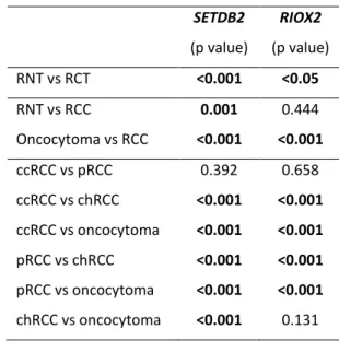
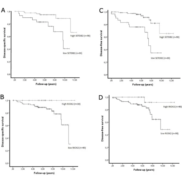
5. Ferreira MJ*, Pires-Luís AS*, Vieira-Coimbra M, Costa-Pinheiro P, Antunes L, Dias PC, Lobo F, Oliveira J, Gonçalves CS, Costa BM, Henrique R, Jerónimo C. SETDB2 and RIOX2 are differentially expressed among renal cell tumor subtypes, associating with prognosis and metastization [submetido para publicação] (*primeiras autoras ex aequo).
Other relevant contributions Outras contribuições relevantes
6.Santos-Silva R, Costa-Pinheiro P, Luís A, Antunes L, Lobo F, Oliveira J, Henrique R, Jerónimo C. MicroRNA profile: a promising ancillary tool for accurate renal cell tumour diagnosis. BJC; 2013; 109(10):2646-53.
Em cumprimento do disposto no referido Decreto-Lei, a candidata declara que participou activamente na definição dos objectivos, bem como na recolha e estudo do material incluído nos trabalhos 2 e 3, bem como na análise e interpretação de resultados, e redacção do artigo, em colaboração com os restantes co-autores. Participou também activamente na definição de objectivos, recolha e estudo do
xi material incluído nos trabalhos 4 e 5 (selecção de casos, extração de RNA, síntese de cDNA, avaliação da expressão de genes por PCR quantitativo em tempo real, avaliação imunohistoquímica), bem como na análise e interpretação de resultados e redação do artigo, em estreita colaboração com a outra primeira autora ex aequo dos referidos artigos, no âmbito das teses de Mestrado das referidas co-autoras.
Colaborou ainda activamente na redação do trabalho 1, e na seleção de casos e recolha de informação clínica dos casos usados no trabalho 6, no âmbito da tese de Mestrado do primeiro autor.
xii
F
UNDING
F
INANCIAMENTO
Este trabalho foi financiado pela Fundação para a Ciência e Tecnologia (FCT) através de uma bolsa individual de interno-doutorando, de referência SFRH/SINTD/94217/2013. Foi ainda financiado por bolsas de investigação do Centro de Investigação do Instituto Português de Oncologia do Porto (CI-IPOP 4-2012) e da Associação Portuguesa de Urologia.
xiv To the loving memory of
Isaura Maria Carreiro
xv
A
CKNOWLEDGEMENTS
A
GRADECIMENTOS
Os meus mais sinceros agradecimentos a todos os que me acompanharam neste percurso. Não há palavras para descrever a gratidão que sinto pela generosidade de me ajudarem a crescer como pessoa e como profissional.
Ao meu Orientador e Diretor de Serviço, Professor Rui Henrique, pela oportunidade de realizar esta Tese de Doutoramento sob a sua orientação, pela integração no Grupo de Epigenética e Biologia do Cancro, pela paciência infindável para questões científicas e burocráticas, por ter sido essencial em todo o processo de realização da Tese e do Internato Médico em paralelo, pela disponibilidade e atenção, por ser uma fonte de inspiração contínua.
À minha co-Orientadora, Professora Carmen Jerónimo, por me ter acolhido e integrado no Grupo de Epigenética e Biologia do Cancro, pelas profícuas discussões científicas e técnicas, por me ter proporcionado um espaço e ambiente científico onde pude aprender metodologias de investigação, pela paciência e compreensão com as vicissitudes de uma médica a aprender a ser, também, cientista.
A todos os colegas do Grupo de Epigenética e Biologia do Cancro, não só os atuais mas também os que já não integram este Grupo, que me acolheram e com quem tanto aprendi, não só procedimentos e protocolos experimentais, mas também formas
xvi diferentes de olhar para as questões. Não consigo enumerar todos aqui, mas recordo todos com enorme gratidão e amizade.
À Dra.Carla Bartosch, minha orientadora de Internato da Formação Específica em Anatomia Patológica e também colega do Programa Doutoral em Medicina e Oncologia Molecular, pelas produtivas discussões científicas, pela generosidade de me permitir aprender com o seu exemplo e conselhos.
A todos os colegas do Serviço de Anatomia Patológica do Instituto Português de Oncologia do Porto e do Centro Hospitalar de Gaia-Espinho. Em fases diferentes deste percurso científico em paralelo com o percurso de formação em Anatomia Patológica, a compreensão e apoio que demostraram foi muito importante.
Aos meus amigos, que permanecem e não desistem, apesar de eu nem sempre conseguir estar presente.
À minha família, pelo apoio, pela compreensão dos horários (ou melhor, da falta de horários) e da menor disponibilidade quotidiana, por todo o acompanhamento e pela inestimável ajuda no “sprint” final.
xvii
A
BSTRACT
Renal cell tumors encompass a heterogeneous group of neoplasms, with distinct clinical, morphological, genetic and epigenetic features. Despite molecular similarities, revealed by large multilevel comparative studies, these molecular differences might portend distinct prognosis and dissimilar response to targeted therapies. Both genetic and epigenetic events may disrupt the main cellular pathways altered in clear cell renal cell carcinoma, papillary renal cell carcinoma, chromophobe renal cell carcinoma and oncocytoma. Despite epigenetic alterations – as gene promoter hypermethylation, histone oncomodification and altered miRNAs expression – have been described as promising biomarkers for detection and discrimination among different renal cell tumors as well as prognostication, validation in clinical series is still lacking, precluding its translation to the clinical setting.
Thus, the main aim of this Thesis was to assess the clinical usefulness of epigenetic-based biomarkers (mainly promoter methylation and histone modifying enzymes expression) in renal cell tumors, especially their diagnostic and prognostic value.
Concerning methylation profile of renal cell tumors, a panel comprising the promoter methylation levels of OXR1 and MST1R was found to be a highly sensitive and specific diagnostic biomarker for renal cell tumors (98% sensitivity, 100% specificity) and for clear cell renal cell carcinoma (90% sensitivity, 98% specificity). Moreover, MST1R promoter methylation was associated with transcription regulation in renal cell tumors, and MST1R expression was associated with prognosis.
xviii Regarding histone modifying enzymes, SMYD2, SETD3, NO66, SETDB2 and RIOX2 (histone demethylases or methyltransferases) were identified as differentially expressed in renal cell tumors and validated in a cohort of 160-cases as well as in TCGA database. Moreover, low SMYD2, SETD3 and NO66 expression levels were associated with shorter disease-specific survival, and low SMYD2, SETD3, NO66 and SETDB2 with shorter disease-free survival (in multivariable analysis). RIOX2 expression level was also associated with the development of metastasis during follow-up.
In conclusion, epigenetic alterations constitute promising diagnostic biomarkers, as promoter methylation and microRNAs, and promising prognostic biomarkers, as histone modifying enzymes, for clinical management of renal cell tumors. Additional assessment in large multicenter trials is needed to support clinical implementation of these biomarkers.
xix
R
ESUMO
Os tumores de células renais compreendem um grupo heterogéneo de neoplasias, com características clínicas, morfológicas, genéticas e epigenéticas distintas. Apesar das semelhanças a nível molecular, reveladas por estudos comparativos, as diferenças moleculares podem condicionar diferentes prognósticos e resposta diferente a terapias dirigidas. Tanto mecanismos genéticos como processos epigenéticos podem alterar as principais vias de sinalização celular desreguladas nos carcinomas de célula renais de tipo célula clara, papilar, cromófobas e em oncocitoma. Apesar de as alterações epigenéticas – como a hipermetilação do promotor de genes, oncomodificação de histonas e expressão alterada de microRNAs – terem sido descritas como biomarcadores promissores para a deteção, discriminação e avaliação de prognóstico em tumores de células renais, não existe validação extensa em séries clínicas, impedindo a sua utilização em contexto clínico.
Assim, o principal objetivo desta Tese foi determinar a utilidade clínica de biomarcadores baseados em alterações epigenéticas (em especial hipermetilação do promotor e alteração da expressão de enzimas modificadoras de histonas) em tumores de células renais, nomeadamente o seu valor como biomarcadores de diagnóstico e prognóstico.
Em relação ao perfil de metilação dos tumores de células renais, um painel constituído pelo nível de metilação do promotor de OXR1 e MST1R mostrou ser um biomarcador de diagnóstico com elevada sensibilidade e especificidade na identificação de tumor de células renais (sensibilidade de 98% e especificidade de 100%) e de carcinoma de
xx células renais de tipo célula clara (sensibilidade de 90% e especificidade de 98%). Adicionalmente, o padrão de metilação do promotor do gene MST1R foi associado à regulação da transcrição em tumores de células renais, e a expressão de MST1R foi associada ao prognóstico.
Relativamente às enzimas modificadoras de histonas, o nível de expressão de SMYD2, SETD3, NO66, SETDB2 e RIOX2 (histonas desmetilases ou metiltransferases) foi distinto em tumores de células renais diferentes, resultado este validado numa série de 160 tumores de células renais e nos casos da base de dados do TCGA. Um nível baixo de expressão de SMYD2, SETD3 e NO66 foi associado a uma menor sobrevivência específica por doença, e nível baixo de expressão de SMYD2, SETD3, NO66 e SETDB2 a uma menor sobrevivência livre de doença (em análise multivariável). O nível de expressão de RIOX2 também foi associado ao desenvolvimento de metastização durante o follow-up.
Em conclusão, as alterações epigenéticas constituem-se como promissores biomarcadores de diagnóstico, como a metilação do promotor e a expressão de microRNAs, e como promissores biomarcadores de prognóstico, como a expressão de enzimas modificadoras de histonas, com potencial de aplicação clínica em tumores de células renais. Será necessária avaliação adicional em grandes ensaios multicêntricos para suportar a implementação clínica destes biomarcadores.
xxi
C
ONTENTS
Statement / Declaração ix
Funding / Financiamento xii
Acknowledgments / Agradecimentos xv
ABSTRACT xvii
RESUMO xix
Contents xxi
Abbreviations list xxiii
Thesis outline xxvi
CHAPTER 1. GENERAL INTRODUCTION 1
1.1. Chapter overview 2
1.2.Intermingling morphological and molecular features – insights
on renal cell tumour classification and pathogenesis 4 1.3.The epigenetics of renal cell tumors: from biology to biomarkers 50
CHAPTER 2. RATIONALE AND AIMS 64
xxii
CHAPTER 4. METHYLATION IN RENAL CELL TUMORS 71
4.1. Chapter overview 72
4.2.Identification of clear cell renal cell carcinoma and oncocytoma
using a three-gene promoter methylation panel 74 4.3.Prognostic significance of MST1R dysregulation in renal cell tumors 84
CHAPTER 5. HISTONE MODIFYING ENZYMES IN RENAL CELL TUMORS 98
5.1. Chapter overview 99
5.2.Expression of histone methyltransferases as novel biomarkers
for renal cell tumor diagnosis and prognostication 101 5.3.SETDB2 and RIOX2 are differentially expressed among renal
cell tumor subtypes, associating with prognosis and metastization 121
CHAPTER 6. GENERAL DISCUSSION 143
6.1. General discussion 144
6.2. Conclusion and perspectives 148
APPENDIXES
xxiii
A
BBREVIATIONS
L
IST
AKT: AKT serine/threonine kinase ALK: ALK receptor tyrosine kinase ARID: AT-rich interaction domain ATM: ATM serine/threonine kinase AUC: area under the curve
BAP1: BRCA1 associated protein 1
CCND1: cyclin D
ccRCC: clear cell renal cell carcinoma CDH1: cadherin 1
CDKN2A: cyclin dependent kinase inhibitor 2A chRCC: chromophobe renal cell carcinoma CI: confidence interval
CNV: copy number variation CTNNB1: catenin beta 1
DFS: disease-free survival
DICER-1: dicer 1, ribonuclease III DNA: deoxyribonucleic acid DNMT: DNA methyltransferases DSS: disease-specific survival
EMT: epithelial to mesenchymal transition
H2A: histone 2A H2B: histone 2B H3: histone 3
H3K27ac: H3 lysine 27 acetylation H3K27me3: H3 lysine 27 trimethylation H3K36me3: H3 lysine 36 trimethylation
H3K4me1/me2: histone 3 lysine 4 methylation/dimethylation H3K4me2/me3: H3 lysine 4 di/trimethylation
H3K79me2: H3 lysine 79 dimethylation H3K9me2/me3: H3 lysine 9 di/trimethylation H4: histone 4
HAT: histone acetyltransferse HDAC: histone deacetylase
xxiv HDM: histone demethylase
HIF: hypoxia inducible factor HMT: histone methyltransferase HOXA9: homeobox A9
HR: hazard ratio
HRAS: HRas proto-oncogene, GTPase
ISUP: International Society of Urologic Pathology
KDM: lysine demethylase
KDM5C / JARID1C: lysine demethylase 5C KDM6A / UTX: lysine demethylase 6A
LAD: lamina-associated domains
LOCK: large organized chromatin K9 modifications LRES: long-range epigenetic silencing domains
MDR1/ABCB1: ATP binding cassette subfamily B member 1 MEK/MAP2K7: mitogen-activated protein kinase kinase 7 MET: MET proto-oncogene, receptor tyrosine kinase miRNA: microRNA
MiT: MET proto-oncogene, receptor tyrosine kinase MST1R: macrophage stimulating 1 receptor
MT-ND: mitochondrially encodedNADH mTOR: mechanistic target of rapamycin
NO66/RIOX1: ribosomal oxygenase 1 NPV: negative predictive value
OXR1: oxidation resistance gene 1
PBRM1: polybromo 1
PCR: polymerase chain reaction
PI3K: phosphatidylinositol-4,5-bisphosphate 3-kinase PPV: positive predictive value
pRCC: papillary renal cell carcinoma PTEN: phosphatase and tensin homolog
QMSP: quantitative methylation specific polymerase chain reaction
xxv RCC: Renal Cell Carcinoma
RCT: Renal Cell Tumor
RIOX2: ribosomal oxygenase 2 RNA: ribonucleic acid
RNT: renal normal tissue
ROBO1: roundabout guidance receptor 1 ROC: receiver operating characteristic
SE: sensitivity
SETD2: SET domain containing 2 SETD3: SET domain containing 3 SETDB2: SET domain bifurcated 2 SFRP: secreted frizzled-related protein SIRT1: sirtuin 1
SLIT2: slit guidance ligand 2
SMARC: switching defective/sucrose nonfermenting (SWI/SNF) related, matrix associated, actin dependent regulators of chromatin SMYD2: SET and MYND domain containing 2
SP: specificity
SWI/SNF complex: switching defective/sucrose nonfermenting
TAD: topologically associating domains TCEB/ELOC: elongin C
TCGA: The Cancer Genome Atlas TERT: telomerase reverse transcriptase TET: tet methylcytosine dioxygenase TGFB: transforming growth factor beta TP53: tumor protein p53
TRBP: TARBP2, RISC loading complex RNA binding subunit TSC: tuberous sclerosis
VEGF: vascular endothelial growth factor VHL: von Hippel-Lindau tumor suppressor
xxvi
T
HESIS
O
UTLINE
This thesis is organized in six chapters and one appendix.
Chapter 1 contains an introduction to the thesis theme, including a review on renal cell
tumor classification, main epigenetic mechanisms, and the most frequent and consistently described alterations in renal cell tumors, both genetic and epigenetic. The second part of this chapter focuses on the potential clinical application of the epigenetic alterations in renal cell tumors.
Chapter 2 defines the aims of this thesis.
Chapter 3 enumerates the experimental procedures performed by the candidate, since
detailed description is depicted in the methods section of each study.
Chapter 4 is composed of two studies on DNA methylation of renal cell tumors. The
first describes the diagnostic significance of a panel including the promoter methylation level of three genes. The second explores the prognostic and functional role of MST1R promoter methylation.
Chapter 5 is composed of two studies focusing on histone modifying enzymes. These
two studies were performed in close collaboration with two distinct Master students (acknowledged as joint first authors of the respective articles) as part of their Master
xxvii Degree project. For the purpose of these studies, I selected cases and I was actively involved in performing RNA extraction from fresh frozen tissue, cDNA synthesis, quantitative real time PCR, data analysis and interpretation, and manuscript writing. The first study identified histone methyltransferases and demethylases differentially expressed among renal cell tumors and assessed the role of three of them as diagnostic and prognostic biomarkers. The second study assessed the prognostic value of other two histone modifying enzymes as prognostic and disease progression biomarkers.
Chapter 6 contains the general discussion of the main results of this thesis, as well as the main conclusion and future perspectives.
The appendix contains a study on microRNAs as diagnostic biomarkers in renal cell tumors, performed by a Master Degree student as part of his Master project. I collaborated by selecting cases, retrieving relevant information from clinical charts and participating in data interpretation and manuscript review.
1
C
HAPTER
1
2
1.1.
C
HAPTERO
VERVIEWThis chapter includes a review paper published in international peer reviewed journal:
Henrique R, Luís AS, Jerónimo C. The epigenetics of renal cell tumors: from biology to biomarkers. Front Genet, 2012; 3:94.
Renal cell tumors encompass a heterogeneous group of neoplasms, with distinct clinical, morphological, genetic and epigenetic features [1, 2]. The relevance of molecular alterations specific for each subtype was reflected by the relatively early inclusion of cytogenetic features in renal cell tumor classification [3, 4]. Despite some molecular similarities, underscored by large multilevel comparative studies [5], these molecular differences might portend distinct prognosis and dismal response to targeted therapies [1, 6].
Over the last years, integrative studies revealed the molecular features of ccRCC [7], pRCC [8], chRCC [9] and oncocytoma [10], revealing the main cell pathways altered in each subtype, mostly by genetic events. These pathways were also frequently altered by epigenetic alterations [2], and thus it was felt that a brief review of the main genetic and epigenetic processes involved in each histotype tumorigenesis, in the first part of this chapter, was essential to highlight the relevance of epigenetic alterations in renal cell tumors.
Moreover, these epigenetic alterations might be useful in renal cell tumor diagnosis and prognostication, which would be promising new tools for renal cell tumor
3 management, and are reviewed in the second part of this chapter, in the published review [11].
REFERENCES
1. Shuch B, Amin A, Armstrong AJ, Eble JN, Ficarra V, Lopez-Beltran A, Martignoni G, Rini BI, Kutikov A: Understanding pathologic variants of renal cell carcinoma: distilling therapeutic opportunities from biologic complexity. Eur Urol 2015, 67:85-97.
2. Morris MR, Latif F: The epigenetic landscape of renal cancer. Nat Rev Nephrol 2017, 13:47-60. 3. Kovacs G, Akhtar M, Beckwith BJ, Bugert P, Cooper CS, Delahunt B, Eble JN, Fleming S, Ljungberg B, Medeiros LJ, et al: The Heidelberg classification of renal cell tumours. J Pathol 1997, 183:131-133. 4. Storkel S, Eble JN, Adlakha K, Amin M, Blute ML, Bostwick DG, Darson M, Delahunt B, Iczkowski K: Classification of renal cell carcinoma: Workgroup No. 1. Union Internationale Contre le Cancer (UICC) and the American Joint Committee on Cancer (AJCC). Cancer 1997, 80:987-989.
5. Chen F, Zhang Y, Senbabaoglu Y, Ciriello G, Yang L, Reznik E, Shuch B, Micevic G, De Velasco G, Shinbrot E, et al: Multilevel Genomics-Based Taxonomy of Renal Cell Carcinoma. Cell Rep 2016, 14:2476-2489.
6. Bhatt JR, Finelli A: Landmarks in the diagnosis and treatment of renal cell carcinoma. Nat Rev Urol 2014, 11:517-525.
7. Cancer Genome Atlas Research N: Comprehensive molecular characterization of clear cell renal cell carcinoma. Nature 2013, 499:43-49.
8. Cancer Genome Atlas Research N, Linehan WM, Spellman PT, Ricketts CJ, Creighton CJ, Fei SS, Davis C, Wheeler DA, Murray BA, Schmidt L, et al: Comprehensive Molecular Characterization of Papillary Renal-Cell Carcinoma. N Engl J Med 2016, 374:135-145.
9. Davis CF, Ricketts CJ, Wang M, Yang L, Cherniack AD, Shen H, Buhay C, Kang H, Kim SC, Fahey CC, et al: The somatic genomic landscape of chromophobe renal cell carcinoma. Cancer Cell 2014, 26:319-330. 10. Joshi S, Tolkunov D, Aviv H, Hakimi AA, Yao M, Hsieh JJ, Ganesan S, Chan CS, White E: The Genomic Landscape of Renal Oncocytoma Identifies a Metabolic Barrier to Tumorigenesis. Cell Rep 2015, 13:1895-1908.
11. Henrique R, Luis AS, Jeronimo C: The epigenetics of renal cell tumors: from biology to biomarkers. Front Genet 2012, 3:94
4
1.2. I
NTERMINGLING MORPHOLOGICAL AND MOLECULAR
FEATURES
–
INSIGHTS ON RENAL CELL TUMOUR
CLASSIFICATION AND PATHOGENESIS
5 1.2.1.KIDNEY CANCER
Kidney cancer incidence is increasing worldwide, with 338000 estimated new cases and 143000 estimated deaths in 2012. Incidence and mortality rates are higher in men and in more developed regions. Although incidence rate has been increasing in the last years, more in men than women, the mortality rate has been stable and even decreasing in most high resource countries (Northern and Western Europe, USA, Australia) since the 90s, more in women than men. The ratio between incidence and mortality is highest in Northern America, indicating a higher survival than in Africa, where this ratio is lowest [1]. The increasing incidence rate has been largely attributed to the growing number of incidental small renal tumours diagnosed due to widespread use of imaging techniques, mostly in high resource countries, in addition to ageing, obesity and smoking, which are known risk factors for the development of kidney cancer. The stabilization or even slightly decrease trend in mortality rate might be related to efficient treatment by surgery in localized cancer, as well as interferon and targeted therapies used mainly in high resource countries. The rising number of small neoplasms incidentally diagnosed, mostly in high resource countries, could also contribute to this mortality trend, not being clear yet if the decreasing mortality corresponds to a survival gain, as the prognosis of these small tumours is generally good and most are cured by partial nephrectomy. A higher proportion of this small tumours diagnosed in high resource countries, in addition to more access to targeted therapies, could contribute to the differences in the ratio between incidence and mortality observed in high resource vs. low resource countries [1].
The vast majority (90%) of kidney cancers are renal cell tumours (RCT), arising from renal parenchyma. The remaining are mostly urothelial carcinomas, arising from renal
6 pelvis. Most RCT are sporadic; however some familiar forms are well documented, including von Hippel-Lindau syndrome, hereditary papillary renal cell carcinoma, familial leiomyomatosis and renal cell carcinoma syndrome and Birt-Hogg-Dubé syndrome [2]. RCTs are a heterogeneous group and include benign tumours, such as oncocytoma (5-9%); and malignant entities, called renal cell carcinomas (RCC), which encompass different tumour types with dissimilar prognosis [3].
The most frequent RCC is clear cell renal cell carcinoma (ccRCC) (70-75%), which also have the worst prognosis among the most prevalent subtypes, followed by papillary renal cell carcinoma (pRCC) (15-20%), with slightly better prognosis than ccRCC. Chromophobe renal cell carcinoma (chRCC) is less frequent (5-7%) and presents a better prognosis than ccRCC and pRCC. For each subtype, the prognosis is worst in tumours with metastasis, which are more frequent in ccRCC and pRCC, and rare in chRCC [3]. Approximately 20 to 30% of all patients present metastases at diagnosis, and approximately 20% develop metastases after nephrectomy [2].
1.2.2. SPORADIC RENAL CELL TUMOURS
1.2.2.1 Conceptual and temporal framework of renal cell tumours classification
The first kidney tumour classifications were based on macroscopic appearance (Koning, in 1826) and on macroscopic and clinical feature (Rayer, in 1841). Later, in the beginning of the 20th century, some classifications based on the histological features were proposed, mostly descriptive, comprising a variable number of entities and mirroring the then accepted idea that renal tumours arose from adrenal remnants, originating the term hypernephroma [4, 5].
7 Although it was initially thought that renal tumours derived from renal tubular epithelial cells, according to Robin (in 1855) and Waldeyer (in 1867) observations, after Grawitz description (in 1884) of small yellow subcapsular renal tumours composed by clear cells as being originated from intrarenal adrenal rests (whereas small papillary tumours originated in renal tubules) the concept of ectopic adrenal rest origin was generalized for all renal tumours. The controversy remained until 1959, when Oberling observed that clear cells from renal tumours presented ultrastructural features similar to renal tubular epithelial cells [5]. Latter classifications further emphasized this renal origin, by designating these neoplasms as “renal cell tumours”.
Noteworthy, these descriptive classifications included benign and malignant entities, despite their distinction was neither straightforward nor uncontroversial, with size being the most widespread criteria to distinguish them, after Bell reported a low metastasization rate for tumours smaller than 3cm in 1928 [4, 6]. Despite reference to papillary tumours and to clear and granular cells was common in these classifications, the first WHO classification of renal cell tumours in 1981 did not include these morphotypes, but acknowledged the existence of large morphological variety in renal cell tumours.
A seminal paper published in 1986 and latter known as “Mainz classification” set the basis for subsequent classification schemes [6]. Tumours were characterized considering cytology and architecture, the main subtypes being clear cell, chromophobe, chromophilic (generally with papillary architecture) and oncocytoma. This was the first study to recognize that granular cells could appear in all the described subtypes, not being an independent subtype as previously thought, and that sarcomatoid change is a manifestation of dedifferentiation of the remaining cancer
8 subtypes. It also included an “unclassifiable renal cell tumour” category, to prevent unclear cases to be classified as a defined tumour morphotype [6]. Subsequent studies expanded the number of identified subtypes and compared these neoplasms to renal tubules epithelial cells, concluding that clear cell and chromophilic cell (papillary) variants originated from proximal tubules, chromophobe cell from intercalated cells of the cortical collecting duct, oncocytoma from cortical collecting duct and Bellini duct variant from principal cells of the medullary collecting duct [7]. This pathomorphological classification was also supported by independent cytogenetical and molecular studies [8-10] that reported specific cytogenetic alterations for each morphological subtype, as depicted bellow.
Thus, the need for integration of these molecular data and morphological criteria in a single classification lead to the Heidelberg/Rochester classification (1997), the first using simultaneously morphological [11] and cytogenetic [12] data to design a new classification scheme for renal cell tumours, including benign tumours as papillary adenoma, oncocytoma and metanephric adenoma, as well as malignant entities as conventional (clear cell), papillary and chromophobe renal cell carcinoma, collecting duct carcinoma, medullary carcinoma and renal cell carcinoma unclassified. One remarkable feature of this classification was the strict morphologic criteria used to define each morphotype, the use of genetic alterations to classify some dubious cases, and the definition of an unclassified category of tumours not meeting the strict morphological features and devoided of the typical genetic features of each subtype [11, 12].
This allowed for a more precise definition and knowledge of the prognosis of the early defined and more frequent histotypes, and contributed for the identification,
9 characterization (clinical, morphological, genetic) and subsequent description of novel RCC subtypes, four of which were included in the 2004 WHO classification [13]: multilocular clear cell renal cell carcinoma, Xp11 translocation carcinomas, renal cell carcinoma associated with neuroblastoma and mucinous tubular and spindle cell carcinoma.
Accumulating evidence on each subtype common and infrequent morphology, prognosis, genetic and epigenetic alterations, in addition to the description of several novel renal tumours, lead the International Society of Urologic Pathology (ISUP) to promote a consensus conference in Vancouver in 2012, to update the classification of renal cell tumours [14]. Some entities were redefined, as multilocular cystic clear cell renal cell neoplasm of low malignant potential and MiT family translocation renal cell carcinoma, that includes Xp11 translocation renal cell carcinoma and t(6;11) renal cell carcinoma; and five novel entities were recognized, including tubulocystic renal cell carcinoma, acquired cystic disease-associated renal cell carcinoma, clear cell (tubulo) papillary renal cell carcinoma and hereditary leiomyomatosis renal cell carcinoma syndrome-associated renal cell carcinoma, as well as three provisional entities. The Vancouver classification set the basis for the 2016 WHO classification [3], which includes the new entities recognized by the Vancouver classification, as well as succinate dehydrogenase-deficient renal cell carcinoma; and also recognizes four provisional entities: oncocytic renal cell carcinoma occurring after neuroblastoma (removed from 2004 WHO and Vancouver classification), thyroid-like follicular renal cell carcinoma, ALK rearrangement-associated renal cell carcinoma and renal cell carcinoma with (angio)leiomyomatous stroma.
10 It should be noted that each subsequent renal cell tumour classification system further describes more precise clinicopathological and molecular features of well established entities, redefines some entities based on newly available data, and incorporates new entities when their morphologic and genetic features are consistently reported as different from the recognized renal cell tumours. So, they are dynamic frameworks for both precise diagnostic and further clinical, genetic and epigenetic research, since a precise definition of tumour entities is the basis of case selection for further studies and allows for the identification of new diagnostic, prognostic and predictive markers, specific for each tumour type and potentially more useful for clinical practice.
Several of such studies have been published, including mutation analysis, gene expression analysis, methylation profiling and microRNA analysis, which are broadening our knowledge about renal carcinogenesis, not only the role of genetic alterations but especially the role of epigenetic alterations.
1.2.2.2 Role of epigenetic alterations in renal tumorigenesis
1.2.2.2.1 General concepts of epigenetic deregulation and carcinogenesis
Epigenetics, first mentioned by Waddington in 1942, was subsequently defined as heritable cellular information, for instance gene expression patterns, not due to changes in the DNA sequence [15, 16]. Epigenetic alterations comprise DNA modifications, histone modifications and microRNA regulation, which might change the gene expression pattern of cells. These alterations are closely interrelated and, unlike genetic changes, might be reversible, upon action of specific enzymes [17]. Epigenetic mechanisms are critical to embryologic development, and some epigenetic alterations, as histone modifications, are involved in kidney development [18].
11 Epigenetic alterations were first recognized in cancer in 1983 [15, 16]. In parallel with the cytogenetic alterations, integrated in renal cell tumour classification since 1997 [11, 12], epigenetic alterations were early recognized as frequent in these tumours [19-21]. It is now widely accepted that epigenetic mechanisms are pivotal to renal cell oncogenesis, and the amount and variety of epigenetic alterations described in RCT is still increasing, some of them carrying diagnostic or prognostic relevance [22].
1.2.2.2.2. DNA modifications
The most well known DNA modification is cytosine methylation in CpG dinucleotides, occurring at the 5th carbon of the cytosine located 5’ of a guanosine (C5-methylcytosine; 5mC), mediated by DNA methyltransferases (DNMT). DNA regions with high proportion of CpG dinucleotides have been called CpG islands, and it was proved by 1980 that DNA methylation at CpG islands located at a gene promoter was associated with gene repression [15, 17]. Other more recently described DNA modifications are N3-methylcytosine (3mC), catalysed chemically; N6-methyladenine (6mA), without known mechanism of modification; and C5-hydroxymethylcytosine (5hmC), C5-formylcytosine (5fC) and C5-carboxylcytosine (5caC), resulting from active demethylation of 5mC by TET enzymes and subsequent oxidation [23].
The first epigenetic alteration described in cancer cells (1983) was global loss of DNA methylation, i.e., hypomethylation [24, 25], which may lead to the activation of gene expression, including that of oncogenes as HRAS or genes associated with drug resistance as MDR1, as well as to chromosomal instability [16], predisposing to abnormal recombination and facilitating deletions, translocations and chromosomal rearrangements [26]. Later on, in the 1990s, hypermethylation of tumour suppressor
12 genes (TSG) promoters, with associated gene inactivation, was consistently described for several tumours [16], including VHL promoter hypermethylation in renal carcinoma, in 1994 [21]. Besides CpG islands in gene promoters, methylation of other DNA regions was associated with gene expression regulation, as CpG shores (low density CpG regions near CpG islands) [27] and gene enhancers. Indeed, enhancer altered DNA methylation was proposed to more closely relate with gene expression changes than promoter methylation itself, which might be in part due to altered transcription factor binding [28]. Additionally, DNA methyltransferases were found to be commonly overexpressed in cancer [29].
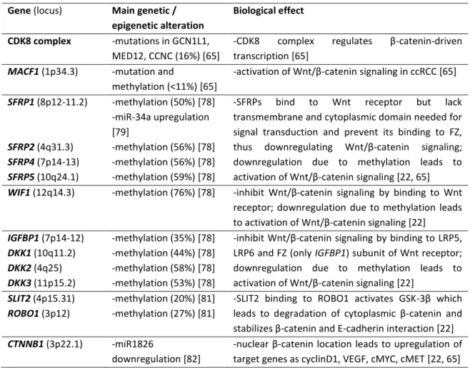
Thus, DNA methylation has emerged as an important epigenetic modification in cancer: both global DNA hypomethylation and locus-specific promoter hypermethylation were reported as early events in tumorigenesis [26], and characteristic hypermethylation profiles of TSG genes were described for some cancer types [20]. Several genes have been reported as hypermethylated in RCC, first identified by a candidate-gene approach, and later by a functional epigenomic approach (comparing gene expression without and after treatment with demethylating drugs to identify genes re-expressed after treatment, which likely correspond to genes silenced by promoter methylation). Genomewide CpG methylation analysis platforms were then used to identify putative new TSG in RCC and to disclose differences in methylation patterns among RCT types (reviewed in [30]). For most reported genes, the frequency of hypermethylation was lower than 70%, and some genes identified in genomewide methylation platforms were not validated by more sensitive and specific techniques [30]. However, it was noted that hypermethylated genes in RCC were involved in some frequently dysregulated pathways (Tables 1-7), including WNT –
β-13 catenin, SLIT-2 – ROBO1, epithelial-to-mesenchymal transition (EMT) and metabolic pathways; as well as in cell cycle regulation, apoptosis and angiogenesis [22, 30].
1.2.2.2.3. Histone modifications
The DNA strand associates with proteins, named histones, to form chromatin. The nucleosome, considered the basic unit of chromatin organization, is formed by a histone octamer (two copies of H2A, H2B, H3 and H4) core, packaging 147 bp of DNA. Each histone has a N-terminal tail, and histone aminoacid residues might be chemically modified, the most frequent being acetylation, methylation or ubiquination of lysine residues, phosphorylation of serine residues and methylation of arginine residues. These modifications, forming the so-called “histone code”, are added and removed by specific enzymes, control gene expression, DNA replication and repair, and chromosome organization [15, 22, 23]. There are currently 12 chemical modifications and at least 130 potential modification sites at the tails of the 4 canonical and 30 histone variants [23]. The “histone code” implies that the combination of histone modifications pattern entail distinct biological outcomes, as gene expression or repression and chromatin remodeling, partially by recruiting downstream effector proteins or protein complexes [17]. For instance, gene expression is associated with histone H3 lysine acetylation and H3 lysine 4 di and trimethylation (H3K4me2 and H3K4me3) in the promoter; with H3 lysines 36 and 79 methylation (H3K36me3 and H3K79me2) in the gene body; and with H3 lysine 27 acetylation (H3K27ac) and lysine 4 methylation (H3K4me1 and H3K4me2) in enhancers. Conversely, gene repression is associated with H3 lysine 27 trimethylation (H3K27me3) and H3 lysine 9 methylation (H3K9me3), in the promoter, gene body (H3K9me2 and H3K9me3) and enhancer
14 (H3K9me2 and H3K9me3) [31]. Globally, histone acetylation is associated with gene expression, and histone methylation could be associated with gene expression or repression depending on the modified aminoacid. Besides these chromatin marks (specific chemical modifications), gene expression and chromatin structure is also related to chromatin features (several linked modifications and complex elements), as nucleosome occupancy, chromatin interactions and chromatin domains [topologically associating domains (TAD), lamina-associated domains (LAD), long-range epigenetic silencing domains (LRES), large organized chromatin K9 modifications (LOCK)] [23]. Several chromatin modifying enzymes have been described, the most extensively studied being histone acetyltransferases (HAT), deacetylases (HDAC), methyltransferases (HMT) and demethylases (HDM), involved in the establishment, maintenance or removal of covalent histone modifications, important for normal cell differentiation; as well as chromatin remodeling protein complexes, like SWI/SNF and BAF-associated complexes, recruited to condense or decondense chromatin, enabling gene silencing or activation, respectively [17, 22, 31].
In neoplastic cells, histone regulating mechanisms might be altered, not only covalent histone marks, but also non-covalent chromatin remodeling mechanisms as nucleosome positioning alterations and histone variants incorporation [17]. Indeed, cancer cells were found to present altered methylation patterns of H3K9 and H3K27 associated with gene repression, and different expression patterns of histone modifying enzymes from normal tissue, which might differ among tumour types and contribute to tumour initiation and progression [15, 32].
It is thought that the deregulation of these mechanisms is relevant for renal carcinogenesis, as mutations in chromatin modification and remodeling genes, like
15 PBRM1, BAP1, SETD2, KDM5C and KDM6A [33-35], which were found to be frequent in RCCs, only surpassed by VHL mutations (Table 2). These enzymes are involved in genomic integrity maintenance, cell-adhesion regulation, HIF signaling and cell cycle control [22].
It should be noted that some histone modifying enzymes can also target non-histone proteins, and their deregulation might affect cell phenotype not only by altering gene activity but also other cellular proteins [23]. Moreover, chromatin modifications and DNA methylation are functionally linked – DNMTs participate in multiprotein complexes with HDACs and HMTs to repress gene expression (Polycomb-mediated silencing), and some CpG islands display high affinity to transcription factors (as CFP1) that recruit activating HMTs and prevent DNA methylation [15, 17].
1.2.2.2.4. microRNAs
The first microRNA (miRNA) was described in 1993, and by 2001, the miRNA regulatory role was broadly, whereas altered miRNA expression in cancer was reported in 2005, and since then the number of altered miRNA and the complexity of miRNA deregulation networks in cancer cells has substantially increased [36-38]. miRNAs are a class of short (19-25bp) non-coding RNAs that bind to complementary sequences in mRNA, targeting them for degradation, and, thus, mediate post-transcriptional gene silencing. Most miRNA are encoded by specific miRNA loci, whereas approximately 30% originate from introns of protein coding genes. The primary miRNA (pri-miRNA) transcribed by RNA polymerase II are cleaved in the nuclei by a protein complex of DROSHA (a double-stranded RNase III enzyme) and its cofactor DGCR8 to a 60-70bp hairpin-shaped precursor miRNA (pre-miRNA), which is exported to the cytoplasm by
16 exportin 5 (XPO5). Pre-miRNA are then processed by DICER1 (a RNase III enzyme), which performs an asymmetrical cleavage of the dsRNA close to the terminal loop sequence, producing the mature miRNA duplex with 2-nucleotide 3’ overhangs. DICER1 associates with TRBP, which binds to dsRNA, enhancing the fidelity of cleavage for a pre-miRNA subset and triggering the formation of iso-miRNAs (1 nucleotide longer than the regular miRNA), and physically bridges DICER1 to the Argonaute proteins. Mature miRNA, DICER1 and other specific proteins form the RNA-induced silencing complex (RISC). In RISC complex, an Argonaute protein binds one strand of the mature miRNA (guide strand) so that its 5’ end (seed region, between nucleotides 2-8) is positioned for interaction with the 3’ untranslated region (3’UTR) of target mRNA. RISC complex binding of complementary target mRNA results in faster mRNA degradation due to accelerated shortening of mRNA poly(A) tail [37, 39]. It is considered that the complementarity between miRNA seed region and target mRNA is crucial for post-transcriptional regulation: perfect complementarity leads to Ago-catalyzed cleavage of target mRNA, whereas imperfect complementarity leads to mRNA translation repression [38].
A single miRNA can target several mRNA, and distinct miRNA can target the same mRNA, creating complex networks. In these networks, as multiple genes can be simultaneously regulated by an individual miRNA, the fine-tuning by regulation distinct sub-networks is facilitated, and, when multiple target mRNA participate in the same signaling pathway, even modest inhibition of multiple targets can generate a stronger response than total inhibition of a single target mRNA. Interestingly, miRNA encoded by the same polycistronic cluster tend to target the same gene or different genes in the same pathway, enhancing its regulatory role. Additionally, some mRNA presenting
17 longer 3’UTRs with more miRNA target sites may constitute relevant regulation points in cell networks, and few miRNA are predicted to account for regulation of the majority of network regulation sites. Thus, it is possible that specific gene expression signatures could be established and maintained by a small number of miRNA [39]. It should also be considered that the biological effect of a specific miRNA is context-dependent, due to differential target gene expression in different cell types, and it is caused not solely by the direct effect on target gene expression, but also by indirect changes in gene expression pattern due to miRNA targeting of transcription factors. Indeed, many miRNA target genes are known to be transcription factors, some transcription factors tend to regulate miRNA more tightly than other genes and are more likely to be regulated by these miRNA [39].It was described that gene expression changes after miRNA disruption were mostly caused by transcription alterations due to transcription factor regulation, more frequent and extensive than post-transcriptional changes [40]. Moreover, it was suggested that some miRNA could directly modulate gene expression, possibly by facilitating RNA polymerase binding and activity in specific genes, and by recruiting chromatin modifiers, crosstalking with other epigenetic players [17, 38].
Cancer cells present distinct miRNA patterns compared to normal cells, which might be due to alterations in miRNA expression and/or to dysregulation of miRNA biogenesis pathway [22, 39]. miRNA expression can be affected by the classic factors that influence gene expression. Indeed, miRNA genes are frequently located at fragile sites or genomic regions subjected to mutations (deletions, amplifications or translocations) in cancer [41, 42], and altered transcription factor activity can also change miRNA expression [37]. Additionally, miRNA may be also regulated by epigenetic mechanisms,
18 especially DNA methylation and histone modification, in a dynamic tissue-specific, miRNA specific and epigenetic effector specific manner [37, 38]. Only about 50% of miRNA genes associate with CpG islands, whereas several miRNA have been reported to be differentially methylated in cancer [43], and histone methylation as well as HDAC overexpression can alter miRNA expression [38]. Moreover, several components of miRNA biogenesis pathway were reported to be dysregulated in cancer: DROSHA and DICER expression levels can be upregulated or downregulated in different cancer models, DGCR8 and Argonaute proteins were reported as upregulated and XPO5 as downregulated in several cancers, and are globally related to an altered miRNA profile, distinct tumour behavior, clinical features or prognosis [37].
In addition to miRNA deregulation, molecular mechanisms to avoid miRNA regulation are also present in cancer cells, as expression of mRNA isoforms with short 3’ UTR and consequently less miRNA binding sites, and single-nucleotide polymorphisms (SNPs) in miRNA genes and in genes required for miRNA biogenesis [39].
Deregulated miRNA in RCTs are involved in key cellular pathways, as VHL-HIF network (for instance, miR-30c-2-3p, miR-30a-3p, miR-210, miR-206), TGFβ and epithelial to mesenchymal transition (EMT) (miR-141, miR-200a, miR-200b, miR-200c), and MET and mTOR pathways (miR-21, miR22, miR-221, miR-222, miR-486, miR-23b-3p) (Tables 1-7). These miRNA are altered by SNPs or promoter methylation, as well as by mutations in DROSHA, DGCR8 and DICER [22, 44]. Interestingly, specific miRNA are associated with distinct RCT subtypes [45, 46], tumour stage and grade [47], and prognosis [48-50], and, thus, their clinical potential in RCTs is diverse and promising [51].
19 1.2.2.3 Malignant tumours – renal cell carcinoma
Because ccRCC is the most frequent RCC subtype, and early studies focused only on ccRCC or included series comprising several RCC subtypes but with ccRCC predominance, the genetic and epigenetic knowledge of this subtype is broader than the remaining RCT subtypes. However, in recent years, an increasing number of publications focusing on less frequent RCT subtypes has been noted. This allows for a deeper understanding on renal carcinogenesis and the comparison of molecular mechanism underlying each RCT morphotype.
1.2.2.3.1 Clear cell renal cell carcinoma
Although it was primordially considered that smaller and circumscribed clear cell tumors were benign [4], it is now well established that all enclose metastatic potential, which increases with the growing size of tumours for kidney confined lesions, and even small clear cell tumours can invade renal sinus or perinephric adipose tissue [52, 53]. However, it was only after the Heidelberg classification in 1997 that the “alveolar” clear cell adenoma was dismissed [12].
Morphologically, ccRCC presents acinar, solid alveolar or cystic pattern, characteristically with a regular network of small and delicate blood vessels. It is composed, in routine hematoxylin/eosin sections, by cells with distinct borders and mostly transparent cytoplasm due to accumulation of glycogen and lipids. Some smaller cells with eosinophilic cytoplasm can be observed, especially in higher grade areas, and an eosinophilic variant is also recognized. Nuclei range from round with homogeneous chromatin and inconspicuous nucleoli in low grade tumours, to larger, polymorphic, with coarse chromatin and prominent nucleoli in higher grade tumours
20 [3, 6, 7]. Ultrastructural features include pinocytotic vesicles, brush-border equivalents and basal infoldings, as proximal tubule epithelial cells [7]. Moreover, large mitochondria with short attenuated cristae and loose matrix are observed in variable number and distribution in tumour cells of the eosinophilic variant [54].
Besides these typical morphological features, chromosomal abnormalities at 3p12-14 (most frequently 3p deletions) have been identified in ccRCC since the late 70s [55]. Subsequent studies reported its absence in other RCC subtypes, especially pRCC [8, 9, 56], and the Heidelberg classification recognized chromosome 3p deletion as a characteristic genetic feature of ccRCC [11, 12]. Additional cytogenetic alterations were consistently described, as chromosome 5q alterations (5q22 trisomy being the second most frequent), and deletion of chromosomes 6q, 8p, 9p and 14q, the last three associated with disease progression, since its frequency is superior in larger and higher grade tumours [10, 12, 57]. Moreover, 1q gain was reported as more frequent in metastatic ccRCC [58]. CGH studies globally confirmed the alterations detected by conventional cytogenetics, additionally identifying loss of 13q and 17p [59].
Interestingly, some genes now known to be altered in ccRCC are located at chromosome 3p, the best characterized and more frequently mutated being von Hippel-Lindau (VHL) gene. VHL is a tumor suppressor gene identified in the familial cancer syndrome von Hippel-Lindau disease [60] and its inactivation is currently recognized as an early and pivotal event in sporadic ccRCCs pathogenesis [61]. Both genetic and epigenetic alterations were described for VHL: nearly 50-75% of sporadic ccRCC display VHL mutation, more than 90% loss of heterozigoty and about 5-20% VHL promoter hypermethylation [61, 62].
21 Comprehensive and integrative ccRCC molecular characterization confirmed VHL mutation as the most frequent in this histotype, and identified frequent mutations in PBRM1, SETD2, KDM5C and BAP1, as well as in PTEN, MTOR and TP53 [63-65]. These approaches have also highlighted frequently deregulated genes and altered cell pathways in ccRCC, and portrayed some contributory genetic and epigenetic mechanisms, integrating and expanding several previous reports.
In addition to VHL-HIF1a-hypoxia pathway (Table 1), the most consistently reported deregulated pathways in ccRCC are the chromatin remodeling machinery, including SWI-SINF complex (Table 2) and PI3K-AKT-MTOR. (Table 3).
For VHL-HIF pathway (Table 1), the most frequent alteration is VHL inactivation mostly by deletion or mutation, leading to HIF accumulation and consequent (over)expression of hypoxia-related genes. Epigenetic modulation of other components of this pathway, mostly by promoter methylation or miRNA altered expression, also contributes to pathway deregulation [22].
Mutations in PBRM1, SETD2 and BAP1, all located at chromosome 3p, were first described in 2010-2011 and confirmed epigenetic alterations as major events in ccRCC carcinogenesis [33-35]. Additionally, some distinct mutations have been described in epigenetic modulators, as SWI/SIFT components, histone modifying enzymes and DNA methyltransferases and demethylases (Table 2), leading to altered gene expression, specific histone modifications and altered DNA methylation patterns.
22 Table 1. More frequently described genetic and epigenetic events affecting the components of the VHL – HIF pathway. Main biological effects of each alteration are also briefly described. Gene
(locus)
Main genetic and / or epigenetic alteration Biological effect VHL (3p25.3) -deletion (78-90%) [63-65] -mutation (50-83%) [63-65] -promoter methylation (5-25%) [22]
-miR-21 and miR-92 upregulation [66, 67]
-component of a protein complex (also including elongin B, elongin C, cullin-2) with ubiquitin ligase E3 activity.
-VHL inactivation leads to absence of ubiquitination and targeting to degradation of target proteins, as HIFs.
HIF-1α
(14q23.2)
-deletion (45%) [63] -mutation (0.7%) [33, 63] -miR-210 [68] and miR-122 [69] upregulation
-miR-20b downregulation [70]
-induces expression of several target genes, for instance VEGF, involved in hypoxic response
-miR-210 upregulation [target genes involved in angiogenesis (EFNA3, PTP1B, HIF-1α), cell cycle and metabolism] [22] -HIF-1α increases miR-210 expression and is also a target of miR-210; HIF-1α targeting by miR-210 contributes to an oncogenic imbalance between HIF-2α and HIF-1α [22].
HIF-2α
(2p21)
-miR-30c-2 and miR-30a downregulation [71]
-miR-30c-2 and miR-30a expression is induced by pVHL; loss of VHL leads to HIF-2α increase due to absence of ubiquitination and loss of miRNA targeting, contributing to an oncogenic imbalance between HIF-2α and HIF-1α [71] -miR-210 upregulation [target genes involved in angiogenesis (EFNA3, PTP1B, HIF-1α), cell cycle and metabolism] [22]
VEGF
(6p21.1)
-miR-206 [22] , miR-106a, miR-20b[70] and miR-126 [72] downregulation -15a, 34a, miR-106b upregulation [70]
- increased expression of VEGF is pro-angiogenic
TCEB1
(8q21.11)
-mutation (1-3.3%) [63, 64] -TCEB1 mutation is
associated with loss of chromosome 8 (bialelic inactivation) [64]
-component of the transcription factor B (SIII) complex, which activates elongation by RNA polymerase II.
-component of the VHL complex; TCEB1 inactivation leads to absence of ubiquitination of VHL-bound HIF proteins [64]. -mutually exclusive mutations in TCEB1 and VHL [64].
Not surprisingly, a strong link between hypoxia and histone modifications has been disclosed [73], paralleling the two more frequently altered cellular mechanisms by genetic and epigenetic changes in ccRCC, and some histone modifications and altered histone modifying enzymes levels have been associated with ccRCC prognosis [74].
23 Table 2. More frequently described genetic and epigenetic events affecting chromatin
remodelers, histone modifying enzymes and DNA methyltransferases. Main biological effects of each alteration are also briefly described.
Gene (locus) Main genetic / epigenetic alteration Biological effect SETD2 (3p21.31) -mutation (4-12%) [33, 63, 64] -deletion (76% ) [65]
-histone methyltransferase, catalyzes trimethylation of lysine-36 of histone H3 (H3K36Me3), associated with active chromatin and reduced CpG methylation [22]
-SETD2 mutation associated with loss of DNA methylation at non-promoter regions [22, 63]
-SETD2 loss could contribute to genomic instability (H3K3Me3 required to ATM and TP53 activation of DNA damage checkpoint) [22]
BAP1
(3p21.1)
-mutation (10%) [63, 64] -deletion (75%)[65] -miR-200b and miR-429 downregulation [70]
-ubiquitin C-terminal hydrolase
-binds BRCA1 (DNA damage response), E2Fs transcription factors, E2F target gene promoters (cell cycle) [22]
-BAP1-mutated tumors present E2F and PRC2 target genes downregulation and increased EZH2 expression [22, 64]
-BAP1 inhibits mTOR activation by AKT; BAP1 inactivation associates with mTOR activation [22]
KDM5C / JARID1C (Xp11.22) -mutation (3.4-7%) [33, 63, 64] -deletion (54%) [65]
-histone demethylase (H3K4Me3 demethylase), inhibits target genes by removing the active chromatin mark H3K4Me3 [22]
-induced by HIF; may be involved in regulating HIF target genes’ expression levels [22]
KDM6A / UTX
(Xp11.3)
-mutation (1-3%) [33, 63] -histone demethylase (H3K27Me3 demethylase), removes the repressed chromatin mark H3K27Me3; KDM6A loss of function might lead to gene downregulation [22]
SIRT1 (10q21.3) -miR-34a upregulation [70] DNMT3B (20q11.21) -miR-148a upregulation [70]
-alteration in DNA methylation
TET2 (4q24) -mutation (6%) [64] -deletion (10%) [64] -miR26a downregulation [75]
-alteration in DNA demethylation
-histone O-GlcNAcylation during gene transcription [64]
SWI/SNF complex (functional interactions between distinct SWI/SNF complexes and associated co-factors regulate
gene-specific transcription; non mutually exclusive mutations in SWI/SNF complex proteins in 58% of ccRCC [64, 76]) PBRM1 (3p21.1) -deletion (73-92%) [63, 65] -mutation (26-33%) [63-65]
-regulates expression of genes involved in cell proliferation (p21), cell adhesion and cell signaling (E-cadherin)
-mutations (mostly inactivating) associated with p21 downregulation, increased cell proliferation and migration [22], and upregulation of hypoxia-related genes [64].
ARID1A
(1p36.11)
-mutation (2-3%) [63, 64] -mutation (3%)[63]
-competition between ARID1A and other ARIDs [76]
ARID1B
(6q25.3)
-deletion (20%) [64] -associated with gene repression, cooperates with ARID2 [76] -represses Wnt/β-Catenin signaling [77]
ARID3B (15q24.1) -miR-125a downregulation [70] SMARCA4 (19p13) -mutation (2%)[63]