UNIVERSIDADE DE LISBOA
Faculdade de Medicina Veterinária
THE FIRST EPIDEMIOLOGICAL STUDY ON THE PREVALENCE OF
CARDIOPULMONARY AND GASTROINTESTINAL PARASITES IN CATS AND DOGS FROM THE ALGARVE REGION OF PORTUGAL USING THE FLOTAC TECHNIQUE
SINCLAIR PATRICK OWEN
CONSTITUIÇÃO DO JURÍ
Doutor José Augusto Farraia e Silva Meireles
Doutor Luís Manuel Madeira de Carvalho
Mestre Telmo Renato Landeiro Raposo Pina Nunes
ORIENTADOR
Doutor Luís Manuel Madeira de Carvalho
CO-ORIENTADOR
Dr. Dário Jorge Costa Santinha
2017
UNIVERSIDADE DE LISBOA
Faculdade de Medicina Veterinária
THE FIRST EPIDEMIOLOGICAL STUDY ON THE PREVALENCE OF
CARDIOPULMONARY AND GASTROINTESTINAL PARASITES IN CATS AND DOGS FROM THE ALGARVE REGION OF PORTUGAL USING THE FLOTAC TECHNIQUE
SINCLAIR PATRICK OWEN
DISSERTAÇÃO DE MESTRADO INTEGRADO EM MEDICINA VETERINÁRIA
CONSTITUIÇÃO DO JURÍ
Doutor José Augusto Farraia e Silva Meireles
Doutor Luís Manuel Madeira de Carvalho
Mestre Telmo Renato Landeiro Raposo Pina Nunes
ORIENTADOR
Doutor Luís Manuel Madeira de Carvalho
CO-ORIENTADOR
Dr. Dário Jorge Costa Santinha
2017
ACKNOWLEDGEMENTS
This dissertation and the research project that underpins it would not have been possible without the support, advice and encouragement of many people to whom I am extremely grateful.
First and foremost, a big thank you to my supervisor Professor Doctor Luis Manuel Madeira de Carvalho, a true gentleman, for his unwavering support and for sharing his extensive knowledge with me. Without his excellent scientific guidance and British humour this journey wouldn’t have been the same.
I would like to thank my co-supervisor Dr. Dário Jorge Costa Santinha, for welcoming me into his Hospital and for everything he taught me. It was a very enriching experience for which I am very grateful.
My heartfelt appreciation goes to my friend now Doctor Ana Margarida Alho, for giving me the opportunity to follow her work towards her PhD and for her constant encouragement, help and for motivating me to finish this dissertation.
A big thank you to Lidia Gomes in the Parasitology lab for helping me identify some parasites, her humour made everything so much more fun.
To all the hardworking staff and volunteers at the shelters I visited, you all do an amazing job.
To Drª Mafalda Andrade a big thank you for sharing your extensive knowledge of ultrasonography and medical know how with me.
To all my friends and colleagues at the Loulé Veterinary Hospital, Drª Joana Soares, Drª Ana Palma, Drª Aline Delgado, Dr. Filipe Pereira, Drª Beatriz Santos, Dr. José Nogueira, Dr. Alexandre Marques, Helena Guerreiro, Manuela Martins, Claudio "Soya", Sandra Medeiros, Sr. Jorge and Dona Leonor Santinha thank you for welcoming me with open arms and paws!
Over the years it took to be finished the course and write this dissertation, I also had the support from my amazing friends and family, thank you all for believing in me.
To Mo Ressler and Michael Joseph, thank you for everything, none of this would have been possible without you.
To my family in Ireland for your encouragement and my uncle Noel Connors for sharing his love and knowledge of horses with me.
To my friend Beatriz Santos, Marie Santos and Guida Azevedo for all your wise words and constant support and encouragement.
To my friends Guilherme Soares, Ana Lopes, Teresa Afonso, Gonçalo Soeiro, Daniel Murta, Pedro Teixeira and Ana Rita Moreira for all the fun and friendship and motivation.
To Carla Motolla and Rui Seixas for your friendship and advice.
To Celeste Oliveira my second mother from Angola, my brother Dean, my sister Nikita, my nieces and nephews, my Godson, Cidalia and all my four legged family members past and present a big thank you for being there for me.
To Carlos Alberto, this is also for you, I am lucky to have you in my life.
I dedicate this dissertation to my parents, Ellen and Arthur Owen, for all the love and encouragement I have received throughout my life to follow my dreams and for helping me do so.
RESUMO
PRIMEIRO RASTREIO DE PARASITAS CARDIOPULMONARES E GASTROINTESTINAIS EM CÂES E GATOS DA REGIÂO DO ALGARVE
UTILIZANDO A TÉCNICA DE FLOTAC
Apesar de nas últimas décadas terem surgido vários trabalhos de rastreio parasitológico em Portugal, atualmente continuam a existir poucos dados sobre a incidência de parasitas gastrointestinais e cardiopulmonares de carnívoros domésticos com relevância clinica no Algarve. Por esta razão foi realizado um estudo entre Fevereiro e Abril de 2016 utilizando a técnica do FLOTAC. No total foram colhidas 142 amostras de canis e gatis da região correspondendo a 66 cães e 76 gatos todos com > 6 meses de idade.
Foram detetadas formas de eliminação fecal de seis grupos de endoparasitas, quatro dos quais com potencial zoonótico. A prevalência global no estudo foi de 31%, com 21.2% das amostras de canídeo positivas e 39.5% de felídeo positivos para pelo menos um parasita. Infeções mistas com dois parasitas foram observadas em 2.6% das amostras de cão e 9.2% das de gato. Toxocara sp. foi o parasita mais frequente em cães evidenciando-se em 13.6% (OPG: 61.3 ± 74.65) das amostras e em 31.6% (OPG 523.92 ± 688.75) das de gato. Nos cães também foram detetadas amostras
positivas para Ancylostomatidae (3%), Taeniidae (6%) e Trichuris spp. (2%) e nos gatos Ancylostomatidae (5.3%), Aelurostrongylus abstrusus (4%), Cystoisospora spp. (6.6%) e Taeniidae (1.3%).
A presença de parasitas com potencial zoonótico na população estudada bem como e elevada prevalência de Toxocara spp. é preocupante e alerta para a necessidade de desparasitação regular e medidas de controlo higio-sanitário adequadas, quer em animais com proprietário, quer em animais de abrigo.
Palavras chave: cão, gato, parasitas gastrointestinais, parasitas cardiopulmonares, zoonoses, FLOTAC.
ABSTRACT
THE FIRST EPIDEMIOLOGICAL STUDY ON THE PREVALENCE OF CARDIOPULMONARY AND GASTROINTESTINAL PARASITES IN CATS AND
DOGS FROM THE ALGARVE REGION OF PORTUGAL USING THE FLOTAC TECHNIQUE
Although over the past decades various parasitologal surveys have been conducted in Portugal, at present scant information is available on the prevalence of clinically-relevant gastrointestinal and cardiopulmonary parasites in the Algarve. This study performed between February and April 2016 using the FLOTAC technique was undertaken to address this. Faecal samples were collected from 66 dogs and 76 cats > 6 months old from shelters in the region.
Faecal forms of six different groups of endoparasites were detected, four of which with zoonotic potential. The overall prevalence in both species was 31%, with 21.2% of the dog samples positive and 39.5% of the cat samples positive for at least one parasite. Concurrent infections with two parasites were detected in 2.6% of the dog samples and 9.2% of the cats. Toxocara spp. was the most common parasite found in 13.6% (EPG: 61.3 ± 74.65) of the dogs samples and 31.6% (EPG 523.92 ± 688.75) of the cats. Other parasites found in dogs were Ancylostomatidae (3%), Taeniidae (6%) and
Trichuris spp. (2%) and in cats Ancylostomatidae (5.3%), Aelurostrongylus abstrusus
(4%), Cystoisospora spp. (6.6%) and Taeniidae (1.3%) were found.
The presence of zoonotic parasites in the population studied as well as the elevated prevalence of Toxocara spp. overall are concerning and highlight the need for regular prophylaxis, hygiene and sanitary control measures in pets as well as shelter animals.
Key words: dog, cat, gastrointestinal parasites, cardiopulmonary parasites, zoonosis, FLOTAC.
INTRODUCTORY NOTE
The present document is the result of the work developed during the author’s traineeship period as part of the requirements for the attribution of the Master’s degree in Veterinary Medicine. The results of the research project were presented in the form of a paper and poster communication at the 7th International EFOMV congress in Lisbon, Portugal held on 26th and 27th of November, 2016). (Appendix IV)
TABLE OF CONTENTS
ACKNOWLEDGEMENTS ... i RESUMO ... iii ABSTRACT ... iv INTRODUCTORY NOTE ... v LIST OF FIGURES ... xLIST OF GRAPHICS... xii
LIST OF TABLES ... xiii
LIST OF SYMBOLS ... xiv
LIST OF ABBREVIATIONS ... xv
CHAPTER I - INTRODUCTION ... 1
CHAPTER II - TRAINING PERIOD ACTIVITIES ... 2
2.1 Hospital Veterinário de Loulé... 2
2.2 Laboratory for Parasitic diseases FMV-ULisboa ... 3
CHAPTER III - REVIEW OF LITERATURE ... 4
3.1 Nematodes ... 4
3.2 Ancylostomatidae ... 4
3.2.1 Life cycle ... 5
3.2.2 Pathogenesis and clinical signs ... 6
3.2.3 Morphology and identification ... 7
3.2.4 Diagnosis ... 8
3.2.5 Treatment and control ... 8
3.2.6 Zoonotic risc ... 9
3.3 Toxocara spp. ... 11
3.3.1 Life cycle ... 11
3.3.2 Pathogenesis and clinical signs ... 14
3.3.3 Morphology ... 15
3.3.4 Diagnosis ... 16
3.3.5 Treatment and control ... 17
3.3.6 Zoonotic risk ... 17
3.4 Trichuris spp. ... 19
3.4.1 Life cycle ... 19
3.4.2 Pathogenesis and clinical signs ... 20
3.4.4 Diagnosis ... 21
3.4.5 Treatment and control ... 21
3.4.6 Zoonotic risk... 22
3.5 Aelurostrongylus abstrusus ... 23
3.5.1 Life cycle ... 23
3.5.2 Pathogenesis and clinical signs ... 24
3.5.3 Morphology ... 25
3.5.4 Diagnosis ... 25
3.5.5 Treatment and control ... 26
3.6 Cestodes ... 26
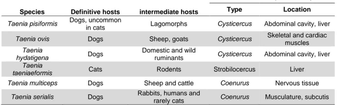
3.7 Taeniidae ... 27
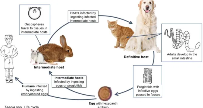
3.7.1 Life cycle ... 28
3.7.1.1 Taenia spp. ... 28
3.7.1.2 Echinococcus spp. ... 29
3.7.2 Pathogenesis and clinical signs ... 29
3.7.3 Morphology ... 30
3.7.3.1 Taenia spp. ... 30
3.7.3.2 Echinococcus spp. ... 31
3.7.4 Diagnosis ... 31
3.7.5 Treatment and control ... 31
3.7.6 Zoonotic risk... 32
3.8 Protozoa ... 33
3.8.1 Cystoisospora spp. ... 33
3.8.2 Life cycle ... 33
3.8.3 Pathogenesis and clinical signs ... 34
3.8.4 Morphology ... 34
3.8.5 Diagnosis ... 34
3.8.6 Treatment and control ... 35
CHAPTER IV - THE FIRST EPIDEMIOLOGICAL STUDY ON THE PREVALENCE OF CARDIOPULMONARY AND GASTROINTESTINAL PARASITES IN CATS AND DOGS FROM THE ALGARVE REGION OF PORTUGAL USING THE FLOTAC TECHNIQUE. ... 36
4.1 Objectives ... 36
4.2.1 Study population ... 36
4.2.2 Sample collection and preparation ... 37
4.2.3 Flotation solutions ... 37
4.2.4 Procedure ... 38
4.2.5 Morphological characterization ... 39
4.2.6 Statistical analysis ... 40
4.2.7 Note regarding safety procedures. ... 41
4.3 Results ... 41
4.3.1 Parasites detected using the FLOTAC technique ... 41
4.3.1.1 Ancylostomatidae ... 41 4.3.1.2 Toxocara spp. ... 42 4.3.1.3 Trichuris spp. ... 43 4.3.1.4 Aelurostrongylus abstrusus ... 44 4.3.1.5 Taeniidae ... 45 4.3.1.6 Cystoisospora sp. ... 45 4.3.2 Dogs ... 46
4.3.2.1 Parasite prevalence in dogs across the region ... 47
4.3.2.2 Prevalence of positive dog samples by municipality... 47
4.3.2.3 Co-infections ... 48
4.3.3 Cats ... 49
4.3.3.1 Parasite prevalence in cats across the region ... 49
4.3.3.2 Prevalence of positive cat samples by municipality ... 50
4.3.3.3 Co-infections ... 50
4.3.4 Quantitative EPG and OPG ... 51
4.3.5 Co-infections in both species ... 52
4.3.6 Parasite families in both species ... 53
4.3.7 Municipal prevalence for both species ... 54
4.3.8 Toxocara prevalence for both species ... 54
4.4 Discussion ... 56
4.4.1 Dogs ... 56
4.4.2 Cats ... 57
4.4.3 General observations ... 58
4.4.4.2 Toxocara spp. ... 61
4.4.4.3 Trichuris spp. ... 63
4.4.5 Pulmonary nematodes ... 64
4.4.6 Gastrointestinal cestodes ... 65
4.4.7 Gastrointestinal protozoa ... 66
4.4.8 Zoonotic risk factors ... 67
4.4.9 Study limitations ... 68
CHAPTER V - CONCLUSION ... 70
REFERENCES ... 71
LIST OF FIGURES
Figure 1 The Loulé Veterinary Hospital ... 3
Figure 2 “Nina”, the Hospital's first client ... 3
Figure 3 Ancylostoma spp. life cycle (original adapted from Prociv & Croese (1996)). ... 6
Figure 4 Toxocara canis life cycle (adapted from CDC, (2017)) ... 14
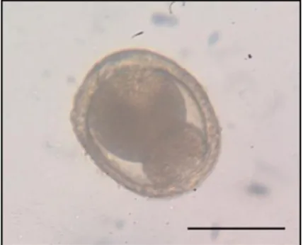
Figure 5 Dividing Toxocara spp. egg, found in cat faeces (original). Scale bar 50μm. ... 15
Figure 6 Trichuris spp. life cycle (original adapted from ESCCAP, (2017)). ... 20
Figure 7 Aleurostrongylus abstrusus life cycle (original adapted from ESCCAP, 2011) ... 24
Figure 8 Taenia spp. life cycle (original illustration) adapted from ESCAAP (2017) .29 Figure 9 General characteristics of adult Taenia spp. (original) ... 30
Figure 10 Taeniidae egg surrounded by embryophore (original) ... 30
Figure 11 Municipalities where samples were collected (original). ... 36
Figure 12 My "assistant" pausing after collection! ... 37
Figure 13 Cats from one of the catteries ... 37
Figure 14 Samples ready for processing ... 37
Figure 15 Filling each falcon tube with the contents from each homogenized container. ... 38
Figure 16 Filling the FLOTAC chambers from the falcon tubes. ... 38
Figure 17 Removal of the screw, key and base. ... 39
Figure 18 Reading the FLOTAC under a microscope ... 39
Figure 19 Ancylostomatidae and Taeniidae co-infection, found in cat faeces 400x (original) ... 42
Figure 20 Ancylostomatidae eggs, found in dog faeces 100x (original) ... 42
Figure 21 Ancylostomatidae eggs, found in cat faeces 400x (original) ... 42
Figure 22 Toxocara spp. eggs with single celled embryos, found in cat faeces (original) Scale bar 50μm. ... 43
Figure 23 Toxocara spp. egg found in dog faeces (original) 100x ... 43
Figure 24 Sample co-infected with Taeniidae and Toxocara spp. found in dog faeces (original) 100x ... 43
Figure 27 A. abstrusus sample co-infected with Toxocara spp. (Original) x100 ... 44
Figure 28 A. abstrusus L1 larvae (original courtesy of Doctor Ana Margarida Alho) Scale bar 100μm. ... 44
Figure 29 A. abstrusus L1 larvae (original courtesy of Doctor Ana Margarida Alho) Scale bar 50μm. ... 44
Figure 30 Taeniidae egg, found in dog faeces (original) x400 ... 45
Figure 31 Taeniidae egg, found in cat faeces (original) x400 ... 45
Figure 32 Taeniidae egg, found in cat faeces (original) x400 ... 45
Figure 33 Sporulated Cystoisospora felis oocysts (original) scale bar 100μm ... 46
LIST OF GRAPHICS
Graphic 1 Prevalence in dogs across the region by parasite ... 47
Graphic 2 Positive dog samples by municipality ... 47
Graphic 3 Co-infections in dogs by parasite ... 48
Graphic 4 Prevalence in cats across the region by parasite ... 49
Graphic 5 Positive cat samples by municipality ... 50
Graphic 6 Co-infections in cats by parasite ... 50
Graphic 7 Descriptive plot comparing mean Toxocara spp. EPG count in dogs and cats. ... 51
Graphic 8 Infection prevalence status, out of the total number of samples tested ... 52
LIST OF TABLES
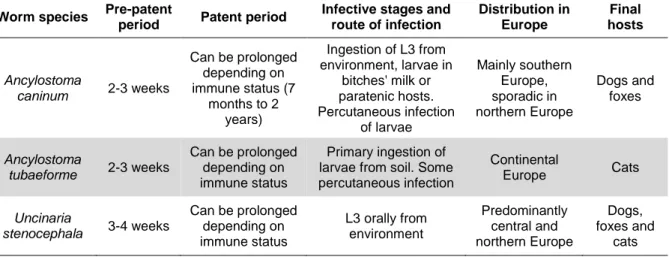
Table 1 Characteristics of the major hookworms affecting cats and dogs in Europe.
Adapted from ESCCAP (2010) ... 5
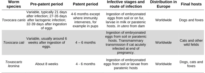
Table 2 Characteristics of roundworms or ascarids found in Europe. Adapted from ESCCAP (2010) with information from (Bowman, 2014) ... 11
Table 3 Trichuris vulpis characteristics. Adapted from ESCCAP (2010) ... 19
Table 4 Characteristics of A. abstrusus - ESCCAP (2010) ... 23
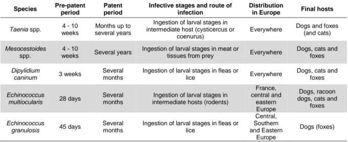
Table 5 Characteristics of tapeworms found in Europe. Adapted from ESCCAP (2010) ... 27
Table 6 Some Taeniidae tapeworm species affecting carnivores (adapted from Ballweber, 2001) ... 28
Table 7 Interactions between different Taenia spp. species that affect either cats or dogs and humans. Data obtained from The Center for Food Security & Public Health (2005). ... 32
Table 8 Characteristics of Cystoisospora oocysts that affect cats in Europe. (ESCCAP, 2011) ... 34
Table 9 Method for preparing each flotation solution (Cringoli et al, 2010). ... 38
Table 10 Positive canine faecal samples by municipality and parasite(s) detected in each sample. [Absolute frequency is presented, followed by the % infected underneath (CI 95%)] ... 46
Table 11 Positive feline faecal samples by municipality and parasite(s) detected in each sample. [Absolute frequency is presented, followed by the % infected underneath (CI 95%)] ... 49
Table 12 Intensity of infections found by species, n positive = nº samples positive for the parasite (CI 95%) ... 51
Table 13 Numbers of EPG or EPG < or > than 100 by species and parasite. ... 52
Table 14 Representation of the types of infection detected by species. ... 53
Table 15 Sample status by municipality ... 54
Table 16 Toxocara spp. prevalence by municipality ... 55
LIST OF SYMBOLS
% – Percentage & – And μm – Micrometer = – equals < – less than / – division + – sum of or addition ± – more or less than x – multiplicationLIST OF ABBREVIATIONS
A. caninum – Ancylostoma caninum A. brazilienze – Ancylostoma braziliense A. ceylanicum – Ancylostoma ceylanicum A. vasorum – Angiostrongylus vasorum
BID – bis in die (twice per day)
C. felis – Cystoisospora felis C. rivolta – Cystoisospora rivolta
CLM – cutaneous larva migrans CNS – central nervous system
E. granulosus – Echinococcus granulosus E. multilocularis – Echinococcus multilocularis
EPG – eggs per gram
ESCCAP – European Scientific Counsel Companion Animal Parasites FLOTAC – Flotation and centrifugation
FMV-UL – University of Lisbon Faculty of Veterinary Medicine FS – Flotation solution GI – Gastrointestinal kg – kilogram L1 – First-stage larva L2 – Second-stage larva L3 – Third-stage larva L4 – Fourth-stage larva L5 – Fifth-stage larva
LRTI - Lower respiratory tract infections mg – milligram
NLM – neural larva migrans OLM – ocular larva migrans OPG – Oocysts per gram
PCR – Polymerase chain reaction PI – post infection
SCT – sedimentation and counting technique s.g. – Specific gravity
sp. – specie spp. – species
TID – ter in die (three times per day)
T. canis – Toxocara canis T. cati – Toxocara cati T. vulpis – Trichuris vulpis
VLM – visceral larva migrans
1
CHAPTER I - INTRODUCTION
Companion animals are responsible for transmitting some of the most significant zoonotic parasitic diseases affecting man (Baneth et al., 2015). Knowledge relating to the epidemiology, modes of transmission, life cycles, pathogenicity, prevention and treatment of these agents is therefore of great importance. Indeed healthy animals not only contribute to physical well-being and mental health, but they are also in part responsible for healthy people (Paul, King, & Carlin, 2010).
In most cases appropriate prophylaxis and good hygiene practices are sufficient to reduce propagation and eliminate infections. However, many factors can influence these including a lack of knowledge on the subject or financial constraints. Indeed a recent study in Portugal found that although the majority of pet owners surveyed do give antiparasitic drugs to their pets, they do so at irregular intervals rendering them ineffective (M. Matos, Alho, Owen, Nunes, & Madeira de Carvalho, 2015).
At present, little information is available on the prevalence and types of cardiopulmonary and gastrointestinal parasites affecting cats and dogs in the Algarve, the most Southern region of Continental Portugal. The epidemiological study described herein, was undertaken to address this and is the first time the FLOTAC technique, a new multivalent technique for qualitative and quantitative copromicroscopic analysis technique developed by Cringoli, Rinaldi, Maurelli, & Utzinger (2010), was used in the region.
It is hoped that the information gathered may ultimately contribute to a better understanding of the health status of the animals in the region and serve as a guide for local authorities, veterinarians and shelters to implement more strategic and targeted prophylactic treatments in the future.
The present dissertation includes an account of the clinical activities undertaken in the first part of the training period, followed by a literature review and discussion of the research project.
2
CHAPTER II - TRAINING PERIOD ACTIVITIES
2.1 Hospital Veterinário de Loulé
The first period supervised by Dr. Dário Santinha took place between the 14th of April and the 28th of August 2015 at the Loulé Veterinary Hospital in the Algarve, Portugal (around 720 hours). The Hospital, founded in 2012, is a full-service modern facility centrally located in the Algarve with a 24 hour emergency service providing routine medical and surgical care for dogs, cats and other companion animals. Great emphasis is directed towards the prevention of disease and this is promoted by encouraging owners to regularly deworm and vaccinate their pets, as well as informing them of the importance of a good diet and exercise.
The facility has a fully equipped laboratory, with modern diagnostic equipment including chemistry and haematology analysers, where routine clinical biochemistry, haemograms and blood smears can take place in house. Diagnostic imaging is catered for with digital x-ray equipment, a modern ultrasound machine with Doppler capabilities for colour flow visualization, as well as endoscopy equipment enabling state of the art internal investigation.
The hospital has comfortable separate dog, cat and isolation ward rooms, as well as an intensive care unit, where constant observation is possible and all needs catered for including the provision of oxygen to high dependency respiratory patients and monitoring using intensive care equipment. Surgery is performed in a modern operating theatre with state of the art anaesthetic monitoring equipment and the highest hygiene and sterilisation standards are adhered to for all procedures.
During the internship period the author had the opportunity to attend the consultations that took place and assist by: helping to restrain animals for examination or treatment, preparing and administering medication, gathering patient history, carrying out supervised physical examinations and position patients for some diagnostic imaging procedures.
The author also assisted in the care of the hospitalized patients by helping to monitor critically ill animals, as well as preparing and administering medication. Other duties included cleaning beds, preparing food, feeding, changing water and taking the patients that were able for walks. There was also the opportunity to help care for some
critically ill patients in the isolation ward which required special attention and care so as not to spread the contagious diseases to the remaining patients.
The number of surgeries performed was large and the author assisted in setting up the surgical suite as well as preparing the patients for surgery which included preparing pre-surgical medications. A variety of procedures were observed from routine spaying and neutering to more complex orthopaedic procedures as well as emergency surgeries including gastropexy for patients with gastric torsion. During less critical procedures the author was tasked with monitoring anaesthesia and post recovery.
Figure 1 The Loulé Veterinary Hospital Figure 2 “Nina”, the Hospital's first client
2.2 Laboratory for Parasitic diseases FMV-ULisboa
The research component, supervised by Prof. Doctor Luis Madeira de Carvalho, took place between February and September 2015 and involved the collection of faecal samples from kennels and catteries in the Algarve followed by processing and analysis which took place at the University of Lisbon, Faculty of Veterinary Medicine’s laboratory for Parasitic diseases (FMV-UL). Work undertaken during the second period formed the basis for the research component in this document.
3
CHAPTER III - REVIEW OF LITERATURE
3.1 Nematodes
Nematodes, otherwise known as roundworms, are a large phylum with over 25,000 species described thus far (Zhang, 2013). These worm-like organisms are diverse and parasite a variety of animals and plants and can be found in a wide range of different environments. Morphologically they are bilaterally symmetrical and have tubular digestive systems with openings on both extremities. Sexual dimorphism occurs with males being smaller than females (Ballweber, 2001)
Most parasitic roundworms do not need an intermediate host for the development of their free-living stages and can therefore infect their final hosts directly. Pregnant females within the host produce thousands of eggs daily which are then excreted in the host faeces into the environment. If conditions are favourable the egg will hatch into a L1 larvae, these then feed on bacteria or other microorganisms. They then develop and molt twice transforming from L1 into L2 and then L3 larvae which for most species is the infective stage. Once ingested by the host the larvae migrate to their preferred site within the host, usually an organ, where they develop into adults and begin reproducing.
Gastrointestinal (GI) infections caused by nematodes are a major threat to human health and currently affect over half of the world’s population, causing hundreds of thousands of deaths annually as well as great morbidity (Stepek, Buttle, Duce, & Behnke, 2006).
3.2 Ancylostomatidae
Family Ancylostomatidae has 7 genera including Uncinaria sp. and Ancylostoma sp. This family belongs to Phylum Nematoda, Class Secernentea, Order Strongylida, Superfamily Ancylostomatoidea and generally designated hookworms (Taylor, Coop, & Coop, 2016).
Adult hookworms are small intestine parasites that can cause varying degrees of blood loss. These small nematodes have large mouthparts at an angle to the rest of the body, hence the name (ESCCAP, 2010a) Ancylostoma spp. species of veterinary importance in Europe (Table 1) include A. caninum (dogs and foxes) which exists worldwide, A.
tubaeforme (cats) worldwide, A. braziliense (dogs and cats) that can be found in
tropical and subtropical environments, A. ceylanicum (dogs and cats) can be found in many parts of Asia and Uncinaria stenocephala which affects dogs, foxes and rarely cats and usually exists in cooler northern temperate regions, including Northern America and Europe (Ballweber, 2001; Zajac & Conboy, 2012).
Table 1 Characteristics of the major hookworms affecting cats and dogs in Europe. Adapted
from ESCCAP (2010)
Worm species Pre-patent
period Patent period
Infective stages and route of infection Distribution in Europe Final hosts Ancylostoma caninum 2-3 weeks Can be prolonged depending on immune status (7 months to 2 years) Ingestion of L3 from environment, larvae in bitches' milk or paratenic hosts. Percutaneous infection of larvae Mainly southern Europe, sporadic in northern Europe Dogs and foxes Ancylostoma tubaeforme 2-3 weeks Can be prolonged depending on immune status Primary ingestion of larvae from soil. Some percutaneous infection Continental Europe Cats Uncinaria stenocephala 3-4 weeks Can be prolonged depending on immune status L3 orally from environment Predominantly central and northern Europe Dogs, foxes and cats 3.2.1 Life cycle
As with most parasitic nematodes A. caninum life cycle is direct. Once the eggs have been excreted into the environment they develop and may hatch into filariform larvae and become infective within 5 to 8 days. Environmental conditions favourable to larval development include warmth (23ºC to 30ºC), humidity, shade and poorly-drained soil (Ballweber, 2001; Bowman, 2014). The host becomes infected either by ingesting the L3 larvae or by percutaneous infection where the larvae penetrate the skin. Once in the host the larvae migrate to the lungs via the bloodstream. Within the trachea and bronchi they then moult into L4 and are then swallowed, allowing them to enter the small intestine where a final moult occurs.
If infective larvae have been ingested, they either burrow into the oral mucosa, and then migrate to the lungsas mentioned previously, or they may progress directly to the intestine where the adult worms burrow their buccal capsules into the intestinal mucosa to obtain nutrients: mainly blood in the case of Ancylostoma spp. and plasma for U.
stenocephala. This burrowing action ultimately damages the intestinal surface. In
infected with Ancylostoma sp. can excrete millions of eggs each day over a period of several weeks.
Although transplacental transmission does not occur, infected bitches can infect their offspring for up to three weeks. This is because Ancylostoma spp. larvae can become arrested at the L3 stage and remain so until the bitch is pregnant. In some susceptible females L3 larvae that have migrated to the lungs may become dormant(Taylor et al., 2016). Transmammary transmission only occurs in dogs infected with A. caninum (Bowman, Montgomery, Zajac, Eberhard, & Kazacos, 2010).
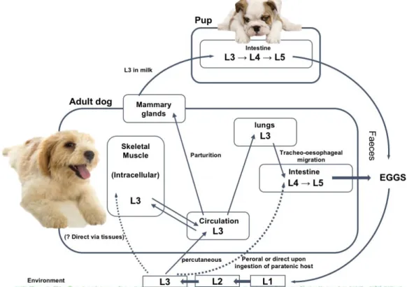
Figure 3 Ancylostoma spp. life cycle (original illustration adapted from Prociv & Croese (1996)). 3.2.2 Pathogenesis and clinical signs
Disease by A. caninum may take one of four forms (Ballweber, 2001; Taylor et al., 2016)
1. Peracute. This form occurs in very young pups due to the ingestion of infective larvae in the dam’s milk. As few as 50 to 100 adult A. caninum may be fatal. The pups are born healthy but their condition deteriorates rapidly. Pups are particularly vulnerable due to their low iron reserves (Taylor et al., 2016). 2. Acute hookworm disease - the sudden exposure of older pups or adults to
respiratory embarrassment (Taylor et al., 2016) . The burden may be so great that clinical signs may precede the presence of eggs in the faeces by about 4 days (Bowman, 2014).
3. Chronic (compensated) - usually occurs in immunocompetent animals that have not been exposed to overwhelming numbers of larvae - clinical signs are not usually apparent. Hookworm eggs are present in the faeces and there are significant reductions in the erythrocyte count, haemoglobin or packed cell volume.
4. Secondary (decompensated) hookworm disease - occurs in older animals and may be secondary to other problems. Severe anaemia usually occurs; the animal may be malnourished or even emaciated.
Infected hosts may experience exhibit respiratory signs either due to lung damage caused by larvae or the anoxic effects secondary to the anaemia caused by blood loss due to the parasites. Ancylostoma caninum, A. tubaeform and A. ceylanicum are more pathogenic than other hookworms like U. stenocephala and A. brasiliense as they cause more blood loss and consequent anaemia (Bowman et al., 2010). If the infection is chronic the host may be emaciated and weak, there may also be a loss of appetite and occasionally pica. Cutaneous lesions, lameness and respiratory embarrassment have also been described (Taylor et al., 2016). Immunity although not complete, does develop following exposure (ESCCAP, 2010a).
3.2.3 Morphology and identification
The eggs are oval shaped and have characteristic thin shells that can be easily seen on flotation. Ancylostoma spp. eggs range from 52–79 × 28–58 μm whereas Uncinaria spp. have dimensions between 71–92 × 35–58 μm. (Zajac & Conboy,
2012). Ancylostomatidae eggs are however indistinguishable morphologically making epidemiological studies complicated (Bowman et al., 2010).
In dogs adult female worms can grow to 14-20mm while males can range from 11-13mm (Prociv & Croese, 1996). Buccal cavities also differ between species, whereas Ancylostoma spp. have sharp teeth, Uninaria spp. have cutting plates (Bowman,
2014). Depending on the species of Ancylostoma spp. the ventral margin of the stoma may have either one (Ancylostoma brasiliense), two (Ancylostoma duodenale) or three
pairs of sharp teeth (A. caninum and A. tubaeforme), (Bowman, 2014). The three teeth enable the differentiation between these and otherwise similar human parasites (Prociv & Croese, 1996).
3.2.4 Diagnosis
When diagnosing hookworm infections ante-mortem, clinical signs and patient history should be considered and upon suspicion, a haematological and faecal examination should be performed. Hookworm eggs can be detected morphologically using faecal flotation techniques (ESCCAP, 2010a). Depending on the technique used, a quantitative assessment of the burden may also be obtained. ESCCAP (2010) guidelines currently recommend the collection of between 3 and 6 g of fresh or fixed faeces for accurate diagnosis.
According to Zajac & Conboy (2012), “In the case of peracute hookworm disease, eggs will not be found on faecal flotation because the profound anaemia occurs before adults begin laying eggs”. Taylor et al. (2016), also noted that “suckled pups may show severe clinical signs before eggs are detected in the faeces.” Although high egg counts may help confirm the health problem is due to hookworm related disease, the presence of small numbers of hookworm eggs in faeces does not necessarily confirm that the underlying problem is hookworm disease (Taylor et al., 2016).
On post-mortem examination Ballweber (2001, p. 142) describes how adult worms are found in the small intestine, especially the jejunum and may be red and up to 2.8 cm in length with the anterior end bent dorsally resulting in a “hook”. The number of teeth in the buccal cavity enables differentiation.
3.2.5 Treatment and control
In acute infection or the chronic (compensated) form, the response to simple anthelmintic therapy is very effective and no further therapy beyond an adequate diet is necessary (Bowman, 2014). Anthelmintics such as mebendazole, fenbendazole or nitroscanate are effective at killing adult and developing intestinal stages. Pyrantel is also effective (Taylor et al., 2016), but may be becoming less so. High-level pyrantel resistance was found in some Brisbane isolates of A. caninum (Kopp, Kotze, McCarthy, & Coleman, 2007). Although macrocyclic lactones are effective, in severe cases anthelmintics alone may not be sufficient and supportive therapy may also be
necessary. In these cases a protein rich diet and parental iron are advised (Taylor et al., 2016) to encourage haematopoiesis and recovery vitamin B12 may also be given. Arrested fourth-stage larvae may be resistant to anthelmintic treatment and therefore anthelmintics must be given again once these larvae mature (Taylor et al., 2016).
To reduce the risk or transmammary infection bitches should be treated with an anthelmintic that is effective against somatic larvae (Taylor et al., 2016). To achieve this, from day 40 of gestation infected bitches can be treated fenbendazole 50mg SID through to day 14 of lactation. Alternatively, 4-9 days before giving birth ivermectin 0.5mg/kg can be given a second treatment should then be followed 10 days later (Ballweber, 2001). To reduce the risk of infection a drug appropriate for use in nursing pups should be given at least twice, once at the 1-2 week mark and then again two weeks later. This also helps to control ascarid infections (Taylor et al., 2016).
As with most parasitic agents, prophylaxis is the best form of action and may take the form of regular anthelmintic therapy which should take place at least quarterly for both weaned pups and adults (ESCCAP, 2010a). Good hygiene practices should also be encouraged (Taylor et al., 2016). In situations where young pups are present, bedding should be changed daily, flooring should be cleaned at least twice a week and kept as dry as possible (Ballweber, 2001). Following the removal of faeces with a shovel, the floor should be hosed with water and then a 1% solution of sodium hypochlorite should be sprayed on the paved surfaces (Taylor et al., 2016). To facilitate the cleaning process kennel floors should be crevice free, a tarmac or concrete flooring is recommended (Stepek et al., 2006; Bowman et al., 2010). In cases where and outbreak may have occurred earth runs can be treated with sodium borate as it is lethal to hookworm larvae (Taylor et al., 2016). The success of any regular hookworm treatment should be confirmed through regular faecal examinations. (Bowman et al., 2010)
3.2.6 Zoonotic risc
Hookworms species believed to be human specific include Ancylostoma duodenale
and Necator americanus. However canine and feline hookworms, including A.
braziliense, A. caninum, A. ceylanicum (Stepek et al., 2006) and Uncinaria stenocephala can cause zoonotic disease most notably cutaneous larva migrans or
penetrate the human’s skin and begin migrating under it. Although these parasites perform complete migrations in their natural hosts, migrate to the lungs from where they are then swallowed and then enter the small intestine where they begin egg production. In paratenic hosts like humans their development generally stops at the lungs.
According to Bauerfeind, Graevenitz, Kimmig, Schiefer, & Schwarz, (2015 p. 431) In the classic form caused by A. braziliense “The burrows appear on the skin surface as elevated alterations, up to 2 mm wide, with surrounding erythema, oedema, and crusts”. This may cause itching and some pain, secondary infections may develop due to the scratching. Symptoms may disappear between 2 to 8 weeks later or persist for up to 2 years if untreated (Bauerfeind et al., 2015).
In the case of A. caninum follicular alterations are more pronounced and pustules may also develop, this species also tends to spread in the organism due to the way it invades human blood vessels. In these cases Eosinophilic infiltrations as well as pneumonia have been described. (Bauerfeind et al., 2015). In Asia, Inpankaew et al., (2014) reported how A. ceylanicum, a hookworm of cats and dogs in Asia, is becoming the “second most common hookworm infecting humans”.
As the main form of infection is via the skin, it is strongly recommended that humans wear appropriate footwear when walking in potentially infected areas. Effective treatments include Albendazole 400mg BID orally for 3 to 7 days or Ivermectin 200 micrograms/kg/ SID orally for two days (Bauerfeind et al., 2015).
3.3 Toxocara spp.
Toxocara spp. are important gastrointestinal helminths affecting domestic animals,
some of which are also zoonotic. These roundworms or ascarids, with worldwide distribution, inhabit the small intestines of dogs, cats and other mammalian hosts and belong to Phylum Nematoda, Class Secernentea, Order Ascaridida, Superfamily Ascaridoidea, Family Ascarididae (Taylor et al., 2016). Common species found in Europe (Table 2) include Toxocara canis (Werner, 1782) which affects dogs and foxes,
Toxocara cati (Schrank, 1788) that mainly infects cats and Toxascaris leonina that also
affects dogs and foxes, as well as cats.
Table 2 Characteristics of roundworms or ascarids found in Europe. Adapted from ESCCAP
(2010) with information from (Bowman, 2014)
Worm
species Pre-patent period Patent period
Infective stages and route of infection
Distribution in
Europe Final hosts
Toxocara canis
Variable, typically 21 days after infection; 27-35 days after lactogenic infection; 32-39 days after ingestion
of eggs 4-6 months except where immunity intervenes, for example in pups Ingestion of embryonated eggs from soil or on fur, larvae in milk or paratenic
hosts. In utero from dam
Worldwide Dogs and foxes
Toxocara cati
Variable, usually around 6 weeks after ingestion of
eggs.
4 – 6 months
Ingestion of embryonated eggs from soil or paratenic
hosts. Transmammary transmission if cat acutely
infected at end of pregnancy.
Worldwide Cats and other wild felids
Toxascaris
leonina About 8 weeks 4 - 6 months
Ingestion of embryonated eggs from soil or larvae from
paratenic hosts
Worldwide Dogs, cats and foxes
3.3.1 Life cycle
The life cycles of both T. canis and T. cati are similar. In this review details of T. canis will be described with additional remarks on the differences to T. cati. There are currently four known pathways for Toxocara spp. infection in a suitable host: ingestion of embryonated eggs from the environment, ingestion of infected paratenic hosts, transplacental transmission and transmammary transmission.
The cycle begins when adults in the hosts’ small intestine shed single-celled eggs which are then excreted into the environment during defecation. The eggs then undergo two moulting steps where structural modifications take place, developing into L3 which is the infective stage. According to Schnieder, Laabs, & Welz (2011) optimal conditions are temperatures between 25 and 30ºC and a relative humidity of 85-95%. If conditions or soil type are not optimal the development can take between 3 to 6
weeks or several months. The embryonated eggs may survive for more than a year in the environment if conditions are optimal (Overgaauw & Knapen, 2008).
Once ingested the eggs hatch and the larvae pierce the hosts gut wall within 2-4 hours, they then use the hosts blood vessels and undergo somatic migration throughout the body, remaining as somatic larvae in the tissues. This form of the disease is known as visceral larva migrans or VLM and according to Overgaauw & Knapen (2008) occurs in most adult dogs and cats with some degree of acquired immunity.
Parasite migration appears to depend on the hosts age as well as the infective load (Schnieder et al., 2011) In younger animals less than 3 months old, tracheal migration is most common. Once the L3 larvae have reached the lungs they then penetrate the alveoli, pass though the bronchioles and migrate up the trachea where they are then swallowed and mature in the intestinal tract as L4 stage larvae. It takes between 7-9 days following infection for larvae to be detected in the trachea and oesophagus (Schnieder et al., 2011). The larvae then mature in the small intestine into L5 adults which can take between 7 to 15 days post infection – PI. Significantly, Lee et al., (2010) describe how a low infective dose of 100 embryonated eggs consistently induces infection in adult dogs and how the risk of re-infection is the same for naïve dogs as it is for those infected transplacentally as puppies.
Although patent Toxocara infections are much more prevalent in young dogs and cats when compared to adults (Overgaauw & Knapen, 2008), dogs and cats of all ages can become infected with their specific Toxocara species through the ingestion of infective eggs or paratenic hosts (Lee et al., 2010). In older puppies and adults once the larvae become trapped in the lung capillaries they undergo somatic migration and re-enter the bloodstream. Somatic larvae then accumulate in tissues where they become arrested for long periods of time. As reviewed by Overgaauw & Knapen (2008) T. canis larvae are more commonly found in the central nervous system, whereas T. cati is more common in the muscles.
Transplacental transmission is the most common source of T. canis infection in dogs, but does not happen in kittens (Overgaauw & Knapen, 2008; Schnieder et al., 2011). It occurs when encysted larvae are reactivated in the final stages of pregnancy thereby enabling the transplacental and transmammary route to puppies. This reactivation is believed to be due to alterations in hormone levels in the final third of the gestation
period or the intrinsic immunosuppression that occurs in the periparturient period. Transplacental migrations cause infection of the foetus' liver and following birth tracheal migrations occur with the puppies shedding eggs within as little as two weeks (Overgaauw & Knapen, 2008; Schnieder et al., 2011)
Transmammary transmission occurs in both dogs and cats (Schnieder et al., 2011; Bowman, 2014). After activation, somatic Toxocara spp. larvae migrate to the milk ducts and are transmitted via the colostrum and the milk. Once ingested the larvae develop, but do not perform tracheal migrations. This route of infection is less significant than the transplacental mode in puppies, but is the primary mode of infection for kittens where faecal egg excretion can be observed 7 weeks following lactogenic transmission (Schnieder et al., 2011).
The ability of some larval nematodes to remain viable in paratenic hosts, namely prey species, enables the parasites continuing survival and distribution. This mode of transmission is effective as the parasite can develop somatically in a variety of vertebrates including rabbits and rodents. Once an infected paratenic host is ingested the larvae develop directly in their intestine. The fact that cats are more avid hunters than dogs may explain why higher rates of infection are found (Overgaauw & Knapen, 2008).
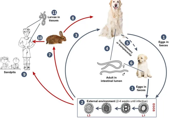
Figure 4 Toxocara canis life cycle (original illustration adapted from https://www.cdc.gov/parasites/toxocariasis/biology.html accessed on 10/05/17.)
(1) Definitive host sheds unembryonated eggs into the environment. (2) over a 2-4 week period the eggs develop into L3 and become infective. (3) Infected eggs ingested by dogs, hatch and penetrate the gut wall. In younger dogs migrations to the lungs, bronchial tree and oesophagus occur. (4) Adult worms mature into L5 and begin laying eggs in the small intestine. (5) During late pregnancy, encysted stages become reactivated and enable the transplacental and transmammary route to puppies. (7) After ingestion by paratenic hosts larvae hatch, penetrate the gut wall and migrate to various tissues where they encyst. (8) Dog ingests paratenic host and larvae develop into egg-laying adults in the small intestine. (9) Humans playing in infected sandpits act as accidental hosts through either the ingestion of infected soil or (10) infected paratenic hosts. Once ingested the eggs hatch and larvae penetrate the gut wall and circulate through the body to various tissues including: liver, lungs, brain, muscle, heart and eyes (11). (original figure) adapted from Toxocariasis life cycle: CDC - US. Department of health and human services - https://www.cdc.gov/parasites/toxocariasis/biology.html accessed on 10/05/17.
3.3.2 Pathogenesis and clinical signs
In the case of mild to moderate infections, during the pulmonary larval migration phase, there is little apparent damage to the tissues and external clinical signs may be absent (Taylor et al., 2016). The presence of adults in the intestine may cause mechanical damage to the small intestine and give the animal a bloated or pot-belly appearance, which may also cause stunted growth as well as occasional vomiting and diarrhoea (Taylor et al., 2016). In heavy infections affecting pups, the larval migrations can cause pulmonary damage and pulmonary oedema which may cause coughing, frothy nasal discharge and increased respiratory rate. (Taylor et al., 2016). The pups and kittens
may also exhibit mild mucoid enteritis, vomiting, diarrhoea and general unthriftiness (Ballweber, 2001). In the gut the parasites may cause mucoid enteritis which in rare cases can cause a partial or complete occlusion. T. canis is most lethal during the pulmonary phase and pups heavily infected transplacentally or lactogenically may die within 2-3 weeks of birth (Ballweber, 2001). Although nervous convulsions have been attributed to toxocarosis, it is not confirmed whether the parasite is responsible.
3.3.3 Morphology
Fresh Toxocara eggs are subspherical with a thick shelled wall, with a markedly pitted surface containing a round, single celled embryo (Uga et al., 2000). Although it is difficult to differentiate the eggs from both species, T. canis eggs are normally more subspherical that T. cati which tend to be more elliptical (Zajac & Conboy, 2012). Morphometric analysis of 442 eggs identified as either T. canis or T. cati by Fahrion, Schnyder, Wichert, & Deplazes (2011) revealed that T. canis eggs had dimensions of 62,3 x 72,7μm (mean values) while T. cati were slightly larger at 74,8 x 86,0μm (mean values). Differentiating both species by microscopy is difficult as described by Uga et al. (2000) who found that although both species have “markedly pitted surfaces” and that the “pitting in T. canis is more coarse than T. cati”, the differences were not absolute. These authors also found that egg measurement wasn’t sufficient to differentiate species as “approximately 90% of eggs measured were of similar size”. In cats, it may also be necessary to differentiate between T. cati and Toxascaris
leonina. Eggs of the latter often are slightly larger and range from 75-85 x 60-75μm,
but similarly to Toxocara eggs they also contain a single celled embryo and a thick shelled wall, however unlike T. cati the embryo is light coloured. Whereas Toxocara eggs have rough outer shell walls, Toxascaris eggs have smoother outer shells.
Adult T. canis worms, most commonly seen in puppies, are cream coloured and can range from 10 to 15cm in length (Bowman, 2014). They have a glandular oesophageal bulb, cervical alae and their mouth openings are surrounded by three prominent lips. In fresh worms reproductive organs can appear white when viewed through the cuticle.
3.3.4 Diagnosis
Centrifugal or simple flotation techniques are indicated to detect eggs, in cases of severe infection a simple faecal smear with a drop of water enables the eggs to be seen. For accurate diagnosis 3 – 5g of faeces are required (ESCCAP, 2010a). The eggs observed are subspherical with a roughly pitted yellow-brown outer shell containing a large dark single cell. However, T. canis and T. cati eggs are not easily distinguished using these methods, suggesting that molecular techniques such as PCR may be more effective in determining the exact species infecting the host. Significantly, although it is assumed that eggs obtained comply with the species from which they are obtained, a study conducted by Fahrion et al., (2011) using PCR demonstrated that out of 35 dogs tested, 11 (31.5%) were in fact infected with T. cati. Notably, out of the 36 cats tested none were found to be infected with T. canis. It is also important to note that some dogs exhibit coprophagia making it difficult, at the time of diagnosis, to determine whether the animal has a patent infection or is simply passing ingested eggs back into the environment (Fahrion et al., 2011).
During the pulmonary phase, where the infection is heavy and larvae are migrating, a tentative diagnosis is possible simply based on the pneumonic signs which may appear in a litter within two weeks of birth (Bowman, 2014). According to Bowman (2014) on post-mortem puppies may appear pot-bellied, cachectic and poorly grown. Large numbers of white worms may be found in the small intestine are sometimes the stomach. The worms range from 3-10cm in length for T. cati and 10-18cm for T. canis. (Ballweber, 2001). The lungs of puppies with migrating T. canis larvae may also have focal haemorrhages.
3.3.5 Treatment and control
Due to the occurrence of transplacental transmission in pups, these should be treated with appropriate anthelmintics starting at 2 weeks of age, followed by 15 day intervals until two weeks after weaning, followed by monthly treatments for six months. Nursing females should be treated alongside their offspring’s first treatment as they may have patent infections (ESCCAP, 2010a).
Monthly treatment is currently recommended by ESCCAP (2010) in high risk situations like where a family may have a pet and small children that share a common area such as a garden. This is because the pre-patent period, from the time of ingestion of larvae, either by paratenic hosts or infective eggs from the environment, is just over 4 weeks and therefore this regimen may minimise the risk. As the parasites are prolific egg layers and the eggs can survive for long periods of time in the environment regular anthelmintic therapy is required in particular if regular diagnostic testing, monthly to three monthly, is not performed (ESCCAP, 2010a). While anthelmintic treatment may be successful at removing adult worms it is not fully effective with juvenile or larval stages (Ballweber, 2001). Also, according to ESCCAP (2010) in the case of patent infections quarterly treatment may not be effective and in this case a monthly regimen may be proposed as it takes into account the parasite’s biology. In all other cases, depending on the risk of multiple worm infections, a broad or narrow spectrum of activity anthelmintic should be given at least 4 times per year.
Effective anthelminthic drugs used to treatment Toxocara infections in dogs and cats include Macrocyclic lactones, Benzimidazoles and Pyrantel (ESCCAP, 2017).
3.3.6 Zoonotic risk
Both T. canis and T. cati can cause significant disease in people. (Lee et al., 2010). Humans may become infected (paratenic hosts) after ingesting embryonated eggs. This can occur for example in children who ingest contaminated objects or soil in areas such as gardens or sandpits. Insects can also act as transport hosts by spreading eggs from faeces they have fed on, to surfaces and foods. As with other mammals, humans may also be infected by ingesting other accidental hosts such as rabbits and chickens, which harbour L3 larval stages in their muscles and organs. The infective process is similar to domestic animals, whereby, upon ingestion the encysted larvae become reactivated and may migrate to human organs including the liver, lungs and eyes,
where tissue necrosis, chronic liver damage, oedema, haemorrhage and eosinophilia may occur. (Stepek et al., 2006)
Although humans infected with larval Toxocara spp. may be asymptomatic, a variety of symptoms may occur including VLM, OLM and neural larva migrans (NLM) (Bowman et al., 2010; Lee et al., 2010). Cases of NLM may be fatal or cause permanent neurological disease to intermediate hosts like man. To avoid infection, it is strictly recommended that high risk foods such as livers and other organs be thoroughly cooked. Regular deworming practices, strict handwashing and discouraging children from putting objects in their mouths are effective ways to reduce transmission.
3.4 Trichuris spp.
Trichuris spp. are intestinal parasites with importance in both veterinary and human
medicine. They belong to Phylum Nematoda, Class Adenophorea, Order Enoplida, Superfamily Trichuroidea, Family Trichuridae (Taylor et al., 2016) and have a worldwide distribution. Commonly known as whipworms due to their characteristic whip shaped body the genus known as Trichuris includes a variety of nematodes affecting a variety of hosts from livestock to humans. For the sake of this review emphasis will be given to Trichuris vulpis, due to its distribution in central and southern Europe.
Table 3 Trichuris vulpis characteristics. Adapted from ESCCAP (2010)
Pre-patent
period Patent period Route of infection
Distribution in
Europe Final hosts
Trichuris
vulpis 8 weeks Up to 18 months
Ingestion of embryonated eggs
Everywhere but mainly in central a southern
Europe
Dogs
3.4.1 Life cycle
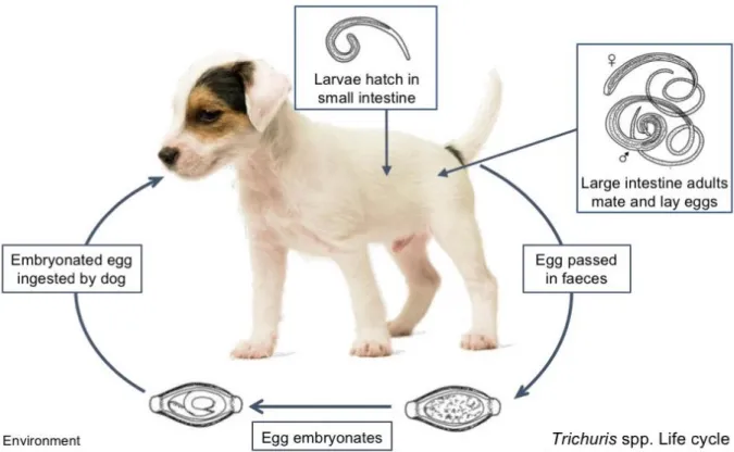
After mating, adult females embedded in the mucosa of the caecum and colon lay non-infective single celled eggs which are then passed into the environment via faeces. Depending on moisture and temperature, the eggs then take up to 8 weeks to develop into their infective embryonated stage (Traversa, 2011). They then remain in the environment and don’t hatch until ingested by a suitable host (Zajac & Conboy, 2012; Bowman, 2014).
After ingestion the egg plugs are lysed releasing the larvae which then penetrate the intestinal glands, during this process which takes up to two weeks the larvae moult and then colonise the large intestine becoming adults (Traversa, 2011). No extra intestinal migrations takes place (Bowman, 2014) and transplacental or transmammary transmission does not occur with this species (Traversa, 2011). The prepatent period takes around 8 weeks (ESCCAP, 2010a).
Figure 6 Trichuris spp. life cycle (original illustration adapted from ESCCAP, (2017)).
3.4.2 Pathogenesis and clinical signs
Most canine infections are unremarkable but heavy infections can cause diarrhoeal faeces, often containing mucus or blood, alternating with periods where normal stools are normal (Bowman, 2014). When less than 200 worms are present they can be found mainly in the cecum, as numbers increase they may also be found in the colon (Bowman, 2014).
As well as bloody diarrhoea, in heavy infections the dog may exhibit weight loss and unthriftiness (Zajac & Conboy, 2012). Trichuris spp. infections are rare in cats and believed to be non-pathogenic (Ballweber, 2001)
3.4.3 Morphology
Freshly passed T. vulpis eggs are brown, lemon-shaped with characteristic plugs which are symmetrical and have rings, the shell is thick and the wall is smooth (Di Cesare, Castagna, Meloni, Otranto, & Traversa, 2012). They measure 72–90 × 32– 40μm (Márquez-Navarro et al., 2012; Zajac & Conboy, 2012) and can be differentiated from human Trichuris trichiura ova as these are typically much smaller measuring 50 to 56 x by 21 to 26 μm, (Dunn, Columbus, Aldeen, Davis, & Carroll, 2002).
Adult worms are whip-shaped and range from 4.5 – 7.5cm in length. The anterior end which embeds into the large intestine is thin and hairline while the posterior tail, which lies freely in the lumen, is thick and broad occupying about ¼ the length of the body (Traversa, 2011; Bowman, 2014).
3.4.4 Diagnosis
Due to the density of the eggs (s.g. = 1.15) detection by centrifugal flotation of faeces, with appropriate flotation solution, is more effective than simple flotation techniques. Current ESCCAP (2010) guidelines suggest collecting between 3 – 5g of fresh or fixed faeces for egg detection.
3.4.5 Treatment and control
As the eggs can survive and remain viable in cold and warm conditions especially wet and shady areas they can persist for long periods of time, thereby becoming a constant source reinfection for dogs living in these contaminated environments (Traversa, 2011). To reduce this risk animals should be treated regularly with effective anthelmintics and removed from the contaminated area until effective decontamination has taken place (ESCCAP, 2010a).
According to Bowman (2014) it is generally believed that parasitic larvae are more resistant than adults to anthelmintics, as T. vulpis larvae take up to 8 weeks to develop, a follow-up dose of anthelmintic should be given three times, at monthly internals after the initial treatment, in order to rid the host of the worms that survived the first treatment.
Due to the limited efficacy of current treatments to treat the human form of the disease caused by Trichuris trichiura, research has been made to find alternatives. A recent study by Partridge et al., (2017) discovered that a new class of compounds, known as dihydrobenzoxazepinones, not previously used as anthelmintics was effective and selective against T. muris and also interrupted the Trichuris life cycle by acting on the eggs. Due to the low toxicity against mouse cells, the authors suggest that these compounds may indeed be a novel pharmaceutical treatment against Trichuris in humans and domestic animals.
3.4.6 Zoonotic risk
Human infections by T. vulpis are rare (Márquez-Navarro et al., 2012; Zajac & Conboy, 2012). However cases have been described in man, these include that of a 49 year old patient with chronic diarrhoea and 5 dogs (Dunn et al., 2002) and a 9-year old Mexican child with symptoms of epistaxis lasting for over two years, with symptoms of rhinitis and a diagnosis of rhinitis (Márquez-Navarro et al., 2012). In the latter case although the child’s pet’s faeces were negative, out of 292 dogs living in the vicinity that were studied, 3,5% were found to be infected with T. vulpis suggesting that the patient may have been infected by dogs living close to her home. Indeed Traversa (2011) states that “its significance for human medicine is its controversial zoonotic ability”.
Unlike Ancylostoma spp. where infections may occur due to skin penetration, as with
Ascaris, Trichuris eggs must be me ingested in order to infect humans. Significantly
although T. vulpis and the human form T. trichiura are morphologically similar, the former is nearly twice as large (Dunn et al., 2002). To control human infections apart from anthelmintic therapy it is essential that the canine sources also be treated in order to avoid reinfection (Dunn et al., 2002).
3.5 Aelurostrongylus abstrusus
Aelurostrongylus abstrusus otherwise known as feline lungworm, is a nematode
belonging to Plylum Nematoda, Class Secernentea, Order Strongylida, Superfamily Metastrongyloidea (Taylor et al., 2016) that infects the terminal respiratory bronchioles and alveolar ducts of domestic cats worldwide (Traversa, Cesare & Conboy, 2010; Di Cesare et al., 2015). Often underdiagnosed the parasite can cause a variety of symptoms and may be fatal in cases of heavy infection (Zajac & Conboy, 2012). Recent studies suggest an increasing geographical distribution of the parasite. (Di Cesare et al., 2015) possibly due to epidemiological and biological factors relating to the gastropod mollusc intermediate hosts and altered development rate due to climate changes (Traversa et al., 2010; Di Cesare et al., 2015).
Table 4 Characteristics of A. abstrusus - ESCCAP (2010)
Worm species Pre-patent
period
Patent period
Infective stages and route of infection
Distribution
in Europe Final hosts
Aelurostrongylus abstrusus 7 - 9 weeks Several years Ingestion of infected intermediate host Everywhere in Europe Cats 3.5.1 Life cycle
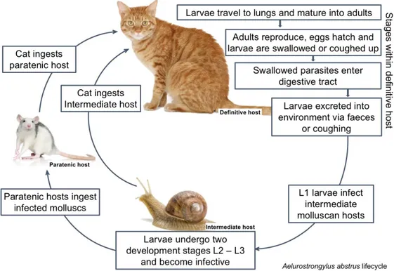
The Aelurostrongylus abstrusus life cycle requires two hosts with cats as final hosts and gastropods as intermediate hosts (Figure 7). The first stage occurs in the environment and involves a variety of terrestrial molluscs such as slugs or snail species, while the second stage (an optional one) involves a variety of paratenic hosts including rodents, amphibians, birds and reptiles (Taylor et al., 2016). Infection begins when a cat either ingests an infected intermediate mollusc or an infected paratenic host such as a rodent or a bird.
On digestion, the infective L3 larvae are released and the larvae migrate to lungs via the lymphatic system or bloodstream. Once in the lungs, they moult a final time and then the adults remain in nodules in the terminal bronchioles, alveolar ducts and pulmonary alveoli (Traversa et al., 2010). After mating the eggs mature and the L1 larvae pass up the bronchial escalator and are either coughed out into the environment or swallowed and released via the faeces (Taylor et al., 2016). The L1 larvae then remain in the environment until they penetrate an intermediate mollusc host, such as a snail or slug, where they develop into infective L3 larvae. They then remain within this host until it is either ingested by a paratenic host or by a cat. The host becomes
infected either by ingesting the paratenic host, the most frequent form of infection, or by ingesting the intermediate host (Taylor et al., 2016). Alternatively L3 from infected molluscs may be released within the snails mucus into the environment where they can potentially contaminate a cat's food or infect other gastropods thereby increasing the number of infected intermediate hosts available to infect other definitive or paratenic hosts (Colella et al., 2015). This alternative method of transmission is known as intermediasis.
The pre-patent period can vary between 7 and 8 weeks (ESCCAP, 2010a). Although larvae may not be present in the faeces patency can last for several years (ESCCAP, 2010a; Taylor et al., 2016).
Figure 7 Aleurostrongylus abstrusus life cycle (original adapted from ESCCAP, 2011)
3.5.2 Pathogenesis and clinical signs
A. abstrusus infections are infrequently diagnosed (Zajac & Conboy, 2012) and
unremarkable which may be due to the worm’s low pathogenicity (Taylor et al., 2016). Consequently, most infections are unremarkable with the majority only diagnosed incidentally during post-mortem examination. Significantly, Foster & Martin (2011) noted that lower respiratory tract infections – LRTI's caused by A. abstrusus may go undiagnosed due to the self-limiting nature of the disease and lack of symptoms which when present mimic feline asthma responding well to symptomatic treatment for feline
Depending on the worm burden, symptoms can vary significantly from asymptomatic to severe. In mild infections, the animal may exhibit a mild chronic cough while resting and during exercise coughing, sneezing, nasal discharge and slight dyspnoea may be observed (Taylor et al., 2016). Severe manifestations include mild to chronic cough, sneezing, dyspnoea, nasal discharge and anorexia and may lead to death (Foster & Martin, 2011; Nabais et al., 2014).
3.5.3 Morphology
First-stage larvae measure 360–400 × 15–20μm (Zajac & Conboy, 2012; Elsheikha et al., 2016) and have a rounded head with a terminal oral opening (Traversa & Di Cesare, 2016). According to Elsheikha et al. (2016) the tail is kinked (S-shaped) and has small finger-like projections at the tip with cuticular spines and a deep dorsal and ventral incision. Adult stages within nodules in the respiratory system measure 5-12mm in length and 54-80μm wide.
3.5.4 Diagnosis
Clinical diagnosis of respiratory parasitosis in cats can be difficult as a variety of other conditions may share similar signs (Traversa & Di Cesare, 2016). According to Foster & Martin (2011) bacteria, fungi and viruses cause lower respiratory tract infections, which complicates diagnosis. Fine-needle-aspiration cytology, broncho alveolar lavage as well as other faecal examination techniques such as centrifugal flotation have been used for diagnosis. Still, the Baermann technique using macerated lung tissue (as a post mortem test) or faeces (as the most common in vivo test) remains the most sensitive method for A. abstrusus detection (Foster & Martin, 2011).
Current ESCCAP (2010) guidelines recommend that samples suspected for lungworms, where L1 larvae may be present, be examined using either the Baermann technique or microscopic detection of bronchial lavage fluid. For coprological methods at least 4g of fresh faeces are required, which should not be collected from kennel floors or runs (ESCCAP, 2010a). In the presence of compatible symptoms, practitioners should remain vigilant for the presence of lungworms especially A.
abstrusus, even in territories traditionally considered free from these parasites, as they
can easily be introduced into new areas due to definitive and intermediate hosts mobility (Traversa & Di Cesare, 2016).