PORTO 2010
Effect of the inoculation of Glycine max
with rhizobia on the metabolic profile and
antioxidant potential of its aerial parts
Efeito da inoculação de Glycine max com
rizóbios no perfil metabólico e potencial
antioxidante das suas partes aéreas
Trabalho de Investigação
Carla de Castro e Couto
Orientadora: Professora Doutora Paula Cristina Branquinho de Andrade Co- orientador: Doutor Luís Manuel Lopes Rodrigues da Silva
Thesis Proposal for obtaining the Undergraduate Degree in Nutritional Sciences
Tese para a obtenção do grau de licenciado em Ciências da Nutrição
CONTENTS Abbreviations i Abstract ii Resumo iii 1. Introduction 1.1 General considerations 1 1.2 G.max leaves 2
1.3 The nodulation process and biological fixation 3
1.4 Environmental and agricultural advantages of root-nodulating rhizobacteria 8 1.5 General characterization of root-nodulating bacteria of G. max 9
2. Aims of the study 10
3. Material and Methods
3.1 Reagents and Standards 10
3.2 Bacterial strains and culture media 10
3.3 Plant material and growing conditions 10
3.4 Volatiles compounds
3.4.1 SPME Fibers 11
3.4.2 Headspace Solid-Phase Microextraction (HS-SPME) 11 3.4.3 Gas Chromatography- Ion- Trap -Mass Spectrometry Analysis
(GC-IT-MS) 12 3.5 Organic acids 3.5.1 Extraction 13 3.5.2 HPLC-UV analysis 13 3.6 Phenolic compounds 3.6.1 Extraction 13
3.6.2 Acid hydrolysis of the methanolic extract 14
3.7 DPPH● scavenging assay 15
3.8 Statistical analysis 15
4. Results
4.1 Nodulation of G. max 16
4.2 Volatiles compounds 16
4.3 Identification and Quantification of Organic Acids by HPLC/UV 19 4.4 Identification and Quantification of Phenolic Compounds by HPLC/DAD 21 4.5 Antioxidant capacity of the methanolic and acidic extracts 24
5. Discussion 26
6. Conclusions 35
ABBREVIATIONS
CHS- chalcone synthase DAD- diode array detector
DPPH●-2,2`-diphenyl-1-picrylhydrazyl radical EI- electron impact
GC-IT-MS- gas chromatography- ion trap- mass spectrometry HPLC- high- performance- liquid chromatography
HS-SPME- headspace solid- phase- microextraction IC25- concentration inhibiting 25 % of the reaction
ISR-induced systemic resistance JA- jasmonic acid
NADH- nicotinamide adenine dinucleotide
NADP- nicotinamide adenine dinucleotide phosphate NADP-ME- NADP-malic enzyme
PAL-phenylalanine ammonia lyase PEP- phosphoenolpryvate
PGPR- plant growth promoting rhizobacteria ROS- reactive oxygen species
SPE- solid phase extraction
VOCs-volatile organic compounds TCA- tricarboxylic acid
ABSTRACT
This study was conducted in order to assess the effect of the inoculation of
Glycine max (soybean) with Bradyrhizobium japonicum and Ensifer fredii on the
metabolic and antioxidant potential of its aerial parts.
Extracts containing phenolic compounds and organic acids were analyzed by HPLC-DAD and HPLC-UV, respectively. Volatiles profile was determined by HS-SPME/GC-IT-MS. The antioxidant activity of both extracts was assessed against DPPH●.
The nodulation caused by B. japonicum led to an increase in total content of organic acids and phenolic compounds, compared with non-inoculated plants. Once the nodulation with E. fredii was not successful the plants grew under severe nitrogen deprivation. This abiotic stress also promoted phenolic compounds accumulation, but total content in organic acids was decreased in the vegetative parts of soybean. With respect to volatile compounds, soybeans nodulated with B.
japonicum showed an increase in these secondary metabolites while the nitrogen
deprivation, in general, decreased or did not change the amount of the several identified compounds. Some terpenes, such as linalyl acetate, menthyl acetate and α-farnesene were only detected in inoculated plants.
The phenolic extracts showed stronger antioxidant capacity than the organic acids ones. Only the phenolic extract from plants nodulated with B.
japonicum exhibited significantly higher antioxidant activity than the other samples,
while both acidic extracts of plants inoculated with rhizobia showed stronger antiradical potential.
These findings suggest that nodulation with B. japonicum and nitrogen deprivation may be employed to manipulate the content of interesting metabolites in G.max aerial parts.
Key-words: Glycine max; Bradyrhizobium japonicum; Ensifer fredii; nodulation;
RESUMO
Este estudo foi desenvolvido para avaliar o efeito da inoculação de Glycine.
max (soja) com Bradyrhizobium japonicum e Ensifer fredii no perfil metabólico e
no potencial antioxidante das suas partes aéreas.
Os extractos contendo compostos fenólicos e ácidos orgânicos foram analisados por HPLC-DAD e HPLC-UV, respectivamente. O perfil de voláteis foi determinado por HS-SPME/GC-IT-MS. A actividade antioxidante de ambos os extractos foi avaliada contra o DPPH●.
A nodulação com B. japonicum aumentou o teor em ácidos orgânicos e compostos fenólicos das partes aéreas de G. max. Atendendo a que a nodulação com E. fredii não foi bem sucedida, as plantas cresceram num meio privado em azoto. Este stresse abiótico promoveu também a acumulação de compostos fenólicos mas o conteúdo total em ácidos orgânicos diminuiu nas partes vegetativas da soja relativamente às plantas controlo. No que respeita aos compostos voláteis, verificou-se o seu aumento na soja nodulada com B.
japonicum enquanto a privação em azoto, no geral, diminuiu ou não causou
alteração nos teores dos vários compostos identificados. Alguns terpenos, como o acetato de linalilo, acetato de mentilo e α-farneseno só foram detectados nas plantas inoculadas.
A actividade antioxidante dos extractos fenólicos revelou-se superior à dos extractos de ácidos orgânicos. Só o extracto fenólico das plantas noduladas com
B. japonicum apresentou actividade antioxidante significativamente mais elevada
que as restantes amostras, enquanto os extractos acídicos das plantas inoculadas demonstraram um potencial anti-radical superior ao das plantas controlo.
Estes resultados sugerem que, quer a nodulação com B. japonicum, quer a privação em azoto, poderão ser usadas para manipular os teores em metabolitos nas partes aéreas de G. max.
Palavras- chave: Glycine max; Bradyrhizobium japonicum; Ensifer fredii;
nodulação; compostos voláteis; ácidos orgânicos, compostos fenólicos; capacidade antioxidante.
1. INTRODUCTION
1.1 General considerations
Soybean (Glycine max L.) is an annual herbaceous plant that belongs to the
Fabaceae, the third largest family of the division Angiospermae, with about 20000
species. This legume is native to China, where it is cultivated for more than 5000 years as both grain and medicinal crop, being considered one of the five sacred grains. Its expansion outside of Asia only occurred during the last three centuries and now it is cultivated in every continent.
The species G. max has now a great diversity of races, resulting from long years of cultivation, selection and further domestication by modern breeding programs. For example, in different ecological regions of China about 23000 cultivars of soybean with distinctive biological characteristics, uses and sowing times are grown(1).
Currently, soybean is the most important grain legume in the world, with an annual production of around 180 million tons and a market value of more than 36 billion euros(2). The United States of America, Brazil and Argentina are, today, the major producers of soybean, covering 75% of the global requirements.
This legume is grown primarily for the production of seed, being an economically important crop for human and animal consumption, providing oil and protein at low-cost. Besides these two macronutrients, the seeds are very rich in daidzein and genistein, that together account for about 0.2 % of dry raw soybean weight(3). Several studies have related the consumption of these isoflavones to some beneficial effects on human health such as antiatherosclerotic(4), decreased risk of coronary disease, improvement of body composition (increase of fat-free mass and decrease of abdominal fat mass)(5) and prevention of
hormone-dependent breast (6) and prostate(7) cancers. Recently, it was shown that genistein decreases malaria liver infection, being a potential prophylactic agent against this disease(8).
1.2 G. max leaves
The world trade market associated to this seed species has been the driving force to several research projects, namely concerning chemical and nutritional composition, influence of several environmental and biotic factors in yield and chemical changes in seeds, impact of metabolites in human health and genetic modification. Unlike the seeds, the vegetative parts of the plant (root, stem and leaf) have been poorly studied since they usually are not part of the human diet only being used as food and dietary supplement sources in the southern province of Korea(9). Besides that, the leaves are used as a detoxifying agent for snake poisoning in Korean folk medicine(9). Ho et al(10) showed, recently, that either soy leaf powder or soy leave ethanol extract modulated the serum lipoprotein profile in hamsters, causing a decrease in the ratio levels of non-HDL-Cholesterol to HDL-non-HDL-Cholesterol.
The few studies made on the chemical composition of the leaves revealed that, in contrast to the seed, this vegetative part of soybean contained trace amounts of malonyl-genistin and genistin, being rich in kaempferol glycosides(11). These differences are explained due by a programmed shift of isoflavonoid to flavonoid metabolism in primary leaf, during the development of soybean seedlings(12). However, Klejdus et al(13) detected, although in low concentrations, other isoflavone aglycones and glycosides in the leaves and stems, such as daidzin, glycitin, daidzein, glycitein and genistein. The flavonols quercetin(14) and
its derivatives(15) and the flavanone naringenin are also present in the leaves as well as several phenolic acids, derived from benzoic and cinnamic acids(14).
The leaves of G. max also present soluble sugars, sugars alcohols, organic acids, mainly tricarboxylic acid (TCA) cycle intermediates, amino acids and phenolic compounds(16). The metabolites are present in high amounts in vacuoles of mature soybean cells leaves, with the exception of amino acids that are very abundant in cytosol(16).
Like most other legumes, G. max is capable to fixate nitrogen (N2) because
this plant can establish symbiotic relationship with some soil nodulating bacteria belonging to the order Rhizobiales. This close association with nitrogen-fixing bacteria turns possible the normal growth and development of legumes in poor nitrogen soil(17).
The symbiotic bacteria inside the nodules reduce atmospheric dinitrogen to ammonia, which is used by the plant. In turn, the plant provides a protective environment to rhizobia and carbon compounds to produce the energy necessary to reduce the atmospheric nitrogen(18).
1.3 The nodulation process and biological nitrogen fixation
Nitrogen is the plant nutrient that is often most limiting to efficient and profitable crop production. Legumes, excepting those of the genus Parasponia, are able to grow in soils depleted in nitrogen, since they have the unique ability to establish symbiotic relationships with rhizobia, as stated above. These symbiotic bacteria lead to the formation of specialized structures in the roots and, in some cases, in stems, called nodules, where biological nitrogen fixation occurs.
The nodulation process requires a mutual controlled secretion and recognition of signal molecules by both host plant and the symbiotic bacteria(19), being initiated by the plant(20).
There is a strong evidence that the root exudates, composed of several organic compounds, initiate and modulate symbiosis with rhizobia(21). Some organic acids (like malic acid), flavonoids and aromatic acids are chemotactic signals, recruiting beneficial bacteria from soil(22-24). Studies on chemotactic response of Bradyrhizobium japonicum, a root-nodulating bacteria of G. max, suggested that isoflavones are not the principal attractants in the exudates(24). Unlike these flavonoids, dicarboxylic and amino acids, such succinate, malonate, malic, glutamate and aspartate, had a strong chemotactic response by this bacteria(24-25).
After microbial attraction, compatible host plant flavonoids, present on root exudates, induce several specific rhizobial genes (nod genes). The transcription of these nod genes occurs after the binding of flavonoids to a bacterial protein. The products of the transcription are lipochitin oligosaccharides (Nod factor), that are secreted by the bacteria and recognized by specific plant receptors. In the case of
G. max, the major molecular inducers of B. japonicum nod genes were
isoflavones(26), such as daidzein, daidzein-7-O-(6’’-O-malonyl) glucoside, genistein, genistein-7-O-glucoside and genistein-7-O-(6’’-O-malonyl) glucoside.
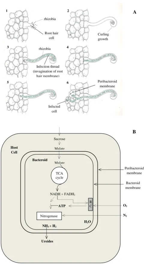
The Nod factor binds to a plant receptor kinase and triggers a complex cascade of developmental and physiological steps in the host plant(17, 27) (Figure 1 A):
- the root cortical-cells divided, leading to the formation of the nodule primordium;
- invasion of the nodule primordium by the bacteria;
- formation of the symbiosomes, which are the result of the enclose of several bacteria by a peribacteroid membrane, that segregates the bacteroids from plant cytoplasm;
- differentiation of the rhizobia into mature bacteroids, which then synthesize nitrogenase, the enzyme responsible for the reduction of N2;
- continuing plant and bacterial division and formation of the mature root nodule.
The biological fixation of N2 by the bacteriods is a process that implies a
relevant expense of energy, which is provided by photosynthetic carbon compounds synthesized by the host plant. Sucrose, delivered by the sieve tubes, is metabolized in the host cell, in phosphoenolpyruvate, by glycolysis, and then is carboxylated to oxaloacetate. The latter is then reduced to dicarboxylic acids, such as malate and succinate, the main carbon compounds used by the symbiotic bacteria(28-30). The dicarboxylic acids are then oxidized by the TCA cycle of the bacteroids in order to obtain energy and reduced equivalents (NADH) to N2
fixation.
The reduction of N2 to ammonium (NH4+) occurs in the cytoplasm of the
bacteroid by nitrogenase a complex, enzyme that requires both a low partial oxygen pressure and a high dose of ATP for its activity. However, this ion cannot cross the membranes, including the peribacteroid membrane and, consequently, cannot reach the host cell. Since NH4+ and ammonia (NH3) are readily
move across the bacteroid and the peribacteroid membrane. On the other hand, Waters et al(31) demonstrated that, in soybean nodule bacteroids, alanine, not NH3,
is excreted as product of nitrogen fixation.
The product of the bacterial nitrogenase complex move across the bacteroid and the peribacteroid membrane, being converted, in the host cell (symbiosome space), mainly into glutamine and asparagine. In some legumes, such as soybean, the fixed nitrogen is delivered to the rest of the plant as ureides, especially allantoin and allantoic acid, where it is incorporated into amino acids and proteins(17) (Figure 1B).
The establishment and maintenance of an effective symbiosis depends on several abiotic factors such as soil pH, soil fertility, drought stress and temperature(32-33). The lifespan of root nodules is relatively short even under optimal growth conditions, occurring over time a process known as nodule senescence(34), which is an irreversible phenomenon. In annual legumes, such as soybean, the nodule senescence occurs in a determined way after pod filling(35).
Figure 1- Infection process of root cells by rhizobia (A) and metabolism of a bacteroid in a root
nodule of soybean (B). RC- respiratory chain; TCA- tricarboxylic acid (Adapted)(17, 36)
rhizobia Root hair cell Curling growth Infection thread (invagination of root hair membrane) rhizobia Infected cell Peribacteroid membrane 1 2 3 4 5 6 O2 N2 Sucrose Malate Malate TCA cycle H2O NADH + FADH2 ATP Nitrogenase NH3 + H2 Ureides Host Cell Bacteroid R C Peribacteroid membrane Bacteroid membrane B A
1.4 Environmental and agricultural advantages of root-nodulating
rhizobacteria
The increase in global food demand from a growing population has pushed the use of additional nutrients inputs, especially mineral nitrogen in the soils, to increase yield of crops.
Farmers are reluctant to use traditional sources of organic nitrogen, because their application implies management problems and higher costs compared with mineral nitrogen fertilizers. However, the excessive application of mineral nitrogen is related to NO3- pollution of groundwater, soil acidification and
increase of denitrification resulting in higher emission of N2O to the atmosphere,
which could impact global warming. In addition, the manufacture of mineral nitrogen fertilizers implies the burning of fossil fuel, which is also a source of hazardous products to human health and to the environment. Besides the environmental and human health impact, the use of synthetic nitrogen fertilizers has high economic costs. For instance, in Brazil is estimated that symbiotic N2
fixation in soybeans reduces the need for nitrogen fertilization, saving $1.8 billion annually(37) while in the U.S.A the modest use of alfalfa in rotation with corn saves $200 to $300 million(38).
Legumes are used in crop rotation for their ability to establish symbiotic relationships with nitrogen-fixing bacteria, an agriculture practice that avoids the decrease in soil fertility. Besides that, the inoculation of legumes with rhizobia improves the proportion of seed yield in relation to the total plant biomass when compared with N-fertilized plants(39).
Therefore, considering all the advantages mentioned above, biological N2
fixation should be promoted.
1.5 General characterization of root-nodulating bacteria of G. max.
As above mentioned, rhizobia refers to a group of bacteria which most of them can fix N2 after establishing symbiotic relationship with legumes, inside the
root nodules. In general, rhizobia are gram-negative motile non-sporulating rods
(27)
.
Most of these bacteria belong to the Bradyrhizobiaceae and Rhizobiaceae families, where the genus Bradyrhizobium and Ensifer are included, respectively.
The microsymbionts B. japonicum, Bradyrhizobium elkanii, Bradyrhizobium
liaoningense, Bradyrhizobium canariense and Bradyrhizobium yuanmingense are
slow growing rhizobia capable of naturally nodulating soybean under field conditions(2). The fast-growers species Ensifer (Sinorhizobium) fredii, Ensifer
xinjiangense and the moderately slow grower Mesorhizobium tianshanense are
also currently known as soybean-nodulating rhizobia(40).
DuTeau et al(41) verified that the root-nodulating bacteria E. fredii was considerably less effective on nitrogen fixation than the classical soybean symbiont B. japonicum.
Although species of the genera Bradyrhizobium, Ensifer and
Mesorhizobium are as widely distributed (indigenous or naturalized) as the
legumes themselves, there are many soils where these rhizobia are absent(42). Since there are advantages for plants in establishing symbiotic relationships with Plant Growth Promoting Rhizobacteria (PGPR), the development of artificial inoculation technology has been the target of several researches. Although the inoculation of soybean seeds with highly effective rhizobia is now a common
practice in agriculture production(43), the success of this technology depends on the establishment and survival of the inoculated rhizobia in the soil environment. 2. AIMS OF THE STUDY
This study was conducted in order to assess the effect of the inoculation of
G. max with B. japonicum and E. fredii on the metabolic profile (organic acids,
phenolic and volatiles compounds) and antioxidant potential of its aerial parts.
3. MATERIALS AND METHODS
3.1 Reagents and Standards. Methanol, formic and hydrochloric acids were
obtained from Merck (Darmstadt, Germany), and sulfuric acid was obtained from Pronalab (Lisboa, Portugal). The water was treated in a Milli-Q water purification system (Millipore, Bedford, MA). DPPH was from Sigma (St. Louis, MO, U.S.A). The standards compounds were purchased from various suppliers: oxalic, citric, ascorbic, malic, fumaric, cis and trans aconitic, succinic and acetic acids, daidzein,
β-cyclocitral, E-2-hexenal were obtained from Sigma- Aldrich (St. Louis, MO, U.S.A); kaempferol, kaempferol-3-O-rutinoside, apigenin and cineole were from Extrasynthése (Genay, France); 1-hexanol was from Fluka (Buchs, Switzerland),
β-ionone and α-ionone were from SAFC (Steinheim, Germany).
3.2 Bacterial strains culture media. Strains of the species Bradyrhizobium
japonicum and Ensifer fredii were isolated from root nodules of Glycine max L.
growing in Argentina. Isolations were made according to the method of Vincent(44) with yeast mannitol agar(45). The cultures used to inoculate G. max plants were purified from a single colony after 2 days of incubation at 28 ºC and cultivated on YMA medium.
3.3 Plant material and growing conditions. Soybean seeds (G. max) were
surface sterilized with a hypoclorite solution (5%) for 15 min., and thoroughly rinsed with sterile distilled water. The seeds were germinated axenically in Petri dishes and then the seedlings were transferred to pots with sterile soil and watered with nitrogen-free Rigaud and Puppo(46) nutrient solution. Ten plant replicates were inoculated with 1 mL bacterial suspension of B. japonicum containing 8 × 10⁸ cells/mL. The same procedure was followed for samples inoculated with E. fredii. The control consisted in ten plants only watered with the same solution with addition of 1.4g/L NH4NO3. After that, inoculated and control
plants were placed for 6 weeks in a plant growth chamber with mixed incandescent and fluorescent lighting (400 microE m-2 s-1; 400 to 700 nm), programmed for a 16-h photoperiod, day-night cycle, under constant temperature (25 to 27ºC) and 50 to 60 % relative humidity. Six weeks after germination, the aerial parts were collected and stored at -20 ºC prior to their lyophilization (Labconco Freezone 4.5 apparatus; Kansas City, MO, US). The ten replicates of each treatment were then mixed and pulverized.
3.4 Volatile compounds.
3.4.1. SPME Fibers.Several commercial fibers can be used to extract volatiles.
According to bibliography, recommendations of supplier (Supelco, Bellefonte, PA, USA) and to our own knowledge based on experimental work(47-48), the fiber chosen was coated with divinylbenzene/polydimethylsiloxane (DVB/PDMS), 65
3.4.2. Headspace Solid-Phase Microextraction (HS-SPME). 1.5 g of fresh aerial
parts of G. max were kept at 40 ºC, under stirring conditions (200 rpm) for 5 min, to promote the compounds’ release. Afterwards the fiber was pulled into the needle sheath and the SPME device was removed from the vial and inserted into the injection port of the GC system for thermal desorption, for 1 min.
3.4.3. Gas Chromatography-Ion Trap Mass Spectrometry Analysis (GC-IT-MS).
The volatiles from G. max were analyzed with a Varian CP-3800 gas chromatograph (USA) coupled to a VARIAN Saturn 4000 mass selective detector (USA) and a Saturn GC/MS workstation software version 6.8. A VF-5 ms 30 m × 0.25 mm × 0.25 µm (FactorFour) column was used. The injector port was heated to 220ºC, and injections were performed in splitless mode. The carrier gas was helium C-60 (Gasin, Portugal), at a constant flow of 1 mL/min. Oven temperature was set at 40ºC (for 1 min), then increasing 2 ºC/min to 220ºC and held for 30 min. All mass spectra were acquired in electron impact (EI) mode. Ionization was maintained off during the first minute. Transfer line, manifold and trap temperatures of the ion trap detector were set at 280, 50, and 180 ºC, respectively. The MS ranged from 40 to 350m/z, with a scan rate of 6 scan/s. The emission current was 50 mA, and the electron multiplier was set in relative mode to auto tune procedure. The maximum ionization time was 25,000 ms, with an ionization storage level of 35 m/z. The analysis was performed in Full Scan mode. Compounds were identified by comparing their retention times with those of authentic compounds analysed under the same conditions, and by comparison of the retention indices (as Kovats indices) with literature data(49-50). The comparison
of MS fragmentation pattern with those of pure compounds and mass spectrum database search was performed using the National Institute of Standards and Technology (NIST 05) MS spectral database, considering fit and retrofit values higher than 70%.
3.5. Organic Acids
3.5.1 Extraction. Each lyophilized powdered (1600 µm) sample (ca 0.2 g), was extracted with H2SO4 0.01N (ca. 50 mL) for 30 minutes under stirring (300 rpm).
The obtained extracts were then filtered and evaporated to dryness under reduced pressure (40ºC) and redissolved in H2SO4 0.01N (1 mL). These extracts were
used for the organic acids analysis and DPPH●assay.
3.5.2 HPLC/UV analysis. The separation and quantification of organic acids was
carried out in a system consisting of an analytic HPLC-UV unit (Gilson) with an ion exclusion column, Nucleogel® Ion 300 OA (300 × 7.7 mm). Elution was performed in isocratic mode with H2SO4 0.01N, under a flow rate of 0.2 mL/min. The
detection was achieved with an UV detector set at 214 nm. Organic acids quantification was achieved by the absorbance recorded in the chromatograms relative to external standards.
3.6 Phenolic compounds
3.6.1 Extraction. Each lyophilized powdered (1600 µm) sample (ca 0.3 g) was mixed with methanol until negative reaction to 20% NaOH. After the extraction, the methanolic extract was filtered through a Büchner funnel, concentrated to dryness under reduced pressure (35 ºC), and redissolved in acidic water (pH=2, with HCl; ca. 50 mL). The aqueous solution obtained was passed through an Solid Phase
Extraction column (SPE) C18 (70 mL/10000 mg; Macherey-Nagel. Duren. Germany), previously conditioned with 30 mL of methanol and 70 mL of acidic water (pH=2, with HCl). The phenolic fraction retained in the column was then eluted with methanol, until negative reaction to 20 % NaOH. The methanolic extract was evaporated to dryness under reduced pressure (35 ºC) and redissolved in methanol (1 mL). These extracts were used for the phenolic compounds analysis and DPPH● assay.
3.6.2 Acid hydrolysis of the methanolic extract. The purified methanolic extract
was evaporated to dryness under reduced pressure (35ºC) and redissolved in 5 mL of HCl 2N. This acid solution was heated at 100ºC during 30 minutes. The obtained hydrolyzed extract was passed through an SPE C18 column, as above. The phenolic fraction retained in the column was eluted with methanol and the methanolic extract was evaporated to dryness under reduced pressure (35ºC).
3.6.3 HPLC/DAD analysis. 20 µL of each methanolic extract were analysed on an analytical HPLC unit (Gilson), using a Spherisorb ODS2 (25,0 × 0,46 cm; 5 µm, particle size) column. The solvent system used was a gradient of water-formic acid (19:1) (A) and methanol (B), starting with 5 % methanol and installing a gradient to obtain 15 % B at 3 min, 25 % B at 13 min, 30 % B at 25 min, 35 % B at 35 min, 45 % B at 39 min, 45 % B at 42 min, 50 % B at 44 min, 55 % B at 47 min, 70 % B at 50 min, 75 % B at 56 min and 80 % B at 60 min, at a solvent flow rate of 0.9 mL/min. Detection was achieved with a Gilson Diode Array Detector (DAD). Spectral data from peaks were accumulated in the range 200–400 nm, and chromatograms were recordedat 280 nm, 320 nm and 350 nm. The data were processed on an Unipoint System software (Gilson Medical Electronics, Villiers le
Bel, France). The compounds in each extract were identified by comparing their retention times and UV–vis spectra in the 200–400 nm range with authentic standards. Phenolic compounds quantification was achieved by the absorbance recorded in the chromatograms relative to external standards. The kaempferol derivatives were quantified as kaempferol-3-O-rutinoside.
3.7 DPPH● scavenging assay. The antiradical activity of the extracts was
determined spectrophotometrically in a Multiskan Ascent plate reader (Thermo Electron Corp.), by monitoring the decrease in absorbance of DPPH● at 515 nm. For each methanolic and acidic extract a dilution series was prepared in a 96 well plate. The reaction mixture in the sample wells consisted of 25 µL of extract and 200 µL of methanolic solution of DPPH● (150 mM). The plate was incubated for 30 minutes at room temperature after addition of DPPH. Ascorbic acid was used as the reference compound, dissolved in methanol and in H2SO4 0.01N. Three
experiments were performed in triplicate. The antiradical activity was expressed in terms of the amount of antioxidants necessary to decrease the initial DPPH absorbance by 25 % (IC25). The IC25 value for each extract was determined
graphically by plotting the percentage of DPPH● scavenging as a function of extract concentration.
3.8 Statistical analysis
All data were recorded as mean ± standard deviation of triplicate measurements. Mean values of control and plants inoculated with the two rhizobia species were compared and separated according to t-test (Graph Pad Prism version 5.00, GraphPad Software, Inc, CA, USA). A p value of < 0.05 was considered to be statistically significant.
4. RESULTS
4.1 Nodulation of G. max
Six weeks after the germination of the seeds, the vegetative parts of the plants were observed, with respect of nodulation, growth and color of the foliage.
All the plants that were inoculated with the slow-growing bacteria B.
japonicum presented visible nodules in the roots (a mean of 19 nodules per plant).
The plants had a healthy appearance, with green foliage. Soybeans inoculated with E. fredii did not nodulate and the leaves were chlorotic, with considerable necrosis and senescence.
The non-inoculated control plants did not possess nodules in their roots. The foliage had a healthy green color and no necrosis was noticed.
4.2 Volatile compounds
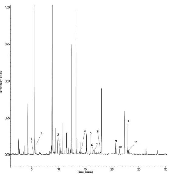
The results of the determination of volatiles in the aerial parts of G. max, by HS-SPME/GC-IT-MS, are displayed in Figure 2 and Table 1. Compounds from several classes were identified: one aldehyde (E-2-hexenal), one alcohol (1-hexanol), five monoterpenes (cineole, menthone, menthyl acetate, carvone and linalyl acetate), one sesquiterpene (α- farnesene), three apocarotenoids (α and β -ionone and β-cyclocitral) and jasmone, a ketone that is a metabolite product of jasmonic acid (JA).
E-2-hexenal in soybean inoculated with B. japonicum and E. fredii was
present in higher amounts than in control plants (p=0.0457 and p=0.6020, respectively). 1-Hexanol was not observed in plants inoculated with E. fredii and,
as for E-2-hexenal, control plants showed lower amounts than did soybean inoculated with B. japonicum (p=0.0859).
Figure 2- Chromatographic profile of volatile compounds of G.max aerial parts by
HS-SPME-GC-IT-MS (fullscan acquisition). 1- E-2-hexenal; 2- 1-Hexanol; 3- Cineole; 4-Menthone; 5- β -Cyclocitral; 6- Carvone; 7- Bergamol; 8- Menthyl acetate; 9- Jasmone; 10- α-Ionone; 11-β-Ionone;
12- α-Farnesene.
The monoterpenes cineole and carvone exhibited behavior similar to that observed for E-2- hexenal (p=0.0078 and p=0.0527 for B.japonicum; p<0.001 and
p=0.0024 for E. fredii). Concerning menthone, although this monoterpene was
present in higher amounts in soybean inoculated with B. japonicum (p=0.023), the opposite was verified for plants inoculated with E.fredii (p=0.0459). Menthyl
acetate and linalyl acetate and α-farnesene were also identified only in plants inoculated with the two symbiotic bacteria, being found in lower amounts in soybean inoculated in E. fredii.
Although in plants inoculated with B. japonicum α− and β-ionone and β -cyclocitral exhibited higher chromatographic area than those in control plants, the differences found were not statistically significant (p=0.8541, p=0.2975 and
p=0.2363, respectively). In opposition, α-ionone and β−cyclocitral in plants inoculated with E. fredii were present in lower amounts than in control plants (p=0.0154 and p=0.0032, respectively)
The aerial parts of soybeans inoculated with B. japonicum and E. fredii showed higher chromatographic area for jasmone (p=0.0002 and p=0.0004).
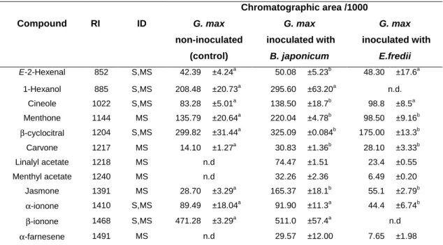
Table 1- Volatile composition of aerial parts of G. max extracts inoculated with B.japonicum and
E. fredii and non-inoculated (expressed as chromatographic area/1000).*
* Results are expressed as a mean standard deviation of three independent analysis. RI- Retention Index; ID-
Identification; S-Identified by comparison with reference compounds; MS-Tentatively identified by NIST05;
n.d.- not detected. Different letters mean statistically significant differences in same row (p< 0.05). Chromatographic area /1000 Compound RI ID G. max non-inoculated (control) G. max inoculated with B. japonicum G. max inoculated with E.fredii E-2-Hexenal 852 S,MS 42.39 ±4.24a 50.08 ±5.23b 48.30 ±17.6a 1-Hexanol 885 S,MS 208.48 ±20.73a 295.60 ±63.20a n.d. Cineole 1022 S,MS 83.28 ±5.01a 138.50 ±18.7b 98.8 ±8.5a Menthone 1144 MS 135.79 ±20.64a 220.04 ±4.78b 98.50 ±9.16b β-cyclocitral 1204 S,MS 299.82 ±31.44a 325.09 ±0.084b 175.00 ±13.3b Carvone 1217 MS 14.10 ±1.27a 30.83 ±1.36b 28.10 ±3.33b Linalyl acetate 1218 MS n.d 74.47 ±1.51 23.4 ±0.55 Menthyl acetate 1240 MS n.d 32.26 ±2.36 6.49 ±0.20 Jasmone 1391 MS 28.70 ±3.29a 165.37 ±18.1b 55.1 ±2.79b α-ionone 1410 S,MS 89.49 ±18.04a 91.90 ±11.3a 44.4 ±6.74b β-ionone 1468 S,MS 471.28 ±3.29a 511.0 ±57.4a n.d α-farnesene 1491 MS n.d 29.57 ±12.00 7.65 ±1.98
4.3 Identification and Quantification of Organic Acids by HPLC/UV.
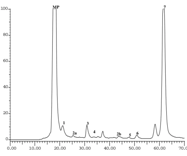
In the present study, the HPLC-UV analysis of the aerial parts of G. max (inoculated and non-inoculated with rhizobia) revealed a profile composed of seven identified organic acids: oxalic, aconitic, citric, malic, succinic, acetic and fumaric acids (Figure 3).
Figure 3. HPLC organic acid profile of G. max aerial parts (detection at 214 nm). Peaks: MP-
mobile phase; 1- oxalic acid; 2a and 2b- aconitic acid;3- citric acid; 4- malic acid; 5- succinic acid;
6- acetic acid; 7- fumaric acid.
As shown in Table 2, with exception of acetic acid, the aerial parts of G.
max inoculated with B.japonicum revealed a tendency to present higher levels of
organic acids than control plants. However, only the concentrations of oxalic, aconitic, malic and fumaric acids were statistically different (p=0.0001, p=0.0001,
p=0.001 and p=0.0240, respectively). 4 0.00 10.00 20.00 30.00 40.00 50.00 60.00 70.00 0 20 40 60 80 100 MP 7 1 3 2a 2b 5 6
The aerial parts of soybeans inoculated with E. fredii had lower amounts of citric, acetic, fumaric (p= 0.0001) and oxalic (p=0.684) acids than those of the control plants. In contrast, aconitic, malic and succinic acids were present in higher concentration (p=0.0018, p=0.001 and p=0.4236, respectively) but only the first two with statistical significance.
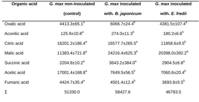
Table 2 –Organic acid composition of aerial parts of G. max extracts inoculated with rizobia and
non-inoculated (mg of organic acid kg-1 of acidic extract dry matter).*
Organic acid G. max non-inoculated
(control) G. max inoculated with. B. japonicum G. max inoculated with. E. fredii Oxalic acid 4413.3±65.1a 6066.7±24.4b 4381.5±107.4a Aconitic acid 125.9±10.8a 274.0±11.3b 180.2±6.6b Citric acid 16201.2±186.4a 16577.7±265.5a 11858.6±9.5b Malic acid 11383.4±721.8a 24216.4±625.3b 20398.0±392.2b Succinic acid 2204.8±10.2a 3643.2±384.0a 2904.5±6.8a Acetic acid 17001.4±168.8a 7649.5±56.5b 7060.6±20.4b Fumaric acid Σ 4424.7±35.4a 51330.0 4501.4±12.4b 58427.6 3693.8±5.5b 46783.5
* Results are expressed as a mean standard deviation of three independent extraction. Σ, sum of the determined organic acids. Different letters mean statistically significant differences in same row (p< 0.05).
It should be noted that the concentration of malic acid was 2.1 and 1.8 fold higher in the aerial parts of soybean inoculated with B. japonicum and E. fredii, respectively, than in the control plants. Moreover, the concentration of acetic acid was 2.2 (B. japonicum) and 2.4 (E. fredii) fold lower than in the aerial parts of the non-inoculated sample.
4.4 Identification and Quantification of Phenolic Compounds by HPLC/DAD.
Previous studies showed that the major compounds in soybean leaf extracts are kaempferol and quercetin glycosides, being richer in the first flavonoid. Ho et al(11) characterized six kaempferol derivatives in G.max leaves that were absent in G. max seeds. The major kaempferol derivative was kaempferol-3-O-rutinoside, followed by kaempferol-3-O-(2,6-di-O-α -rhamnopyranosyl)-β-galactopryranoside, kaempferol-3-O-α -L-rhamnopyranosyl-(1-6)-β-D- galactopryranoside, digalactopyranoside, kaempferol-3-O-diglucopyranoside and kaempferol-3-O-α-L-rhamnopyranosyl-(1-2)-β -D-glucopyranosyl (1-6)-β-D-galactopryranoside.
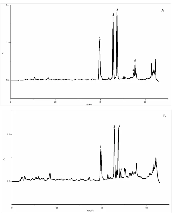
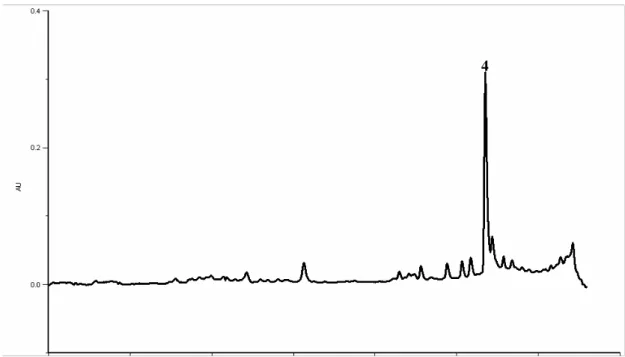
In the present study kaempferol-3-O-rutinoside was also identified as the most abundant compound in the three samples of aerial parts of soybean (Figure 4). Compounds 1 and 2 are supposed to be kaempferol derivatives according to their UV spectra and considering that this type of compounds were previously found in high amounts in the leaves, as mentioned above(11). Thus, in order to confirm this hypothesis, acid hydrolysis was performed to cleave the O-glycosyl bond, releasing the aglycone. After this treatment compounds 1-3 were no longer observed in the chromatogram and, at the same time, the peak corresponding to kaempferol was increased (Figure 5). This indicates that compounds 1 and 2 are kaempferol derivatives, putatively kaempferol-3-O-(2,6-di-O-α
-rhamnopyranosyl)-β-galactopryranoside and kaempferol-3-O-α-L-rhamnopyranosyl-(1-6)-β-D- galactopryranoside according to the results obtained by Ho et al(11).
In addition, kaempferol (a flavonol), apigenin (a flavone), and the isoflavone daidzein were also identified (Figure 4).
Figure 4- HPLC-DAD phenolics profile of G. max aerial parts. Detection at 350 nm (A) and at 280
nm (B). Peaks: 1- kaempferol derivative; 2- kaempferol derivative; 3- Kaempferol 3- O- rutinoside;
4- kaempferol; 5- apigenin; 6- daidzein
A 1 2 3 4 5 6 B 1 2 3
Figure 5- HPLC-DAD phenolics profile of G. max aerial parts after the acid hydrolysis. Detection at
350 nm. Peak: 4- kaempferol
Table 3 –Phenolic composition of aerial parts of G. max extracts inoculated with rhizobia and
non-inoculated (mg of phenolic compound kg-1 of methanolic extract dry matter).*
Phenolic compound G. max
non-inoculated
G. max inoculated
with. B. japonicum
G. max inoculated
with. E. fredii
Kaempferol derivative (peak 1) 552.1±5.5a 1481.4±1.4b 1201.5±4.2b
Kaempferol derivative (peak 2) 772.0±3,3a 1565.3±3,4b 1114.0±0.2b
Kaempferol 3- O-rutinoside 787.6±2.2a 1703.1±5.2b 1251.1±7.0b Daidzein Kaempferol Apigenin 9.3±0.0a 58.0±1.4a 39.7±0.7a 4.0±0.3b 24.6±1,2b 44.2±0.9b 6.9±0.3b 19.1±0.2b 40.7±5.3a Σ 2218.8 4822.6 3633.4
* Results are expressed as a mean standard deviation of three independent extraction. Σ, sum of the determined phenolic compound. Different letters mean statistically significant differences in same row (p< 0.05).
The methanolic extracts of the aerial parts of soybean inoculated with
B.japonicum and E. fredii showed higher levels of the three kaempferol derivatives
than control plants (p<0.0001). Apigenin content was also higher in inoculated materials, but the differences were not statistically significant (p=0.1914 and
p=0.5089). On the other hand, kaempferol was present at higher concentration in
control plants than in the inoculated soybeans (p<0.0002 and p< 0.0001), as it happened for daidzein (p< 0.0026 and p=0.0460, respectively; Table 3).
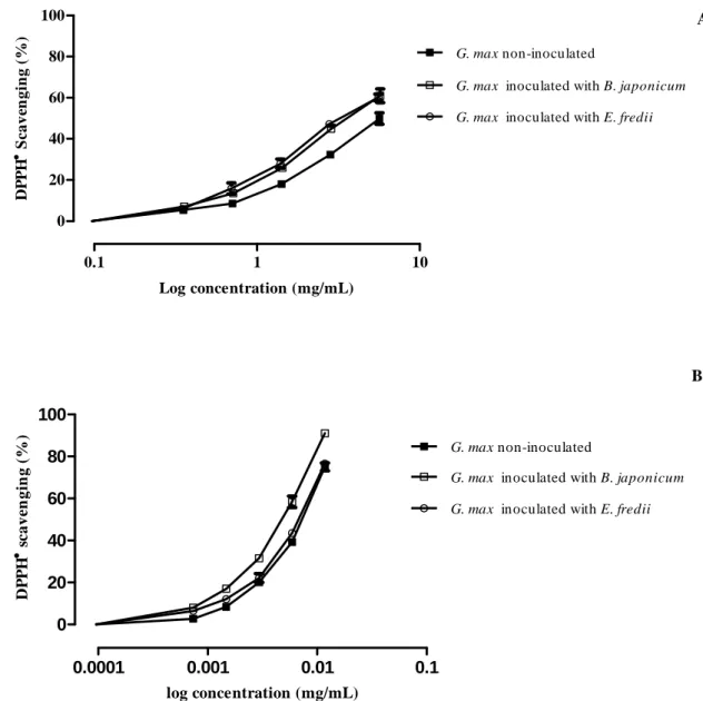
4.5 Antioxidant capacity of the methanolic and acidic extracts.
The antioxidant ability of the methanolic and acidic lyophilized extracts of soybean aerial parts was screened by the DPPH● assay (Figure 6).
Since both methanolic and acidic extracts (H2SO4 0.01N, pH=2) were used,
it was necessary to determine if the scavenging capacity against DPPH radical was affected or not by the solvent. In this study we tested methanolic and H2SO4
0.01N solutions of ascorbic acid, and IC25 values of 0.0024 and 0.0026 mg. mL -1
were found, respectively. The results obtained were not considered statistically different (p>0.05).
The methanolic extracts displayed a strong concentration-dependent antiradical activity (figure 6B). As shown in Table 4, the antioxidant capacity of the methanolic extract from the plant inoculated with B. japonicum was significantly higher (p=0.0004) than that observed for the other samples.
The three acidic extracts from the aerial parts of soybean showed weak scavenging capacity against DPPH● (Figure 6A, Table 4). Nevertheless, plants inoculated with B. japonicum and E. fredii exhibited stronger antiradical potential than the extracts from control plants (p=0.0015 and p=0.0013, respectively).
0.1 1 10 0 20 40 60 80 100
G. max inoculated with E. fredii G. max inoculated with B. japonicum G. max non-inoculated Log concentration (mg/mL) D P P H •••• S c a v en g in g ( % ) 0.0001 0.001 0.01 0.1 0 20 40 60 80 100
G. max inoculated with B. japonicum G. max inoculated with E. fredii G. max non-inoculated log concentration (mg/mL) D P P H •••• s c a v e n g in g ( % )
Figure 6- DPPH scavenging ability of acidic (A) and methanolic (B) extracts of G. max aerial parts.
Values show mean ± SE from three experiments performed in triplicate
Table 4 – IC25 values (mg. mL -1
) of methanolic and acidic extracts of the aerial parts of G. max inoculated with rhizobia and non-inoculated*.
Methanolic extract Acidic extract
Samples IC25
G.max non-inoculated 0.0036a 2.1a
G. max inoculated with B. japonicum 0.0023b 1.4b
G. max inoculated with E. fredii 0.0034a 1.2b
* Results are expressed as a mean of three independent assays. Different letters mean statistically significant
differences in same column (p< 0.05).
B A
5. DISCUSSION
Plants are sessile organisms which makes them unable to escape from a constantly wide spectrum of biotic and abiotic stresses. Thus, they have developed a broad range of strategies to cope with environmental stresses, which are translated into biochemical changes, metabolic adjustments and physiological alterations. Consequently, plant development is reprogrammed(51-54).
Changes in secondary metabolism have been the target of research on plant defense against biotic and abiotic stresses and only very recently the primary metabolism has received more attention(52, 54).
Although rhizobia constitute a group of non-pathogenic bacteria, their uptake into the host plant is considered a controlled infection. It is documented that some of them induces a phenomenon called Induced Systemic Resistance (ISR) on plants, a type of resistance characterized to be activated by benign root-colonizing bacteria, that stimulate and prime the defense mechanisms to resist a broad spectrum of plant pathogens(55). ISR induced by rhizobacteria triggers the salicylic acid-independent ISR pathway, which is dependent on jasmonic acid (JA) and ethylene signaling(56). Jasmonates (JA and related compounds, such as methyl jasmonate and cis-jasmone), are also involved in plant signaling to abiotic stress factors(57), including nitrogen deficiency(58).
In the present study, it was found that the aerial parts of soybeans nodulated with B. japonicum are richer in jasmone than of the control plants (p=0.0002) suggesting that this bacteria has induced an ISR, triggering JA and/or related compounds such as jasmone as signaling molecules. The aerial parts of soybeans inoculated with E. fredii also showed higher chromatographic area of jasmone than control plants (p= 0.004; Table 1). This rhizobial species did not
nodulate soybean, so these plants grew in a deficient nitrogen environment, which triggers the biosynthesis of jasmonates. In fact, the symptomatology presented by the plants (chlorosis, necrosis and senescence of the leaves) is indicative of deprivation of this macronutrient(36).
In addition to jasmonates, terpenes and C6 compounds have been identified
as potential signals molecules for plants and others organisms(59-60).
In this work it was found that plants inoculated with B. japonicum presented higher chromatographic area of E-2- hexenal, cineole, carvone and menthone than the control soybeans. In addition, linalyl acetate, menthyl acetate and α-farnesene were only identified in plants inoculated with rhizobia (Table 1). Some studies showed that the exogenous treatment of plants with jasmonates induced the systemic release of a blend of Volatiles Organic Compounds (VOCs), due to the activation of the multiple biosynthetic pathways of these secondary metabolites
(61-62)
. This seems to support the hypothesis that in this study jasmonates were responsible for the enhancement and/or induction of C6 compounds and terpenes
verified in soybeans inoculated with B. japonicum and E.fredii. However, in the last case, some VOCs showed a decrease, or no change, in their levels when compared with control plants probably due to the decrease in primary metabolism.
This is the first report of cineole, menthone, β-cyclocitral, carvone, α- and β -ionone, linalyl acetate and menthyl acetate in the aerial parts of G.max.
The emissions of some VOCs by soybean, induced by B. japonicum and by nitrogen deprivation, might protect the plants against phytopathogenic bacteria and insects. It was already described that E-2-hexenal is highly bactericidal (63) and reduces the aphid and spider-mite fecundity(64-65); cineole and β-cyclocitral are
repellents of insects and oviposition(66-67) and carvone and linalyl acetate are toxic to some insect species(68-69).
Besides the biological and agricultural importance, the VOCs induced and/or with enhanced biosynthesis in soybean nodulated with B.japonicum could have potential pharmaceutical/medicinal value. The jasmonates are now a possible novel family of anti-cancer agents, since some of their synthetic derivatives exhibit anti-cancer activity in vitro and in vivo(70), linalyl acetate might be a potential anti-inflammatory agent(71) and bactericidal against Staphylococcus
aureus and Escherichia coli(72). In food technology, the use of E-2-hexenal as a
natural compound to improve the shelf-life and the safety of minimally processed fruits is now a target of research(73).
The symbiotic relationship with root-nodulating rhizobia implies higher spending of carbon skeletons and energy by the plant, to maintain the growth, activity and reserves of the bacteroids. Kaschuk et al(39) showed that these
symbionts stimulate the rate of photosynthesis in 28%, when compared to N-fertilized legumes, to compensate the carbon costs. Therefore, the nodulated plants can increase their respiration rate in order to obtain more energy and reducing equivalents. The high photosynthetic rate also allows higher carbon skeletons channeling to others metabolic pathways, namely for phenolic and volatiles compounds.
As shown in Table 2, the intermediates of the TCA cycle, namely oxalic, aconitic, malic, succinic fumaric and citric acids are in higher amounts in the aerial parts of soybean nodulated with B.japonicum than in the sample treated with NO3/NH4 (control). This result suggests that the flux through the TCA cycle is
Besides energy production, organic acids are involved in others biochemical pathways, such as in the formation of precursors for amino acid and lipid biosynthesis(36).
Fluctuations in the levels of malic acid, and changes in the activity of malic-enzymes (ME), in response to biotic and abiotic stress suggest that this primary metabolite has also a role in plant defense metabolism(74). Curzi et al(75) found that the leaves of rice plants inoculated with the PGPR Herbaspirillum seropedicae have higher amounts of malic acid. It has been suggested that the non-photosynthetic cytosolic isoform of nicotinamide adenine dinucleotide phosphate malic enzyme (NADP-ME) is related to plant defense response, since this enzyme has an increased activity after infection with phytopathogens(52, 76-78).
Malic acid, that at physiological pH is present as malate, besides an intermediate in the Krebs cycle, is an intermediate of the glyoxylate cycle, a metabolic pathway that occurs in peroxisomes. Cots et al(79) found that the
pathogenic attack increases fatty acid β-oxidation, inducing a carbon reallocation mechanism based on the reinitiation of the glyoxylate cycle. In this cycle malate, could be metabolized by NADP-ME in the cytosol, an enzyme widely distributed in different metabolic pathways in prokaryotes and eukaryotes. More specifically, NADP-ME, catalyses the oxidative descarboxylation of malate to yield pyruvate, CO2 and NADPH. The last is necessary for the biosynthesis of specific defense
compounds, such as flavonoids(78), or act as a cofactor for antioxidant enzymes(80). The pyruvate may serve as a precursor for the synthesis of phosphoenolpyruvate (PEP), a molecule that is used in the shikimate pathway, leading to the synthesis of aromatic amino acids, like phenylalanine, the common substrate for lignin and flavonoid synthesis(78) (Figure 7).
The results of the studies above mentioned suggest that probably the malate content of the aerial parts of G. max inoculated with B. japonicum are probably due to the increase of the flux through the TCA cycle and also to the reinduction of the glyoxylate cycle.
Zhao et al(81) verified that nitrogen deficiency decreased the leaf area, chlorophyll content and leaf photosynthetic and respiration rates in sorghum plants, resulting in lower dry matter accumulation. Thus, since soybeans inoculated with E. fredii grew in nitrogen deficiency, which affects the photosynthetic rate, it was expected a decrease in the flux through the TCA cycle and, consequently, a decrease in the amounts of the several organic acids intermediates of this cycle. However, the amount of malic acid was 1.8 fold higher than in control plants (Table 2). Natural or induced senescence also reinduces the glyoxylate cycle in plants(79) which might explain this result in the soybeans grown under nitrogen deficiency. This organic acid can be metabolized by NADP-ME, an enzyme that can also be induced by abiotic stresses. It is known that nitrogen starvation may increase the production of Reactive Oxygen Species ROS(82), so a higher production of NADPH and pyruvate is important for the biosynthesis of compounds with scavenging properties.
With respect to phenolic compounds, the highest total amounts that were found in soybean inoculated with the two rhizobia species (Table 3), might be related with enhanced transcription of genes that codified enzymes of the phenylpropanoid pathway. In fact, previous studies have shown that JA and /or related compounds enhance the transcription of genes encoding for phenylalanine ammonia lyase (PAL)(83-84) and chalcone synthase (CHS)(85); and that the increase of phenolic compounds, due to nitrogen deficiency, is correlated with enhanced
PAL activity(86). Furthermore, probably the high concentrations of malic acid, and the enhanced activity of NADP-ME in these plants, might also contribute to the biosynthesis of these secondary metabolites, as explained above.
Curiously, soybeans grew under nitrogen deficiency, a macronutrient essential to the biosynthesis of amino acids, proteins, including enzymes, and nucleic acids, expending energy and carbon skeletons in the biosynthesis of secondary metabolites, such as phenolic compounds. Some authors proposed that the enhanced PAL activity will release nitrogen for amino-acid metabolism, whereas the carbon skeletons are shunted via 4-coumaroyl-CoA into the flavonoid biosynthetic pathway(87) (Figure 7).
Soybeans inoculated with B. japonicum and grew under nitrogen deficiency, presented lower amounts of kaempferol and higher contents of kaempferol glycosides than the control plants (Table 3). This result indicates that kaempferol biosynthesis is higher in these plants, but the majority of this flavonoid undergoes glycosylation.
Ramos-Solano et al(88) found that the isoflavone levels in different parts of
G. max var. Osumi can change after inoculation with PGPR. These authors found
that some bacterial strains increased the amounts of daidzein in the shoots and others had the opposite effect, when compared to control plants. In our study, the inoculation with B. japonicum leads to the decrease in the amount of daidzein in the aerial parts of soybean (Table 3).
The higher phenolic compounds content could have physiological importance in plants that establish symbiotic relationship with bacteria and in plants developed under nitrogen deprivation, since these compounds have antioxidant activity. In the first case, there is higher accumulation of ROS due to
the nitrogen fixing symbiosis(89) and due the higher rate of photosynthesis. In the second case, the nitrogen deficiency affects the photosynthetic membranes due to starch accumulation, which increases sensitivity to high radiation levels. The production of photoprotective pigments, such as flavanols, might afford protection against light-induced oxidative damage(90).
Agricultural strategies leading to an increase of phenolic compounds in plants is particularly important for consumers, since their consumption is associated with many potential health benefits. There is now emerging evidence that phenolic compounds and their metabolites exert modulator effects in different components of intracellular signaling cascades vital for cellular functions such as growth, proliferation and apoptosis, which explains the role of these nutrients in preventing degenerative pathologies like cardiovascular diseases, diabetes, cancer and stroke(91).
Although the total amount of phenolic compounds in plants grown under nitrogen deprivation was higher than in control plants (Table 3), the antioxidant activity of their methanolic extracts was not statistically different (Table 4, Figure 6A). These results might be explained by the antagonistic and/or synergistic and/or additive interactions between all compounds present in these methanolic extracts, including phenolics and other non- identified ones. Concerning the plants nodulated with B. japonicum, their methanolic extracts showed stronger scavenging capacity against DPPH● than control plants (Table 4; Figure 6A). These plants presented the highest total phenolic compounds content (Table 3) which, in part, might explain the stronger antioxidant capacity. Antagonistic and/or synergistic and/or additive interactions between the several compounds present in these extracts might also contribute for this result.
The acidic extracts of the aerial parts of plants inoculated with rhizobia showed stronger scavenging capacity against DPPH● than control plants (Table 4; figure 6B). However, the three samples exhibit a weak antioxidant activity when compared with the methanolic extracts. It should be noticed that, although the extract from the plants inoculated with E. fredii and nodulated with B. japonicum presented, respectively, the smallest and the highest total amount of organic acids (Table 2), their scavenging activities were similar (Table 4; Figure 6B). These results could be explained by the same reasons presented above for the methanolic extracts. With respect to human health, due their antioxidant activity, these primary metabolites can exert protection against diseases that involve oxidative damage. Besides that, the increase in organic acids content might be interesting since, in addition to an antioxidant action, they can enhance the bioavailability of polyphenols(92).
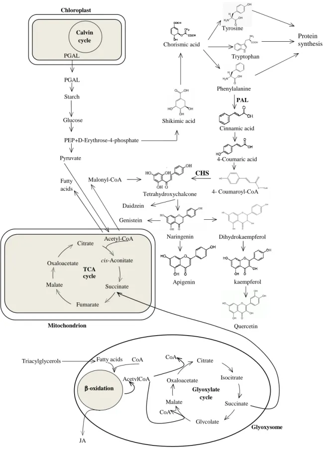
Figure 7- General and simplified biosynthetic pathway of primary and secondary metabolites in
plants. JA- jasmonic acid; CHS- chalcone synthase; PAL- phenylalanine ammonia lyase; PEP- phosphoenolpyruvate; PGAL- glyceraldehyde 3-phosphate; TCA- tricarboxylic acid.
Genistein Tetrahydroxychalcone Naringenin Malonyl-CoA Apigenin kaempferol Quercetin PEP Triacylglycerols ββββ-oxidation Oxaloacetate CoA AcetylCoA Citrate Glyoxylate cycle Malate Glycolate Isocitrate Succinate CoA
Fatty acids CoA
Glyoxysome JA Mitochondrion Pyruvate Fatty acids Oxaloacetate Citrate Malate Fumarate cis-Aconitate Succinate TCA cycle Acetyl-CoA D-Erythrose-4-phosphate Shikimic acid Chorismic acid Tyrosine Phenylalanine Tryptophan Protein synthesis PAL Cinnamic acid 4-Coumaric acid 4- Coumaroyl-CoA CHS Dihydrokaempferol Starch PGAL Calvin cycle PGAL + Daidzein Chloroplast Glucose
6. CONCLUSIONS
The results from this study showed that the nodulation with symbiotic bacteria B. japonicum and the deprivation of nitrogen (caused by the inoculation with E. fredii) changed the primary and secondary metabolism of G. max.
Data suggest that soil nodulating rhizobacteria and nutritional status might be employed to manipulate the chemical composition of the vegetative plant tissues, to improve nutritional and health benefits.
In addition, some secondary metabolites, whose production was induced and/or enhanced in G.max by symbiotic bacteria B. japonicum, besides being beneficial to their host, could be harnessed for potential use in medicine, pharmacy or food industry. Another area of research for future studies is the evaluation of VOCs as potential control agents of plant pathogens.
7. REFERENCES
1. Carter TE, Hymowitz T, Nelson RL. Biogeography, local adaptation, vavilov, and genetic diversity in soybean. Heidelberg: Springer Verlag; 2004.
2. Vinuesa P, Rojas-Jimenez K, Contreras-Moreira B, Mahna SK, Prasad BN, Moe H, et al. Multilocus sequence analysis for assessment of the biogeography and evolutionary genetics of four Bradyrhizobium species that nodulate soybeans on the asiatic continent. Appl Environ Microbiol. 2008; 74:6987-96.
3. Franke AA, Custer LJ, Cerna CM, Narala K. Rapid HPLC analysis of dietary phytoestrogens from legumes and from human urine. Proc Soc Exp Biol Med. 1995; 208(1):18-26.
4. Cavallini DC, Abdalla DS, Vendramini RC, Bedani R, Bomdespacho LQ, Pauly-Silveira ND, et al. Effects of isoflavone-supplemented soy yogurt on lipid parameters and atherosclerosis development in hypercholesterolemic rabbits: a randomized double-blind study. Lipids Health Dis. 2009; 8:40. 2765949.
5. Riesco E, Aubertin-Leheudre M, Maltais ML, Audet M, Dionne IJ. Synergic effect of phytoestrogens and exercise training on cardiovascular risk profile in exercise-responder postmenopausal women: a pilot study. Menopause. 2010.
6. Barnes S. The biochemistry, chemistry and physiology of the isoflavones in soybeans and their food products. Lymphat Res Biol 2010; 8:89-98.
7. Majid S, Dar AA, Shahryari V, Hirata H, Ahmad A, Saini S, et al. Genistein reverses hypermethylation and induces active histone modifications in tumor suppressor gene B-Cell translocation gene 3 in prostate cancer. Cancer. 2010; 116(1):66-76.
8. Cunha-Rodrigues M, Portugal S, Prudencio M, Goncalves LA, Casalou C, Buger D, et al. Genistein-supplemented diet decreases malaria liver infection in mice and constitutes a potential prophylactic strategy. PLoS One. 2008; 3:e2732. 2443290.
9. Lee JH, Ha TJ, Baek I-Y, Han W-Y, Cho KM, Park K-Y, et al. Evaluation of Isoflavones from the Leaves of Soybean (Glycine max L.) Cultivars. J Appl Biol Chem. 2008; 51:172-75.
10. Ho HM, Leung LK, Chan FL, Huang Y, Chen ZY. Soy Leaf Lowers the Ratio of Non-HDL to HDL Cholesterol in Hamsters. J Agric Food Chem. 2003; 51:4554-58.
11. Ho HM, Chen RY, Leung LK, Chan FL, Huang Y, Chen ZY. Difference in flavonoid and isoflavone profile between soybean and soy leaf. Biomed Pharmacother. 2002; 56:289-95. 12. Graham TL. Flavonoid and isoflavonoid distribution in developing soybean seedling tissues and in seed and root exudates. Plant Physiol. 1991; 95:594-603. 1077573.
13. Klejdus B, Mikelova R, Petrlova J, Potesil D, Adam V, Stiborova M, et al. Evaluation of isoflavone aglycon and glycoside distribution in soy plants and soybeans by fast column high-performance liquid chromatography coupled with a diode-array detector. J Agric Food Chem. 2005; 53:5848-52.
14. Porter PM, Banwart WL, Hassett JJ. Phenolic acids and flavonoids in soybean root and leaf extracts. Environmental and Experimental Botany. 1986; 26:65-73.
15. Romani A, Vignolini P, Galardi C, Aroldi C, Vazzana C, Heimler D. Polyphenolic content in different plant parts of soy cultivars grown under natural conditions. J Agric Food Chem. 2003; 51:5301-6.
16. Benkeblia N, Shinano T, Osaki M. Metabolite profiling and assessment of metabolome compartmentation of soybean leaves using non-aqueous fractination and GC-MS analysis. Metabolomics. 2007; 3:297-305
17. Heldt H-W. Plant biochemistry. 3rd ed. Amsterdam: Elsevier; 2005. p. 275-324.
18. Krishnan HB, Kim WS, Sun-Hyung J, Kim KY, Jiang G. Citrate synthase mutants of Sinorhizobium fredii USDA257 form ineffective nodules with aberrant ultrastructure. Appl Environ Microbiol. 2003; 69:3561-8. 161545.
19. Limpens E, Bisseling T. Signaling in symbiosis. Curr Opin Plant Biol. 2003; 6:343-50. 20. Lang K, Lindemann A, Hauser F, Gottfert M. The genistein stimulon of Bradyrhizobium japonicum. Mol Genet Genomics. 2008; 279:203-11.
21. Badri VD, Vivanco JM. Regulation and function of root exudates. Plant, Cell and Environment. 2009; 32:666-81.
22. Rudrappa T, Czymmek KJ, Pare PW, Bais HP. Root-secreted malic acid recruits beneficial soil bacteria. Plant Physiol. 2008; 148:1547-56. 2577262.
23. Caetano-Anolles G, Crist-Estes DK, Bauer WD. Chemotaxis of Rhizobium meliloti to the plant flavone luteolin requires functional nodulation genes. J Bacteriol. 1988; 170:3164-69. 211264.
24. Barbour WM, Hattermann DR, Stacey G. Chemotaxis of Bradyrhizobium japonicum to soybean exudates. Appl Environ Microbiol. 1991; 57:2635-39. 183632.
25. Parke D, Rivelli M, Ornston LN. Chemotaxis to aromatic and hydroaromatic acids: comparison of Bradyrhizobium japonicum and Rhizobium trifolii. J Bacteriol. 1985; 163:417-22. 219138.
26. Subramanian S, Stacey G, Yu O. Distinct, crucial roles of flavonoids during legume nodulation. Trends Plant Sci. 2007; 12(7):282-5.
27. Madigan MT, Martinko JM, Parker J. Brock Biology of Microorganisms. 10th ed. New Jersey: Prentice Hall; 2003.
28. Streeter JG. Transport and metabolism of carbon and nitrogen in legume nodules. Adv Bot Res. 1991; 18:129-87.
29. Udvardy MK, Day DA. Metabolite transport across symbiotic membranes of root nodules. Annu Rev Plant Physiol. 1997; 48:493-523.
30. Lodwig E, Poole P. Metabolism of Rhizobium Bacteroids. Critical Reviews in Plant Sciences. 2003; 22:37-78.