CÁRMEN LÚCIA DE VASCONCELOS NÓBREGA
Mice urinary bladder chemical carcinogenesis:
Evaluation of the therapeutic effect of
gemcitabine, sirolimus and everolimus
Advisor: Professora Doutora Paula Alexandra Martins Oliveira
Universidade de Trás-os-Montes e Alto Douro Vila Real, 2012
This work was prepared as an original dissertation for the degree of Doctor in Veterinary Science
This work was funded by Fundação Para a Ciência e a Tecnologia, Ministério da Educação e Ciência (Portugal): (SFRH/BD/40896/2007)
v To my parents
vii To Prof. Dra. Paula Alexandra Martins de Oliveira
ix
ACKNOWLEDGEMENTS
This PhD thesis would not have been possible without the help and support of many people and institutions to whom I would like to express my gratefulness.
First I would like to overstate my gratitude to my advisor Prof. Dra. Paula Alexandra Martins de Oliveira with whom I had the honor and the pleasure of working. With her enthusiasm, inspiration, and her wonderful ability to explain things clearly and simply, she has taught me, both consciously and unconsciously, how good experimental work is done. The joy and enthusiasm she has for her research was contagious and motivational. Throughout all this period, she provided sound advice, encouragement, good teaching, good company, friendship and lots of good ideas. I would have been lost without her.
To Prof. Dra. Aura Colaço, who opened me the door for this wonderful team, for her dedication, support, encouragement and her kind words in every situation.
To Prof. Dra. Maria dos Anjos Pires, who provided conditions in the Laboratory of Pathological Anatomy of UTAD for some of the experimental work, for her kindness and for such a lovely companion in some of the long journeys by the microscope.
To Prof. Dr. Bruno Colaço, who I am please to meet for a long time, since we both were child, and who for the first time talk to me about the possibility of doing my PhD in our university, UTAD.
To Dra. Regina Arantes, my companion on this PhD journey, for all the help, all the talks, suggestions and all the laughs. Thank you for Florence, Siena, Venice, Pisa and those still to come.
x
To Andreia Henriques, a passionate for science, for all the help through this period and to be an example of perseverance.
To Dra. Céu Costa, from Universidade Fernando Pessoa, for her important collaboration in the immunohistochemistry technique and for her kindness.
To Dra. Rosário Pinto-Leite for her important collaboration in the cellular lines studies, and for the way how we were received in her laboratory.
To Prof. Dra. Rita Ferreira from Universidade de Aveiro, for her kindness and important collaboration that made possible immunobloting analysis and the determination of mTOR and Akt expression in cell lines.
To Mrs. Lígia Bento, from the Laboratory of Pathological Anatomy of UTAD, a special acknowledgment for all of her work regarding histological material fundamental for this thesis.
To Mrs. Ana Maria, for her kind help in cleaning all the material used during the experimental phase of this thesis, and for her ever-present smile.
To Agrarian School of Viseu (Polytechnique Institute of Viseu) and in particular to Zootechnic, Rural Engineering and Veterinary Department, for the encouragement to finish this academic step.
To Fundação da Ciência e Tecnologia, we gratefully acknowledge the funding sources that made this PhD work possible (SFRH/BD/40896/2007 and PTDC/DES/114122/2009).
xi Lastly, I would like to thank my family and friends. Family is a gift of God. You don’t choose it, but I have the best I could ever ask. Friends are the family you choose, and I consider myself fortunate for all the friends I have collect in life. They were always beside me and gave me strength, encouragement and happy moments all along this period. For my parents, a special word of gratitude for their love, encouragement and life example and for being always there for me. Without them, everything would be much difficult. To Lara and Néné, next to whom life is always fun and simple.
xiii
RESUMO
O cancro é uma doença que afeta milhões de pessoas em todo mundo. De acordo com a Organização Mundial de Saúde uma em cada oito mortes é provocada por cancro. Numa escala global, esta doença provoca mais mortes do que a combinação de doenças como a síndrome de imunodeficiência adquirida, a tuberculose e a malária.
O cancro da bexiga é o sétimo tumor mais comum no homem e o segundo quando nos concentramos no trato urogenital, sendo precedido apenas pelo cancro da próstata. Exibe predisposição sexual, sendo o homem cerca de quatro vezes mais afetado do que a mulher. Em termos histológicos, a maioria dos tumores da bexiga são superficiais, limitados à mucosa, submucosa ou lâmina própria. O carcinoma invasivo é menos frequente, no entanto assume uma grande importância clínica, dado o seu difícil tratamento, a elevada possibilidade de metastização e em consequência, ter um prognóstico reservado.
O modelo animal de carcinoma invasivo da bexiga induzido pela administração prolongada na água de bebida da N-butil-N-(4-hidroxibutil)nitrosamina (BBN) a murganhos é excelente para a investigação em oncologia urológica, porque permite avaliar a ação de novos agentes terapêuticos ou profiláticos e estudar os mecanismos biopatológicos associados ao crescimento, invasão e metastização dos tumores. Foram objetivos desta tese de doutoramento avaliar o efeito terapêutico de alguns fármacos, isolados ou combinados (gencitabina, sirolimus, everolimus), no modelo invasivo do cancro da bexiga em murganhos; avaliar a ação in vitro do everolimus em linhas celulares humanas de tumores de bexiga (T24; HT1376; 5637); avaliar o stresse oxidativo hepático e mitocondrial induzido pela BBN e ainda estudar a expressão da caderina E e da β catenina nas lesões uroteliais induzidas pela BBN.
Para a realização da componente prática foram utilizados 106 murganhos macho da estirpe ICR, 86 dos quais foram expostos à BBN durante 12 semanas. Destes, 60 foram divididos em 4 grupos de 15 animais, para avaliar o efeito terapêutico dos fármacos acima mencionados. Os restantes 26 animais fizeram parte do grupo controlo. A gencitabina e o sirolimus foram administrados por via intraperitoneal e o everolimus foi administrado por via oral. Todos os animais foram sacrificados no final do trabalho experimental e submetidos a necrópsia.
Os resultados obtidos permitem-nos afirmar que o uso isolado da gencitabina e do sirolimus diminui a incidência das lesões uroteliais quimicamente induzidas, no entanto, a sua associação, no mesmo modelo, não teve qualquer efeito. O everolimus
xiv
não teve efeito significativo na diminuição da incidência das lesões uroteliais quimicamente induzidas pela BBN. Os resultados do ensaio in vitro obtidos com o everolimus foram variáveis, havendo resultados promissores apenas na linha celular 5637. No que diz respeito às alterações hepáticas induzidas pela BBN podemos afirmar que histologicamente não foram observadas lesões, no entanto, foi detetado um aumento do stresse oxidativo mitocondrial que poderá favorecer a hepatotoxicidade quando a BBN é associada a fármacos sujeitos a biotransformação hepática. Neste modelo, as informações relativas à expressividade da caderina E e β catenina, moléculas de adesão celular, são escassas. Nesse sentido, ambas as moléculas foram avaliadas em cortes histológicos do urotélio normal, de lesões pré-neoplásicas e pré-neoplásicas. Na hiperplasia simples e nodular, predominou o padrão identificado no urotélio normal (marcação membranar). Nas lesões displásicas predominou o padrão citoplasmático. Nos carcinomas invasivos observámos um padrão heterogéneo, com a coexistência de marcação citoplasmática e nuclear e por vezes também membranar. A análise da expressividade destas moléculas permitiu-nos concluir que, à semelhança do que está descrito no Homem, também em murganhos estas podem ser consideradas como indicadoras do grau de agressividade e da evolução do tumor.
Palavras-chave: carcinogénese química; N-butil-N-(4-hidroxibutil)nitrosamina; carcinoma invasivo; murganho; modelos animais; sirolimus; gencitabina; everolimus; caderina E; B-catenina; hepatotoxicidade.
xv
ABSTRACT
Cancer is a devastating disease affecting millions of people all over the world. According with World Health Association, worldwide one in eight deaths is due to cancer. In a global scale, cancer causes more deaths than acquired immunodeficiency syndrome, tuberculosis, and malaria combined.
Bladder cancer is the seventh most common tumour in man, and the second most common tumour of the genitourinary system, being preceded only for prostate cancer. It has sexual predisposition, with males being up to four times more affected than females. Histologically, the majority of bladder tumours are superficial, confined to mucosa, submucosa or lamina propria. Invasive carcinoma is less frequent, however it assumes great clinical importance, since it is difficult to treat, metastasis is a strong possibility and the prognosis is less favourable.
Mice model of invasive bladder cancer, induced by oral administration of N-butyl-N-(4-hydroxybutyl)nitrosamine (BBN) in drinking water, is a good and widely used research model for urologic oncology. It allows the evaluation of new therapeutic or prophylactic agents and also to study the basic mechanisms of tumour growth, invasion and metastasis.
In this thesis we evaluate the therapeutic efficiency of several drugs (gemcitabine, sirolimus, everolimus) alone or in combination in chemically induced bladder cancer in male mice. Everolimus was also analysed in vitro, in three different bladder cancer cell lines, two invasive (T24; HT1376) and one superficial (5637). Hepatic and mitochondrial oxidative stress induced by BBN and the analysis of E-cadherin and β-catenin were also executed.
106 male ICR mice were used during experimental work. 86 of them were exposed to BBN for 12 weeks, and from these, 60 were divided into four groups of 15 animals each, to analyse the therapeutic effects of drugs. The remaining 26 mice were used as control animals. Gemcitabine and sirolimus were administered by intraperitoneal route. Everolimus was administered by gavage. All animals were euthanized at the end of the experimental work and submitted to a complete necropsy. According to our results, gemcitabine and sirolimus used as single agents, reduce the incidence of chemically induced urothelial lesions in mice. However, their combination did not add any improvement regarding urothelial incidence. Everolimus showed no significant effect in decreasing urothelial lesions incidence. In vitro, its effect on proliferation and apoptosis across different bladder cancer cell lines was heterogeneous. Promissory results were seen only in cell-line 5637.
xvi
BBN is metabolized mainly in liver. Regarding hepatic changes induced by BBN, we can say that liver histological evaluation did not show any microscopic changes compatible with liver stress, however mitochondrial oxidative stress was detected. This can lead to hepatotoxicity, when BBN is combined with drugs with liver biotransformation.
E-cadherin and β–catenin are adhesion molecules that were evaluated in control mice and in mice exposed to BBN. In simple and nodular hyperplasia, membrane staining was dominant. In dysplasia, cytoplasmic pattern was prevalent and in invasive carcinomas a heterogeneous staining was observed (cytoplasmic, nuclear and membrane staining). Like in man, also in mice these adhesion molecules can be considered as indicators of tumour aggressiveness and evolution.
Key-words: chemical carcinogenesis; N-butyl-N-(4-hydroxybutyl)nitrosamine; invasive carcinoma; mouse; animal models; sirolimus; gemcitabine; everolimus; E-cadherin; β–catenin; hepatotoxicity.
xvii
LIST OF PAPERS, POSTERS AND ORAL PRESENTATIONS RELATED WITH THIS EXPERIMENTAL WORK
1.1–FULL PAPERS PUBLISHED IN SCI INDEXED JOURNALS
- Vasconcelos-Nóbrega C, Costa C, Arantes-Rodrigues R, Henriques A, Vala H, Colaço A, Santos L, Lopes C, Oliveira PA. E-cadherin and β-catenin expression during urothelial carcinogenesis induced by N-butyl-N-(4-hydroxybutyl)nitrosamine in mice (accepted for publication in Urologia Internationalis).
- Vasconcelos-Nóbrega C, Colaço A, Lopes C, Oliveira PA (2012). BBN as a urothelial carcinogen. In vivo, 26(4):727-39 (IF1.159; Q3).
- Vasconcelos-Nóbrega C, Pinto-Leite R, Arantes-Rodrigues R, Ferreira R, Brochado P, Cardoso ML, Palmeira C, Salvador A, Guedes-Teixeira CI, Colaço A, Palomino LF, Lopes C, Santos L, Oliveira PA (2011). In vivo and in vitro effects of RAD001 on bladder cancer. Urol Oncol-Semin Ori, Dec 9 (IF3.172; Q2; Times cited: 1).
- Oliveira MM, Teixeira JC, Vasconcelos-Nóbrega C, Félix LM, Sardão VA, Colaço AA, Oliveira PA, Peixoto FP (2011). Mitochondrial and liver oxidative stress alterations induced by N-butyl-N-(4-hydroxybutyl)nitrosamine: relevance for hepatotoxicity. J Appl
Toxicol, Nov 16 (IF2.322; Q2).
- Vasconcelos-Nóbrega C, Colaço A, Santos L, Vala H, Palomino LF, Lopes C, Oliveira PA (2011). Experimental study of the anti-cancer effect of gemcitabine combined with sirolimus on chemically induced urothelial lesions. Anticancer Research, 31(5):1637-1642 (IF1.656; Q3; Times cited: 1).
1.2-ABSTRACTS PUBLISHED IN SCI INDEXED JOURNALS
- Vasconcelos-Nóbrega C, Costa C, Arantes-Rodrigues R, Henriques A, Vala H, Colaço A, Santos L, Lopes C, Oliveira PA (2012). E-cadherin in mice: expression in normal urothelium, pre-neoplastic and neoplastic urothelial lesions. Virchows Archiv (accepted for publication).
xviii
- Vasconcelos-Nóbrega C, Colaço A, Santos L, Vala H, Guedes-Teixeira CI, Palomino LF, Lopes C, Oliveira PA (2011). In vivo activity of rapamycin in a mice model of bladder cancer. Tumour Biol 32 Suppl 1:S69.
- Vasconcelos-Nóbrega C, Arantes-Rodrigues R, Colaço A, Santos L, Vala H, Palomino LF, Lopes C, Oliveira PA (2011). Mice: an animal model for bladder cancer.
Virchows Archiv 495: S288-S289.
- Vasconcelos-Nóbrega C, Colaço AA, Santos LL, Vala H, Pinto-Leite R, Palomino LF, Lopes C, Oliveira PA (2010). mTor inhibitors Rapamycin and RAD001 in chemically induced bladder cancer. International Journal of Molecular Medicine 26:S42-S42.
- Vasconcelos-Nóbrega C, Colaço AA, Santos LL, Vala H, Palomino LF, Lopes CS, Oliveira PA (2010). Gemcitabine vs sirolimus in chemically induced bladder tumours: preliminary results. Virchows Archiv 457: 245.
- Vasconcelos-Nóbrega C, Colaço A, Santos L, Vala H, Palomino LF, Lopes C, Oliveira PA (2009). The effect of gemcitabine on urothelial lesions induced by N-butyl-N-(4-hydroxybutyl)nitrosamine in ICR male mice. International Journal of Molecular Medicine 24 (Suppl 1): S27.
- Vasconcelos-Nóbrega C, Colaço A, Santos L, Vala H, Palomino LF, Lopes C, Oliveira PA (2009). The effect of RAD001 on urothelial lesions induced by N-butyl-N-(4-hydroxybutyl)nitrosamine in ICR male mice. Virchows Archiv 455: 236.
1.3-POSTER PRESENTATIONS IN INTERNATIONAL CONFERENCES
- Vasconcelos-Nóbrega C, Costa C, Arantes-Rodrigues R, Henriques A, Vala H, Colaço A, Santos L, Lopes C, Oliveira PA (2012). E-cadherin in mice: expression in normal urothelium, pre-neoplastic and neoplastic urothelial lesions. 24th European Congress of Pathology. September 8th - 12 th, Prague, Czech Republic.
- Vasconcelos-Nóbrega C, Costa C, Vala H, Colaço A, Santos L, Lopes C, Oliveira PA (2012). Β cathenin in mice: expression in normal urothelium and in chemically induced pre-neoplastic and neoplastic lesions. 30th Meeting of the European Society of
xix
- Vasconcelos-Nóbrega C, Colaço A, Santos L, Vala H, Guedes-Teixeira CI, Palomino LF, Lopes C, Oliveira PA (2011). In vivo activity of rapamycin in a mice model of bladder cancer. 39th Meeting of the ISOBM. October 15th-19th, Florence, Italy.
- Vasconcelos-Nóbrega C, Arantes-Rodrigues R, Colaço A, Santos L, Vala H, Palomino LF, Lopes C, Oliveira PA (2011). Mice: an animal model for bladder cancer. 23rd European Congress of Pathology. August 27th – September 1st,Helsinki, Finland. - Vasconcelos-Nóbrega C, Colaço AA, Santos LL, Vala H, Pinto-Leite R, Palomino LF, Lopes C, Oliveira PA (2010). mTor inhibitors Rapamycin and RAD001 in chemically induced bladder cancer. 15th WorldCongress on Advances in Oncology and 13th International Symposium on Molecular Medicine. October 7th - 9 th, Loutraki, Greece. - Vasconcelos-Nóbrega C, Colaço AA, Santos LL, Vala H, Palomino LF, Lopes CS, Oliveira PA (2010). Gemcitabine vs. Sirolimus in chemically induced bladder tumours: preliminary results. Intercongress Meeting of the European Society of Pathology. August 31st - 3rd September, Krakow, Poland.
- Vasconcelos-Nóbrega C, Colaço A, Santos L, Vala H, Palomino LF, Lopes C, Oliveira PA (2009). The effect of gemcitabine on urothelial lesions induced by N-butyl-N-(4-hydroxybutyl)nitrosamine in ICR male mice. 14th WorldCongress on Advances in Oncology and 12th International Symposium on Molecular Medicine. October 15th -17th, Loutraki, Greece.
- Vasconcelos-Nóbrega C, Colaço A, Santos L, Vala H, Palomino LF, Lopes C, Oliveira PA (2009). The effect of RAD001 on urothelial lesions induced by N-butyl-N-(4-hydroxybutyl)nitrosamine in ICR male mice. European Congress of Pathology. September 4th -9th, Florence, Italy.
1.4-POSTER PRESENTATIONS IN NATIONAL CONFERENCES
- Vasconcelos-Nóbrega C, Arantes-Rodrigues R, Henriques A, Colaço AA, Ginja MD, Santos LL, Vala H, Palomino LF, Lopes CS, Oliveira PA (2009). Urethral tumour in mice induced by N-butyl-N-(4-hydroxybutyl) nitrosamine. Sociedade Portuguesa de Patologia Animal. Vila Real, 15 de Maio.
xx
1.5-ORAL PRESENTATIONS IN NATIONAL CONFERENCES
- Vasconcelos-Nóbrega C, Arantes-Rodrigues R, Henriques A, Colaço AA, Vala H, Oliveira PA (2012). Welfare of rodents in chemically induced bladder cancer. II SPCAL Meeting. Lisboa, 24-25 de Maio.
- Vasconcelos-Nóbrega C, Colaço AA, Vala H, Oliveira PA (2010). Bladder carcinogenicity of N-butyl-N-(4-hydroxybutyl)nitrosamine. XV Encontro da Sociedade Portuguesa de Patologia Animal. Viseu, 12 de Março.
xxi
Table of Contents
ACKNOWLEDGEMENTS ... ix
RESUMO ... xiii
ABSTRACT... xv
LIST OF PAPERS, POSTERS AND ORAL PRESENTATIONS RELATED WITH THIS EXPERIMENTAL WORK ... xvii
LIST OF FIGURES ... xxvi
LIST OF TABLES ...xxviii
LIST OF ABREVIATURES ... xxix
CHAPTER 1 1. General introduction ...3
1.1- Bladder cancer ...3
1.2. Research in bladder cancer ...5
1.2.1. Cell lines ...5
1.2.2. Animal models and novel drugs for bladder cancer treatment ...6
1.3. References ...9 CHAPTER 2 2. Aims ... 17 CHAPTER 3 3. BBN as a urothelial carcinogen ... 21 3.1. Introduction ... 21
3.2. Experimental models of bladder cancer ... 21
3.3. Bladder tumour cell lines ... 25
3.4. Nitrosamines ... 25
3.4.1. N-butyl-N-(4-hydroxybutyl)nitrosamine (BBN) ... 25
3.4.1.1. Urothelial lesions induced by BBN ... 28
3.4.1.2 Extra urinary bladder tumours ... 31
xxii
3.6. Genetic alterations in BBN-induced urothelial tumours ... 34 3.7. Other chemicals ... 34 3.8. Conclusions ... 35 3.9. References ... 36
CHAPTER 4
4. Experimental study of the anti-cancer effect of gemcitabine combined with sirolimus on chemically induced urothelial lesions ... 49
4.1. Introduction ... 49 4.2. Material and Methods ... 50 4.2.1. Chemicals ... 50 4.2.2. Animals ... 50 4.2.3. Animal experiments ... 51 4.2.4. Evaluation of treatment ... 52 4.2.5. Statistical analysis ... 53 4.3. Results ... 53 4.3.1. General findings ... 53 4.3.2. Effects of BBN on urothelial tumorigenesis ... 54 4.3.3. Effects of gemcitabine, sirolimus and their association on urothelial tumorigenesis in mice. ... 55
4.3.4. Non-urothelial lesions ... 57 4.4. Discussion... 57 4.5. References ... 58
CHAPTER 5
5. In vivo and in vitro effects of everolimus on bladder cancer ... 63 5.1. Introduction ... 63 5.2. Material and Methods ... 64 5.2.1. In vivo studies ... 64 5.2.1.1 Chemicals ... 64
xxiii 5.2.1.2. Animals ... 64 5.2.1.3. Animal experiments ... 64 5.2.1.4. Evaluation of treatment ... 65 5.2.1.4.1-Histology ... 65 5.2.1.4.2-Immunohistochemistry ... 66 5.2.1.4.2-1: Immunohistochemical evaluation ... 66 5.2.2. In vitro studies ... 66 5.2.2.1. Chemicals ... 66 5.2.2.2. Tumour cell lines and culture conditions ... 66 5.2.2.3. Drug exposure ... 67 5.2.2.4. Cell viability assay ... 67 5.2.2.5. TUNEL assay ... 68 5.2.2.6. Cell-cycle analysis ... 68 5.2.2.7. mTOR and Akt expression analysis ... 69 5.2.3. Statistical analysis ... 69 5.3. Results ... 70 5.3.1. In vivo results ... 70 5.3.1.1. Animal growth and water and food consumption ... 70 5.3.1.2. Macroscopic evaluation ... 71 5.3.1.3. Microscopic evaluation... 71 5.3.1.3-1: Histology ... 71 5.3.1.3-2: Immunohistochemistry ... 72 5.3.2. In vitro results ... 74 5.3.2.1. Cytotoxic effect of everolimus against bladder-cancer cell lines ... 74 5.3.2.2. TUNEL assay ... 75 5.3.2.3. Cell cycle and apoptosis analysis... 76 5.3.2.4. Effects of everolimus on mTOR and Akt activation ... 77
xxiv
5.4. Discussion... 78 5.5. References ... 80
CHAPTER 6
6. Mitochondrial and liver oxidative stress alterations induced by N-butyl-N-(4-hydroxybutyl) nitrosamine – relevance for hepatotoxicity ... 87 6.1. Introduction ... 87 6.2. Materials and Methods ... 88 6.2.1. Chemicals ... 88 6.2.2. Animals ... 88 6.2.3. Animal Experiments ... 88 6.2.4. Isolation of mouse liver mitochondria and mitochondrial respiratory rates 89 6.2.5. Mitochondrial membrane potential measurements ... 90 6.2.6. Mitochondrial enzyme assays ... 90 6.2.7. Western Blot analysis ... 91 6.2.8. Calcium-induced mitochondrial permeability transition pore ... 91 6.2.9. Hydrogen peroxide generation ... 92 6.2.10. Lipid peroxidation ... 92 6.2.11. Reduced glutathione quantification ... 92 6.2.12. Antioxidant enzymatic activities ... 93 6.2.13. Statistical analysis ... 93 6.3. Results ... 94 6.3.1. Animal growth, water and food consumption ... 94 6.3.2. Macroscopic and microscopic evaluation ... 94 6.3.3. Mitochondrial Bioenergetics ... 96 6.3.4. Mitochondrial complex protein levels ... 98 6.3.5. Measurement of the mitochondrial permeability transition (MPT) ... 99 6.3.6. Effect of BBN treatment on mitochondrial and liver oxidative stress... 100
xxv 6.4. Discussion... 102 6.5. References ... 105
CHAPTER 7
7. E-cadherin and β-catenin expression during urothelial carcinogenesis induced by N-butyl-N-(4-hydroxybutyl) nitrosamine in mice. ... 113 7.1. Introduction ... 113 7.2. Material and methods ... 114 7.2.1. Chemicals ... 114 7.2.2. Animals ... 114 7.2.3. Animal experiments ... 114 7.2.4. Histological evaluation ... 115 7.2.5. Immunohistochemistry evaluation ... 116 7.2.6. Statistical analysis ... 117 7.3. Results ... 117 7.3.1. E-cadherin ... 117 7.3.2. β-catenin... 120 7.4. Discussion... 122 7.5. References ... 123 CHAPTER 8 8. General discussion ... 127 8.1 References... 131 CHAPTER 9 9. Final Considerations ... 137 9.1. References ... 138
xxvi
LIST OF FIGURES
CHAPTER 2
Figure 1: Animal models of bladder cancer. ...7
CHAPTER 3
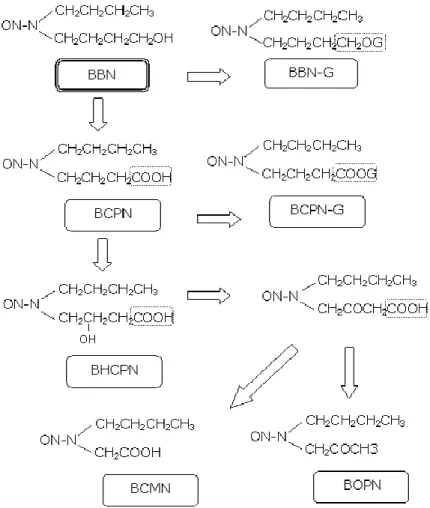
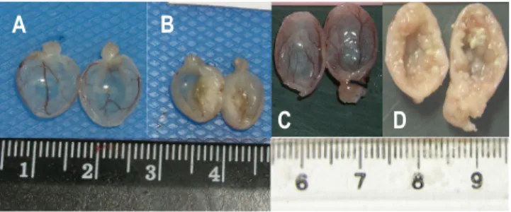
Figure 1: Metabolism of N-butyl-N-(4-hydroxybutyl)nitrosamine (BBN) ... 27 Figure 2: Macroscopic appearance of the mouse (A, B) and rat (C, D) bladder. B and D were exposed to N-butyl-N-(4-hydroxybutyl)nitrosamine. A: Normal bladder; B: Invasive carcinoma; C: Normal bladder; D: Papillary neoplasm. ... 28
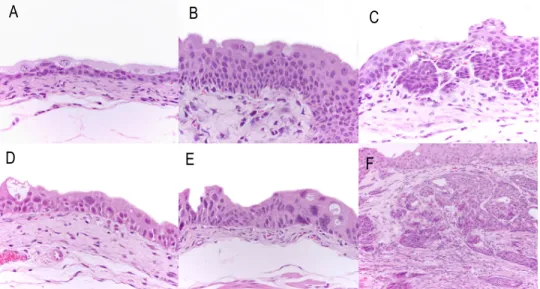
Figure 3: Histopathological evaluation of lesions identified in mouse urothelial carcinogenesis induced by BBN. A: Normal urothelium (H&E, x400). B: Simple hyperplasia (H&E, x600). C: Nodular hyperplasia (H&E, x400). D: Dysplasia (H&E, x600). E: Carcinoma in situ (H&E, x400). F: Invasive carcinoma with squamous differentiation (H&E, x400). ... 30
Figure 4: Histopathological evaluation of lesions identified in rat urothelial carcinogenesis induced by BBN. A: Normal urothelium (H&E, x400). B: Simple hyperplasia (H&E, x200). C: Nodular hyperplasia (H&E, x200). D: Papilloma (H&E, x100). E: Invasive carcinoma (H&E, x200). F: Squamous metaplasia (H&E, x100). ... 31
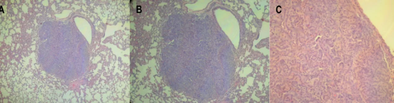
Figure 5: Macroscopic metastasis in the lymph node of a female Wistar rat exposed to N-butyl-N-(4-hydroxybutyl)nitrosamine. ... 32 Figure 6: Lung metastasis in a male ICR mouse exposed to N-butyl-N-(4-hydroxybutyl)nitrosamine with invasive urothelial carcinoma (H&E) (A: x100; B: x200; C: x400). ... 32 Figure 7: Histopathological evaluation of lesions identified in mouse urethra after BBN treatment. A: Normal urethra (transverse section) (H&E, x100). B: Dysplasia (H&E, x600). C: Nodular hyperplasia (H&E, x200). D: Invasive carcinoma (H&E, x100x). E: Invasive carcinoma (D) with higher magnification (H&E, x400). F: Squamous metaplasia (H&E, x100). ... 33
CHAPTER 4
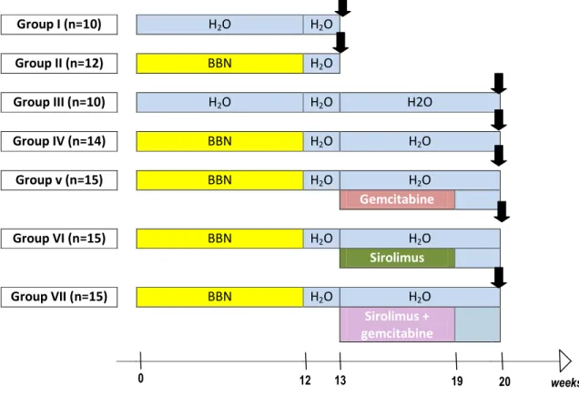
Figure 1: Experimental design. ... 51
CHAPTER 5
Figure 1: Experimental design. ... 65 Figure 2: (A) mTOR expression in normal mouse urothelium (x400); (B) mTOR expression in simple hyperplasia (x400); (C) mTOR expression in dysplasia (x400); (D) mTOR expression in squamous metaplasia (x400); (E) absence of mTOR expression in invasive carcinoma (x400); (F) mTOR expression in invasive carcinoma (x400). ... 73
xxvii Figure 3: Cell proliferation of bladder cancer cell lines (T24; HT1376; 5637), treated with different doses of everolimus. ... 75
Figure 4: Cell-cycle analysis of the T24, 5637 and HT1376 bladder-cancer cell lines after exposure to different concentrations of everolimus.. ... 76
Figure 5: Sub-G0/G1 fraction on bladder-cancer cell lines T24, 5637 and HT1376, after
exposure to different concentration of everolimus. ... 77 Figure 6: Effect of 2mM everolimus on the expression of mTOR, phospho-mTOR, Akt and phospho-Akt in bladder cell lines (T24, HT1376 and 5637). ... 78
CHAPTER 6
Figure 1: A: Normal mouse liver from the control group (H&E, x200); B: mouse liver from the N-butyl-N-(4-hydroxybutyl)nitrosamine (BBN) group, without any histological change (H&E, x200). ... 95
Figure 2: Effect of in vivo treatment with N-butyl-N-(4-hydroxybutyl) nitrosamine (BBN) (0.05%) on respiratory rates of liver mitochondria.. ... 97
Figure 3: Effect of N-butyl-N-(4-hydroxybutyl)nitrosamine (BBN) treatment on the protein levels of mitochondrial subunits from respiratory complexes, assessed by western blotting. (A) Complex I subunit (NDUSF3 nuclear); (B) complex IV subunit (MTCO 1 mitochondrial).. ... 99
Figure 4: Effect of treatment with N-butyl-N-(4-hydroxybutyl)nitrosamine (BBN; 0.05%) on the mitochondrial permeability transition pore induced by calcium.. ... 100
Figure 5: Rate and extent of mitochondrial oxidation induced by ADP/Fe2+ evaluated as
oxygen consumption. ... 101
CHAPTER 7
Figure 1: Experimental design. ... 115 Figure 2: Schematic drawing representing staining patterns of E-cadherin and β-Catenin in the urothelia of mice submitted to BBN’s action. ... 117
Figure 3: E-cadherin staining. A- Normal urothelium (x600); B- Simple hyperplasia (x600); C- Nodular hyperplasia (x600); D- Dysplasia (x400); E- Invasive carcinoma with squamous differentiation (x200); F- Squamous metaplasia (x400). ... 118
Figure 4: E-cadherin staining in different urothelial lesions (%).. ... 119 Figure 5: β-catenin staining. A- Normal urothelium (x600); B- Simple hyperplasia (x600); C- Nodular hyperplasia (x600); D- Dysplasia (x600); E- Invasive carcinoma (x200); F- Squamous metaplasia (x600)... 120
xxviii
List of tables
CHAPTER 3
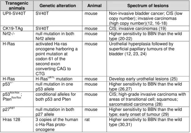
Table I - Genetically altered animals used in carcinogenesis studies. ... 22 Table II - Spectrum of urothelial lesions induced by N-butyl-N-(4-hydroxybutyl)nitrosamine ... 29
CHAPTER 4
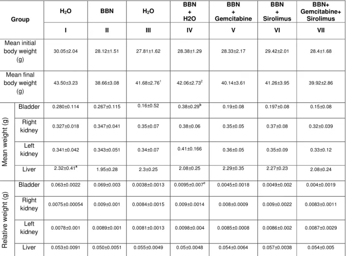
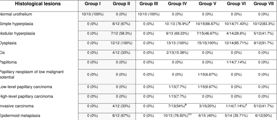
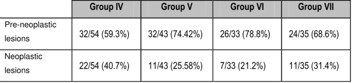
Table I - Initial and final body weights and mean and relative weights of liver, kidney and bladder at the end of the study. ... 54 Table II - Incidence values of histological lesions. ... 56 Table III - Incidence of pre-neoplastic and neoplastic lesions in groups IV to VII... 57
CHAPTER 5
Table I - Mean and relative weight of bladder, kidney and liver (value ±SD). ... 70 Table II - Incidence of urothelial lesions in ICR mice exposed to N-butyl-N-(4-hydroxybutyl)
nitrosamine and treated with RAD001 ... 72 Table III - Cytoplasm immunoreactivity of urothelial lesions with mTOR antibody in ICR mice
exposed to N-butyl-N-(4-hydroxybutyl) nitrosamine and treated with RAD001. ... 74 Table IV - Apoptotic index values (%) in the three bladder-cancer cell lines after exposure to
RAD001. ... 75
CHAPTER 6
Table I - Body weight, mean and relative weight of bladder, kidney and liver ... 94 Table II - Incidence of urothelial lesions in ICR mice exposed to N-butyl-N-(4-hydroxybutyl)
nitrosamine and control animals. ... 95 Table III - Effects of BBN treatment on mitochondrial membrane potential membrane and
respiratory indexes. ... 97 Table IV - Effect of BBN-treatment on the activities of liver mitochondrial complex I, II-III and IV,
expressed as % of control values. ... 98 Table V - Effect of BBN treatment on mitochondrial H2O2 production, GSH and MDA contents. 101
Table VI - Effect of BBN treatment on antioxidant enzymes in mice liver. ... 102
CHAPTER 7
Table I - E-cadherin expression pattern in normal urothelium and urothelial lesions (%). ... 119 Table II - β-catenin expression pattern in normal urothelium and urothelial lesions (%). ... 121
xxix
List of abreviatures
∆Ψ - Electrical potential
Akt - serine/threonine-specific protein kinase BBN - N-butyl-N-(4-hydroxybutyl) nitrosamine BCG - Bacillus Calmette-Guérin BCMN - N-butyl-N-(carboxymethyl)nitrosamine BCPN - N-butyl-N-(3-carboxypropyl)nitrosamine BHCPN - N-butyl-N-(2-hydroxy-3-carboxy-propyl) nitrosamine BOPN - N-butyl-N-(2-oxopropyl)nitrosamine
BSA - Bovine serum albumin
CAT - Catalase
CIS - Carcinoma in situ
DMN - Dimethylnitrosamine
DMSO - Dimethylsulfoxide
EDTA - Ethylenediaminetetraacetic acid EGFR - Endothelial growth factor receptor
EGTA - Ethyleneglycol-bis(-aminoethyl ether) N,N,N’,N’-tetraacetic acid ELISA - Enzyme-Linked Immunosorbent Assay
FANFT - [4-(5-nitro-2furyl)-2-thiazolyl]formamide GC - Gencitabine and cisplatin
GR - Glutathione reductase GSH - Reduced glutathione GSSG - Oxidized glutathione GST - Glutathione S-transferase
Gy - Gray
HER2 - Human EGF (Epidermal Growth Factor) Receptor 2 (Proto-oncogene)
HRP - Horseradish peroxidase
HVA - Homovanilic acid
KCl - Potassium cloride
KCN - Potassium cyanide
KH2PO4 - Potassium di-hydrogen phosphate
xxx
MDA - Malondialdehyde
MNU - N-methyl-N-nitrosourea
MPT - Mitochondrial permeability transition MPTP - Mitochondrial permeability transition pore mTOR - Mammalian target of rapamycin
MTT - 3-[4,5-dimethylthiazol-2-yl]-2,5-diphenyl tetrazolium bromide MVAC - Methotrexate, vinblastine, adriamycin and cisplatin
Na2S2O4 - Sodium hydrosulfite
NBT - Nitrotetrazolium blue chloride NDEA - N-nitrosodiethylamine
NNK - 4-(N-methyl-N-nitrosamino)-1-(3-pyridyl)-1-butanone PI3K - Phosphatidylinositol 3 kinase
PI3K/Akt/mTOR - Intracellular signalling pathway PVDF - Polyvinylidene difluoride PMS - Post Mitochondrial Supernatant RCR - Respiratory control ratio
SD - Standard deviation
SDS - Sodium Dodecyl Sulfate
SE - Standard error
SOD - Superoxide dismutase
TPP+ - Tetraphenyl phosphonium-selective UCC - Urothelial cell carcinoma
VDAC - Voltage-dependent anion channel
C
HAPTER
1:
3
1. General introduction
1.1- Bladder cancer
Bladder cancer is the second-most common tumour of the genitourinary tract, with prostate cancer the leader in terms of morbidity and mortality among urogenital tumours (1, 2). An estimated 386,300 new cases and 150,200 deaths from bladder cancer occurred worldwide in 2008, with males more frequently affected than females (1). The highest rates of bladder cancer are found in Europe, North America and Northern Africa, and the lowest in Melanesia and Middle Africa (1). In Europe, bladder urothelial cancer accounts for 7% of all cancers (3), but since de 1990s, a decline in the mortality rate (particularly in men) has been observed, perhaps due to the reduction in smoking prevalence in western countries along with reductions in occupational exposures known to cause bladder cancer (4). Tobacco smoke and other chemicals, like specific industrial chemicals, dietary nitrates and arsenic represent the most important exogenous risk factors associated with bladder cancer, particularly in developed countries (1, 5-7). It has been shown that smoking increases the risk of bladder cancer by two- to fivefold (5, 6). Occupational hazards related to the exposure to certain chemicals, include hairdressers and allied occupations and workers in the petroleum industry (8-10). Individuals living in areas with residential exposure to trihalomethanes in treated water have an increased risk of bladder cancer. Bladder cancer risk was also significantly elevated among those reporting longer duration showers or baths as well as among individuals who “ever” swam in swimming pools (9). ortho-Toluidine used as a biological stain and also in the production of dyes, pigments and rubber chemicals and benzidine-based dyes, used mainly for colouring paper, textiles, and leather, are some examples of carcinogenic chemicals associated with bladder cancer (8-11). In developing countries, in the Middle East and Africa,
Schistosoma haematobium is the main etiological agent for bladder cancer (1, 6). Urinary bladder tumours remain one of the deadliest and expensive human tumours, with regard to both cost per patient per year and lifetime cost per patient (12, 13). Early detection of bladder cancer, through the use of cystoscopy, radiographic imaging and urine cytology, improves patient prognosis since it enables the identification of lesions at an early stage (14). The staging, treatment and prognosis of bladder cancer all depend on how deeply it has invaded the bladder wall. Surgery is the standard treatment, both in the form of transurethral endoscopic resection - mainly for superficial incidence of the disease, and in the form of radical cystectomy with
4
urinary diversion when the disease is muscle-invasive (2, 14-17). Intravesical administration of Bacillus Calmette-Guerin (BCG) has been employed after endoscopic resection as the most effective agent for both the prophylaxis of disease recurrence and preventing its progression from superficial to invasive disease (5).
Systemic therapy for bladder cancer can be seen in four contexts: neoadjuvant, adjuvant, bladder sparing with radiation and chemotherapy, and metastatic disease:
- Neoadjuvant therapy is considered to be a standard of care in muscle-invasive bladder cancers planned for surgical intervention (18, 19). The integration of chemotherapy in the pre-surgical phase offers early treatment of systemic micrometastases, potential down staging of the primary and regional disease, and an in
vivo assessment of chemosensitivity. This approach also avoids a potential delay in systemic treatment due to postoperative complications, which are frequent in bladder cancer (18, 20);
- Adjuvant chemotherapy is a systemic treatment option for patients still with muscle-invasive bladder cancer after being submitted to surgery. Surgical intervention allows the removal of as much of a tumour as possible and the relief of symptoms. Furthermore, it allows the pathologic staging that more accurately assesses risk, since clinical staging is often innacurate (18). There are several protocols being used in this modality, like cisplatin as a single agent or in association with methotrexate, or methotrexate, vinblastine, adriamycin and cisplatin in association (MVAC) or an association of methotrexate, vinblastine, epirubicin and cisplatin (MVEC). However, while available data suggest a beneficial effect from adjuvant therapy, further studies and trials are needed;
- Bladder preservation may be accomplished in appropriately selected patients with muscle-invasive urinary bladder carcinoma, without compromising outcomes, using a trimodality approach: surgery (transurethral resection), chemotherapy and radiation therapy. Chemotherapy has been used in two phases of treatment: as radiosensitizers, given concurrently with radiation treatment, and as adjuvant treatment, recognizing that survival will only be improved by the successful treatment of micrometastases (21). Continued surveillance with cystoscopy both during and after chemoradiotherapy is mandatory (18).
- When metastatic disease is established, conventional cytotoxic chemotherapy, in the form of MVAC or gemcitabine and cisplatin (GC), is commonly used. However, the frequent occurrence of drug resistance and serious side effects has made treatment outcomes unsatisfactory (22, 23). Along with these, the median overall survival rates at 5 years in patients with metastatic urinary bladder cancer treated with
5
chemotherapy (15.2 months for MVAC and 14 months for GC) and the 5-year overall survival rates (15.3% for MVAC and 13% for GC) are poor (23).
1.2. Research in bladder cancer
Intensive research is being carried out in bladder cancer, to elucidate the reasons for the development of tumours, and to find out which factors determine the tumour progression. Bladder cancer is a recurrent and very prevalent cancer whose treatment involves high economic costs. This situation forces more investigation in the urological field, with the objective to do early diagnostics, to obtain better treatments and fewer side effects. The combination of in vitro and in vivo studies will provide insights into the biology of the tumour and is essential for the implementation of new therapeutic and/or preventive modalities (24, 25).
1.2.1. Cell lines
Tissue culture was first introduced in 1907 by Harrison. Since then, cell lines have evolved and have come to be widely used for research in several areas like biochemistry, virology, immunology and oncology.
Regarding cancer, cell lines have been very important in order to build our understanding of the disease’s molecular pathophysiology and its treatment. The first continuous cancer cell line was HeLa cells, isolated from the aggressive glandular cervical cancer of a young woman approximately 60 years ago (26, 27). Nowadays, cancer cell lines are routinely used for various kinds of biomedical research, from drug-sensitivity tests to identifying potential therapy targets and pharmacologically useful compounds (27-29).
For the study of bladder cancer, several cell lines have been established. T24, HT1376, 5637, UM-UC-3 are some examples of human bladder cancer cell lines (27). Some have origin in superficial tumours but the majority are from invasive and metastatic ones (27, 28). Cell lines from experimental bladder cancer are fewer than those of human origin. AY-27 and NBT-II are bladder cancer cell lines derived from rats (27, 30-32). BTT-T739 and MB49 are examples of bladder cancer cell lines with origin in mice (27, 33).
In vitro studies with cell lines demand a great amount of care concerning the origin of cell lines. It is crucial to ensure that they are reliable, because cell
cross-6
contamination in cell cultures is a common problem during cell culturing and use. Cross-contamination provides misleading research results leading to unusable therapeutic products. The unwitting use of misidentified cell lines may, ultimately, expose patients to inappropriate, or even harmful, treatments.
Masters and co-workers (2001) (35), found a total of nine cell lines that were cross-contaminated with T24, a line started from human urinary bladder carcinoma in 1970. Four of the tested cell lines were 100% identical to T24, but traded under a different name. Van Bokhoven et al. (2001) (36), also tested two cell lines frequently used as models in prostate cancer (TSU-Pr1 and JCA-1) and concluded that they appear to be derivatives of the bladder carcinoma cell line T24 and not of prostatic origin, whereby they could no longer be used as models for the study of prostate cancer.
In urological research, cell lines, particularly human urothelial cell lines are well-established tools for preclinical trials. For most cytotoxic agents, if it does not work in
vitro, it will most certainly not work in vivo. If it works in vitro, then there is the possibility it may be effective in vivo. They are a cost-efficient method of searching for drug activity and can further our understanding of drugs’ action on several tumours. Other advantages in using cell lines can also be highlighted: they are easy to handle and can be replicated almost infinitely. If anything goes wrong, such as contamination or death of the cell culture, it can be easily replaced from frozen stocks. Additionally, they exhibit a relatively high degree of homogeneity. Cell lines, however, have some disadvantages. They are prone to genotypic and phenotypic drift during their continual culture. Subpopulations may arise and cause phenotypic changes over time by the selection of specific, more rapidly growing clones within a population (37).
1.2.2. Animal models and novel drugs for bladder cancer treatment
Animal models have greatly contributed to the understanding of bladder carcinogenesis.They are at the centre of experimental research and, at the same time, are the connection between experimental and clinical research (38). Animal models allow the investigation of aspects that cannot be studied under clinical conditions, such as the evaluation of new chemotherapeutic, immunotherapeutic or prophylactic agents, drug regimens, or other treatment methods and can also provide further information on basic mechanisms of tumour growth and spread (39, 40).
Although there is the possibility to use several animal species, rodents (rats and mice) are those most often used in animal experimentation (41-55).
7
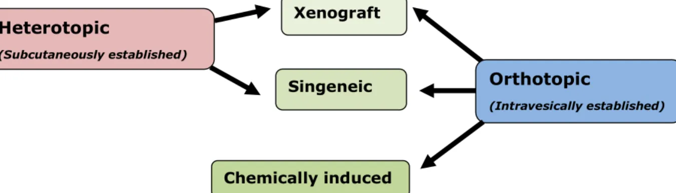
Bladder tumours in rodents can be established subcutaneously (heterotopically) by transplantation of tumour cells, or intravesically (orthotopically) either by transplantation of tumour cells or by chemical induction (Figure 1) (33, 39, 47).
In the heterotopic model, the tumour is usually located in the flank or hind leg of the animal. This model can be syngeneic, when rodent urothelial cell carcinomas (UCC) tumour fragments or cells are inoculated via a small incision into an immunocompetent host of the same strain from which the tumour was originally derived. Or it can be a xenograft model, when human UCC fragments or cells are used in immunodeficient hosts (33). Heterotopic tumour models have been widely used, since tumour evolution is easily assessed. These models allow urinary tumour development to be studied in vivo, since the tumour tissue is developing within a healthy environment. However, it is questionable if the tumour development at these sites parallels the development of the tumour in the organ of its origin. Technically, they are easier to establish than orthotopic ones (33, 39, 47).
The orthotopic model can be divided into three different types: chemically induced bladder cancer models, the xenograft model (transplantation of human UCC into immunodeficient hosts) and the syngeneic tumour model (transplantation of carcinogen-induced bladder cancer into syngeneic immunocompetent hosts) (33, 48). Orthotopic tumour models have the great advantage of simulating the local cancer environment and resembling the behaviour of the disease in humans (33, 39, 47). One disadvantage of the xenograft model is that the immune response of intravesical therapy cannot be evaluated in mice with immune deficiencies (39, 48). An orthotopic model in immunocompetent animals has advantages over models of bladder cancer that either use immunodeficient rodents and/or heterotopic models. This is because the influence of the immune system and the anatomical and physiological factors of the tissue of origin, which undoubtedly influence the metastatic process, are not affected (49).
Figure 1: Animal models of bladder cancer (33, 39, 47). Heterotopic (Subcutaneously established) Orthotopic (Intravesically established) Xenograft Singeneic Chemically induced
8
The mouse model of chemically induced bladder cancer was the model used in this study, and it is widely utilized by investigators all over the world in bladder cancer research. It has enormous applications. It can be used to test prophylactic drugs, to test therapeutic drugs and also to define the impact of chemical carcinogens on other organs. There are several chemical carcinogens that can be used to induce bladder cancer in experimental animals. Nitrosamines, N -[4-(5-nitro-2furyl)-2-thiazolyl]formamide (FANFT) and N-methyl-N-nitrosourea (MNU), are some examples (50-54).
Apart from these models, there are also transgenic models for bladder cancer, that are genetically altered animals (lacking genes, expressing genes or having mutated genes) (54-58) that exhibit increased carcinogenicity (59).
The use of animal models, allowed a growing understanding of tumour molecular biology, and also enabled the identification of the signalling pathways involved in bladder tumorigenesis and progression. This important knowledge opened the door for the discovery of new therapeutic pathways. The PI3K/AKT/mTOR pathway and the RAS-MAPK pathway, involved in bladder cancer, are now identified as important targets for the development of new therapeutic drugs (60-62). Endothelial growth factors, that promote angiogenesis and tumorigenesis, are also possible targets for therapeutic antiangiogenic intervention in cancer (63, 64).
Among therapeutic agents targeting the PI3K/AKT/mTOR pathway, mTOR inhibitors are the most developed. Rapamycin, also known as sirolimus, and its derivatives such as everolimus, tensirolimus and deforolimus, have antineoplastic activity when it comes to a variety of solid tumours - including bladder cancer - and are being used for clinical trials. Other agents like endostatin, bevacizumab and cetuximab that can target vascular endothelial growth factor and, therefore, tumour angiogenesis are also being subjected to clinical trials to analyze the action they have against bladder cancer (65, 66). To target EGFR and HER2/neu, two of the generally overexpressed genes, there are monoclonal antibodies such as cetuximab and panitumumab (35, 65). Tipifarnib, a farnesyltransferase inhibitor, is able to inhibit farnesylation, an important step in activating Ras oncogene. Sorafenib (BAY43-9006) is an oral, dual inhibitor of Raf and vascular endothelial growth factor receptors (VEGFR). Other agents that have shown promising results regarding bladder cancer (vinflunine, celecoxib, TKI-258 and AEZS-108), are currently under clinical trials (15, 66, 67). This entire therapeutic advance would not be possible without animal models;
9
however, more data concerning drugs action within monotherapy or politherapy regimens and any eventual side effects are still needed.
1.3. References
1 Jemal A, Bray F, Center MM, Ferlay J, Ward E, Forman D: Global cancer statistics. CA Cancer J Clin 61(2):134, 2011.
2 Winer E, Gralow J, Diller L, Karlan B, Loehrer P, Pierce L, Demetri G, Ganz P, Kramer B, Kris M, Markman M, Mayer R, Pfister D, Raghavan D, Ramsey S, Reaman G, Sandler H, Sawaya R, Schuchter L, Sweetenham J, Vahdat L, Schilsky RL. Clinical Cancer Advances 2008: Major research advances in cancer treatment, prevention, and screening—a report from the American Society of Clinical Oncology. J Clin Oncol 27:812–26, 2009.
3 Stein JP, Skinner DG. Radical cystectomy for invasive bladder cancer: Long-term results of a standard procedure. World J Urol 3:296 –304, 2006.
4 American Cancer Society. Global Cancer Facts & Figures 2nd Edition. Atlanta: American Cancer Society; 2011.
5 Braud F, Maffezzini M, Vitale V, Bruzzi P, Gatta G, Hendry WF, Sternberg CN: Bladder cancer. Critical Reviews in Oncology/Hematology 41:89–106, 2002. 6 Fadl-Elmula I: Chromosomal changes in uroepithelial carcinomas. Cell Chromosome
4:1, 2005.
7 Volanis D, Kadiyskaa T, Galanisb A, Delakasa D, Logothetic S, Zoumpourlisc V: Environmental factors and genetic susceptibility promote urinary bladder cancer. Toxicol Lett 193: 131–137, 2010.
8 Baan R, Straif K, Grosse Y, Secretan B, El Ghissassi F, Bouvard V, Benbrahim-Tallaa L, Cogliano V (International Agency for Research on Cancer Monograph Working Group): Special Report: Policy - Carcinogenicity of some aromatic amines, organic dyes, and related exposures. The Lancet Oncoly 9: 322-323, 2008.
9 Clapp RW, Jacobs MM, Loechler EL: Environmental and occupational causes of cancer new evidence, 2005–2007. Rev Environ Health 23(1): 1–37, 2008. 10 Takkouche B, Regueira-Méndez C, Montes-Martínez A: Risk of cancer among
hairdressers and related workers: a meta-analysis. Inter J Epidemiol 38:1512– 1531, 2009.
10
11 Pira E, Piolatto G, Negri E, Romano C, Boffetta P, Lipworth L, McLaughlin JK, La Vecchia C: Bladder cancer mortality of workers exposed to aromatic amines: a 58-year follow-up. J Natl Cancer Inst 102:1096–1099, 2010.
12 Lotan Y, Kamat AM, Porter MP, Robinson VL, Shore N, Jewett M, Schelhammer PF, White RV, Quale D, Lee CT: Key Concerns About the Current State of Bladder Cancer. Cancer 115(18): 4096-4103, 2009.
13 Stenzl A, Hennenlotter J, Schilling D: Can we still afford bladder cancer? Curr Opin Urol 18(5):488-492, 2008.
14 Crawford JM: The origins of bladder cancer. Laboratory Investigation 88: 686–693, 2008.
15 Wallerand H, Bernhard JC, Culine S, Ballanger P, Robert G, Reiter RE, Ferrière JM, Ravaud A: Targeted therapies in non-muscle-invasive bladder cancer according to the signaling pathways. Urol Oncol 29(1):4-11, 2011.
16 Youssef RF, Raj GV: Lymphadenectomy in management of invasive bladder cancer. Int J Surg Oncol 2011, 9pp.
17 Turker P, Bostrom PJ, Wroclawski ML, van Rhijn B, Kortekangas H, Kuk C, Mirtti T, Fleshner NE, Jewett MA, Finelli A, Kwast TV, Evans A, Sweet J, Laato M, Zlotta AR: Upstaging of urothelial cancer at the time of radical cystectomy: factors associated with upstaging and its effect on outcome. BJU International, 2012: doi: 10.1111/j.1464-410X.2012.10939.x. Epub ahead of print.
18 Costantini C, Millard F: Update on chemotherapy in the treatment of urothelial carcinoma. ScientificWorldJournal 11: 1981-1994, 2011.
19 Feifer AH, Taylor JM, Tarin TV, Herr HW: Maximizing cure for muscle-invasive bladder cancer: integration of surgery and chemotherapy. Eur Urol 59(6):978-84, 2011.
20 Karam JA, Kamat AM: Optimal timing of chemotherapy and cystectomy. F1000 Med Rep 23(2): pii: 48, 2010.
21 Kauffman DS: Challenges in the treatment of bladder cancer. Annals of Oncology 17(s5): v106-v112, 2006.
22 Trosko JE: The Role of Stem Cells and Gap Junctional Intercellular Communication in Carcinogenesis. Journal of Biochemistry and Molecular Biology 36(1): 43-48, 2003.
23 Karam JA, Huang S, Fan J, Stanfield J, Schultz RA, Pong R-C, Sun X, Mason RP, Xie X-J, Niu G, Chen X, Frenkel EP, Sagalowsky AI, Hsieh J-T: Upregulation of TRAG3 gene in urothelial carcinoma of the bladder. International Journal of Cancer 128: 2823–2832, 2011.
11
24 Eiján AM, Lodillinsky C, Sandes EO: Chapter 19. Animal Models for Basic and Preclinical Research in Bladder Cancer. In: Bladder Cancer – From Basic Science to Robotic Surgery. Abdullah EC (Ed). Croatia. 383-398, 2012.
25 Vasconcelos-Nóbrega C, Pinto-Leite R, Arantes-Rodrigues R, Ferreira R, Brochado P, Cardoso ML, Palmeira C, Salvador A, Guedes-Teixeira CI, Colaço A, Palomino LF, Lopes C, Santos L and Oliveira PA: In vivo and in vitro effects of RAD001 on bladder cancer. Urol Oncol, 2011 Dec 9. [Epub ahead of print]. 26 Masters JR: HeLa cells 50 years on: the good, the bad and the ugly. Nature
Reviews: Cancer 2: 315-318, 2002.
27 Gabriel U, Bolenz C, Michel MS: Experimental Models for Therapeutic Studies of Transitional Cell Carcinoma. Anticancer Research 27: 3163-3172, 2007. 28 Hatina J, Huckenbeck W, Rieder H, Seifert HH, Schulz WA:
Harnblasenkarzinomzelllinien als Modellsysteme zur Pathobiologie des Harnblasenkarzinoms. Der Urologe A 47(6): 724-734, 2008.
29 Tsuji K, Kawauchi S, Saito S, Furuya T, Ikemoto K, Nakao N, Yamamoto S, Oka M, Hirano T, Sasaki K: Breast cancer cell lines carry cell line-specific genomic alterations that are distinct from aberrations in breast cancer tissues: Comparison of the CGH profiles between cancer cell lines and primary cancer tissues. BMC Cancer 10:15, 2010.
30 Nishi, M: A cell line derived from BBN (N-butyl-N-[4-hydroxybutyl]-nitrosamine)-induced rat bladder cancer: establishment and scanning electron microscopic cell surface characteristics. Acta Med Okayama32(3): 181-205, 1978.
31 Chen JJ, Ye ZQ, Koo MWL: Growth inhibition and cell cycle arrest effects of epigallocatechin gallate in the NBT-II bladder tumour cell line. BJUInternational 93: 1082 – 1086, 2004.
32 Hojo H, Kaneko A, Kayagaki N, Saki M, Hashimoto Y: Subcellular localization and characterization of interleukin-1α produced by rat bladder cancer cells. Immunology Letters 43(3): 215-220, 1994.
33 Arentsen HC, Hendricksen K, Oosterwijk E, Witjes JA: Experimental rat bladder urothelial cell carcinoma models. World J Urol 27:313–317, 2009.
34 Chen F, Zhang G, Cao Y, See WA: the MB49 murine urothelial carcinoma: a molecular and phenotypic comparison to human cell lines as a model for studying the direct tumor response to BCG. The Journal of Urology 181(4s): 412, 2009.
35 Masters JR, Thomson JA, Daly-Burnsa B, Reidd YA, Dirks WG, Packer P, Toji LH, Ohno T, Tanabe H, Arlett CF, Kellandk LR, Harrison M, Virmani A, Ward TH,
12
Ayres KL, Debenham PG: Short tandem repeat profiling provides an international reference standard for human cell lines. PNAS 98(14):8012-8017, 2001.
36 van Bokhoven A, Varella-Garcia M, Korch C, Miller GJ: TSU-Pr1 and JCA-1 cells are derivatives of T24 bladder carcinoma cells and are not of prostatic origin. Cancer Research 61: 6340–6344, 2001.
37 Burdall SE, Hanby AM, Lansdown MRJ, Speirs V: Breast cancer cell lines: friend or foe? Breast Cancer Res 5:89-95, 2003.
38 Reis LO, Pereira TC, Favaro WJ, Cagnon VHA, Lopes-Cendes I, Ferreira U: Experimental animal model and RNA interference: a promising association for bladder cancer research. World Journal of Urology 27(3): 353-361, 2009. 39 Günther JH, Jurczok A, Wulf T, Brandau S, Deinert I, Jocham D, Böhle A:
Optimizing syngeneic orthotopic murine bladder cancer (MB49). Cancer Research 59, 2834–2837, 1999.
40 Steele VE, Lubet RA: The use of animal models for cancer chemoprevention drug development. Seminars in Oncology 37(4): 327-338.
41 Okajima E, Hiramatsu T, Hirao K, Ijuin M, Hirao Y, Babaya K, Ikuma S, Ohara S, Shiomi T, Hijioka T, Ohishi H: Urinary bladder tumors induced by N-butyl-N-(4-hydroxybutyl)nitrosamine in dogs. Cancer Res 41(5): 1958-1966.
42 Cohen AE, Weisburger EK, Weisburger JH, Ward JM, Putnam CL: Cystoscopy of chemically induced bladder neoplasms in rabbits administered the carcinogen dibutylnitrosamine. Invest Urol 12: 262–6, 1975.
43 Cohen SM: Cell proliferation and carcinogenesis. Drug Metab Rev 30: 339–57, 1998.
44 Hueper WC: Aniline tumors of the bladder. Arch Pathol; 25: 858, 1938.
45 Kunze E, Chowaniec J. Pathology of tumours in laboratory animals. Tumours of the rat. Tumours of the urinary bladder. IARC Sci Publ 99: 345–97, 1990.
46 Clayson DB, Fishbein L, Cohen SM: Effects of stones and other physical factors on the induction of rodent bladder cancer. Food Chem Toxicol 33(9):771-784, 1995.
47 Chan ESY, Patel AR, Smith AK, Klein JB, Thomas AA, Heston WD, Larchian WA: Optimizing Orthotopic Bladder Tumor Implantation in a Syngeneic Mouse Model. The Journal of Urology 183(6): 2926-2931, 2009.
48 Chan E, Patel A, Heston W, Larchian W: Mouse orthotopic models for bladder cancer research. BJU International 104(9): 1286-1291, 2009.
13
49 Levett D, Flecknell PA, Rudland PS, Barraclough R, Neal DE, Mellon JK, Davies BR: Transfection of S100A4 produces metastatic variants of an orthotopic model of bladder cancer. Am J Pathol 160(2):693-700, 2002.
50 Lijinsky W: Chemistry and biology of N-nitroso compounds. In: Cambridge Monographs on Cancer Research. Cambridge University Press, Cambridge UK, 1992.
51 Druckrey H, Preussmann R, Ivankovic S, Schmidt CH, Mennel HD and Stahl KW: Selective induction of bladder cancer in rats by dibutyl- and N-butyl-N-butanol(4)-nitrosamine. Z Krebsforsch 2(66): 280-90, 196457
52 Swaminathan S and Bryan GT: Biotransformation of the bladder carcinogen N-[4-(5-nitro-2-furyl)-2-thiazolyl]formamide in mice. Cancer Res 44: 2331-2338, 1984. 53 Cui L, Shi Y, Daib G, Panb H, Chen J, Song L, Wang S, Chang HC, Sheng H and
Wang X: Modification of N-methyl-N-nitrosourea initiated bladder carcinogenesis in Wistar rats by terephthalic acid. Toxicol Appl Pharmacol 210: 24-31, 2006.
54 Reis LO, Fávaro WJ, Ferreira U, Billis A, Fazuoli MG and Cagnon VHA: Evolution on experimental animal model for upper urothelium carcinogenesis. World J Urol 28: 499-505, 2010.
55 Martin-Sanz P, Mayoral R, Casado M, Bosca L: COX-2 in liver, from regeneration to hepatocarcinogenesis: what we have learned from animal models? World J Gastroenterol 16(12): 1430-1435, 2010.
56 Zhang Z-T, Pak J, Shapiro E, Sun T-T, Wu X-R: Urothelium-specific Expression of an Oncogene in Transgenic Mice Induced the Formation of Carcinoma in Situ and Invasive Transitional Cell Carcinoma. Cancer Res 59(14): 3512-3517, 1999.
57 Wu X-R: Biology of urothelial tumorigenesis: insights from genetically engineered mice. Cancer and Metastasis Reviews 28(3-4): 281-290, 2009.
58 Gollapudi BB, Stott WT, Yano BL, Bus JS: Mode of action considerations in the use of transgenic animals for mutagenicity and carcinogenicity evaluations. Toxicol Lett 102-103:479-84, 1998.
59 Oliveira PA, Colaço A, De La Cruz P LF, Lopes C: Experimental bladder carcinogenesis-rodent models. Exp Oncol 28(1): 2-11, 2006.
60 Ching CB, Hansel DE: Expanding therapeutic targets in bladder cancer: the PI3K/Akt/mTOR pathway. Laboratory Investigation 90: 1406–1414, 2010. 61 Zachos I, Tzortzis V, Konstantinopoulos PA, Karatzas A, Gravas S, Melekos M,
14
cancer: implications for novel targeted therapies. Curr Mol Med 11(8):623-32, 2011.
62 Netto GJ: Molecular biomarkers in urothelial carcinoma of the bladder: are we there yet? Nat Rev Urol 9: 41–51, 2012.
63 Mitra AP, Cote RJ, Path FRC: Searching for novel therapeutics and targets: Insights from clinical trials. Urologic Oncology: Seminars and Original Investigations 25: 341–343, 2007.
64 Bagley RG, Rouleau C, Weber W, Mehraein K, Smale R, Curiel M, Callahan M, Roy A, Boutin P, St. Martin T, Nacht M, Teicher BA: Tumor endothelial marker 7 (TEM-7): A novel target for antiangiogenic therapy. Microvascular Research 82: 253–262, 2011.
65 Ma WW, Adjei AA: Novel Agents on the Horizon for Cancer Therapy. Ca Cancer J Clin 59:111-137, 2009.
66 Stephenson JJ, Gregory C, Burris H, Larson T, Verma U, Cohn A, Crawford J, Cohen RB, Martin J, Lum P, Yang X, Amado RG: An Open-Label Clinical Trial Evaluating Safety and Pharmacokinetics of Two Dosing Schedules of Panitumumab in Patients with Solid Tumors. Clinical Colorectal Cancer 8(1): 29-37, 2009.
67 Schally AV, Engel JB, Emons G, Block NL, Pinski J: Use of analogs of peptide hormones conjugated to cytotoxic radicals for chemotherapy targeted to receptors on tumors. Curr Drug Deliv 8(1):11-25, 2011.
15
C
HAPTER
2:
17
2. Aims
Over the years, bladder cancer research has contributed to a better knowledge in bladder cancer biology, treatment and prevention. Although most superficial bladder cancers can be transurethrally removed with excellent clinical outcomes, muscle-invasive disease can represent a major clinical challenge since it is associated with postoperative recurrence, metastasis and a worse prognosis. Regardless of all the investigation, current treatments are not good enough. To improve survival, it is mandatory to search for new therapeutic options, using new drugs or old drugs in new regimens.
With this thesis we had the ultimate goal of bringing new contribution to bladder cancer basic research. This was supported in four lines of action:
1) The first one was to gather all the information concerning N-butyl-N-(4-hydroxybutyl)nitrosamine (BBN) as a urothelial carcinogen, sharing the knowledge gained over many years of research using this chemical in experimental urinary bladder cancer research with rodents.
2) The second one was to test the therapeutic effect of several drugs, in vivo and in vitro:
- In vivo: Using the mice model of bladder cancer chemically induced by BBN that mimics the human invasive bladder cancer in a normal microenvironment, we intended to test the efficacy of gemcitabine, sirolimus and their association, and also to test the efficacy of everolimus;
- In vitro: to evaluate everolimus, in three different cell lines (T24; HT1376; 5637).
3) The third one was to determine any eventual liver damage induced by N-butyl-N-(4-hydroxybutyl)nitrosamine;
4) The fourth objective was to study E cadherin and β catenin expression in normal mice urothelium and in preneoplastic and neoplastic lesions induced in ICR mice by N-butyl-N-(4-hydroxybutyl)nitrosamine.
19
C
HAPTER
3:
BBN
AS A UROTHELIAL CARCINOGEN
The content of this chapter was presented/ published in:
- Vasconcelos-Nóbrega C, Colaço A, Lopes C, Oliveira PA (2012). BBN as a urothelial carcinogen. In vivo, 26(4):727-39 (Full paper).
- Vasconcelos-Nóbrega C, Colaço AA, Vala H, Oliveira PA (2010). Bladder carcinogenicity of N-butyl-N-(4-hydroxybutyl)nitrosamine. XV Encontro da Sociedade Portuguesa de Patologia Animal. Viseu, 12 de Março (Oral presentation).
21
3. BBN as a urothelial carcinogen
3.1. Introduction
Bladder cancer is among the most common malignancies worldwide, and it assumes greater prominence in developed countries. It is the second-most common tumour of the genitourinary tract and the second-most common cause of death in patients with genitourinary tract malignancies (1). In humans, the most common cell type of bladder cancer is transitional cell carcinoma, although adenocarcinomas, squamous cell carcinomas and sarcomas can also occur (2). The majority of bladder-cancer patients, 75-85%, are diagnosed with superficial tumours while the remaining 15% to 25% are invasive ones (2, 3). The superficial ones are low-grade, well-differentiated papillary tumours that do not invade or metastasize. Histological evolution in this case starts as hyperplasia, evolving to papilloma, papillary urothelial neoplasms of low malignant potential and papillary carcinoma (low- and high-grade) (4). The invasive ones originate as flat lesions like hyperplasia, dysplasia and carcinoma in situ (CIS) that then evolve into invasive carcinomas (5). These are high-grade tumours that frequently have areas of squamous cells or glandular differentiation or areas of undifferentiated tumour, including small-cell or spindle-cell variations. Invasive tumours are devastating, since over 50% of the patients will die from metastatic disease (2).
3.2. Experimental models of bladder cancer
The need for experimental models of tumours that are as similar as possible to human tumours has prompted researchers to attempt various approaches. Animals such as dogs, rabbits, guinea pigs and hamsters have all been used to induce and study urinary bladder carcinogenesis (6). However, for ethical questions and due to the conditions in animal facilities, dogs cannot be used in experimental carcinogenesis models. Animal facilities to maintain guinea pigs and rabbits also require special equipment, making models difficult to apply. For these reasons, small rodents such as mice and rats are the most common species used. These animals have a lower urinary tract that is comparable to humans, and neoplasms in their bladders are morphologically very similar (7). On the other hand, in most strains of rodents, bladder cancer is not commonly found unless a chemical or other treatment is applied (8). Another advantage is associated with the fact that these are small animals and there is