Genetic analysis of reciprocal differences in the inheritance of
in vitro
characters in pearl millet
Valluri V Satyavathi
1,*, V. Manga
1, Muktinutalapati V. Subba Rao
1,and Malladi Chittibabu
2 1Department of Botany, Andhra University, Visakhapatnam, India.
2Department of Statistics, Andhra University, Visakhapatnam, India.
Abstract
Reciprocal differences persist in nature because of the unequal contribution of cytoplasmic determinants from male and female gametes to the zygote. The inheritance of genetic differences is an important factor that influences vari-ous traits, including somatic embryogenesis and regenerationin vitro. In this report, we estimate the cytoplasmic and maternal effects in pearl millet and their adequacy in describing the observed reciprocal differences based on an in depth study of the parents, F2s and reciprocal backcross progenies needed for fitting genetical models. Our study
re-vealed that of the two characters examined, embryogenic callus quantity and regeneration frequency, the former showed a greater proportion of cytoplasmic nuclear interaction whereas the latter showed a greater role of nuclear factors. Additive-maternal effects influenced total callus quantity and dominance-maternal effects influenced total callus quantity, embryogenic callus quantity and regeneration frequency. Dwarfing was associated with the produc-tion of large quantities of embryogenic callus that had visually recognizable characteristics. The phenotypic nature of dwarf parents (green dwarf with long narrow leaves) with a genetic basis for a given character controlled by nuclear and cytoplasmic determinants can be exploited for other breeding programs.
Keywords: cytoplasmic inheritance, d2dwarf,in vitroregeneration, pearl millet, reciprocal differences.
Received: January 20, 2015; Accepted: May 18, 2015.
Introduction
In vitrocharacters are often used in combination with other agronomic traits for crop improvement programs. The availability of an efficientin vitroplant regeneration system is a prerequisite for the successful application of any biotechnological tool in plant breeding. Ikeuchiet al. (2013) provided an overview of callus and plant formation in vitroand described in detail the genetic and epigenetic mechanisms that induce or repress callus induction. An ob-vious challenge for tissue culture researchers is to develop in vitrotechnologies for faster, more predictable produc-tion of embryogenesis and plant regeneraproduc-tion. Despite all numerous technological advances, the successful applica-tion of plant tissue culture techniques to crop improvement is still dependent on understanding the genetic basis for ‘in vitroaptitude’ that will help to predict thein vitroresponses of genotypes for further selection. The influence of geno-type onin vitrocharacters and the genetic basis of thein vi-troresponse have been analyzed in several plant systems,
including pearl millet (Pueschelet al., 2003; Satyavathiet al., 2006; Dodig et al., 2008; Chakravarthi et al., 2010; Etedalet al., 2012; Wanget al., 2013).
The study of the inheritance of genotypic differences is often complicated because of the occurrence of recipro-cal differences (Powell and Caligari, 1987). The analysis of cytoplasmic factors in understanding the underlying modes of transmission ofin vitrotraits is more complicated than for nuclear factors. Various studies have examined the in-fluence of cytoplasmic and/or maternal inheritance and the interaction between nuclear and cytoplasmic factors onin vitrosomatic embryogenesis and morphogenesis in wheat (Ekiz and Konzak, 1991; Taylor and Veilleux, 1992; Be-beli, 1995; Ben-Amer and Boner, 1997), maize (Tomes and Smith, 1985; Willmanet al., 1989; Geet al., 1994), barley (Foroughi-Wehret al., 1982; Powell and Caligari, 1987; Houet al., 1994), rice (Abe and Futsuhara, 1991), red clo-ver (Keyes et al., 1980), Brassica (Narasimhulu et al., 1989), tomato (Singsit and Veilleux, 1989), Medicago (Walton and Brown, 1988; Wan et al., 1988), Allium ampeloprasum L. (Silverstand et al., 1995) and Desmodium(Krottjeet al., 1996).
The basis of the reciprocal differences inin vitro re-sponses may be cytoplasmic factors, the maternal plant ge-notype or the segregation of nuclear factors. Discrimination between the first two phenomena becomes necessary in the Send correspondence to Valluri V. Satyavathi. Department of
Bot-any, Andhra University, Visakhapatnam 530003, India. E-mail: [email protected]; [email protected]
*Current address: Laboratory of Molecular Genetics,
APEDA-CDFD, Centre for DNA Fingerprinting and Diagnostics, Nampally, Hyderabad 500001, India.
analysis of reciprocal differences. When the female parent influences the phenotype of the progeny regardless of the latter’s genotype, it is considered as maternal effect, whereas in cytoplasmic inheritance the mitochondrial DNAs transmitted to the progeny will have an effect on the phenotype of the organism (Henry et al., 1994). Powell (1988), using the biometrical analysis of Hayman (1954a), analyzed reciprocal differences in barley cultures attributed to component ‘c’ (average reciprocal differences) to deter-mine the percentage of green and albino plant production. This group also estimated component ‘d’ (further recipro-cal differences that were not accounted for byc) to assess the percentage corresponding to anthers and green plant production.
Among dicots, Narasimhulu et al. (1989) reported significant differences caused by cytoplasmic factors dur-ing shoot morphogenesis inBrassica carinata. Pueschelet al.(2003) analyzed the genetic control of thein vitro re-sponse ofCyclamen persicumgenotypes with and without embryogenic potential. Based on the F2segregation ratio,
these authors postulated two major dominant genes with epistatic interaction for regeneration capacity.
Among monocots, Li et al. (2007) attempted to clarify the inheritance pattern of callus browning using reciprocal segregating F2populations and two BC1
popu-lations derived from crosses between two inbred rice lines. Genetic analysis of callus browning and a map-based cloning strategy were used to locate the browning gene.
In wheat, Dodig et al. (2008) used correlation and path coefficient analyses to study associations between tis-sue culture and agronomic traits. Path coefficient analysis revealed that grain yield had the highest positive direct ef-fect on regenerative calli and plant number per embryo. These authors observed a strong influence of genotype on tissue culture traits evaluated from immature embryos of wheat genotypes.
Mythiliet al.(1997) suggested the presence of recip-rocal differences for in vitro responses in pearl millet. However, no detailed analysis of such reciprocal differ-ences has been reported. Subsequently, Satyavathiet al. (2006), in a study of the genetics ofin vitrocharacters in pearl millet, reported the occurrence of reciprocal differ-ences for all four of thein vitrocharacters they examined in reciprocal hybrids involving two inbred lines, P3 (d2
dwarf) and P4 (purple pigmentation in plant parts) with
phenotypic marker characters. This finding prompted use of the F2 and backcross progenies of these particular
crosses to establish the cause of the reciprocal differences based on the method by Hayman (1954a). The aim of the present study was to investigate the underlying genetic ba-sis of reciprocal differences for callus characters andin vi-tromorphogenesis using biometrical genetical analyses in pearl millet.
Material and Methods
Plant material
Five inbred lines of pearl millet,Pennisetum glaucum (L.) R.Br., with different phenotypic marker characters were grown in the experimental farm of the Botany Depart-ment at Andhra University, Visakhapatnam, and have been maintained through selfing and sib mating for at least 8-10 generations. The five inbred lines were crossed in a diallel manner and the genetics of theirin vitroresponse was stud-ied (Satyavathi VV, 1998, PhD thesis, Andhra University, Visakhapatnam, India). Two parents, P3(d2dwarf
measur-ing 80-120 cm) and P4(IP 3128 with purple pigmentation in
the internodes, leaf sheath, midrib and margin) showing re-ciprocal differences for all of thein vitrocharacters studied, were crossed reciprocally and the F1 hybrids were
back-crossed reciprocally with the respective parents. The F1s
were selfed to produce eight F2populations.
Explant type and culture conditions
For callus initiation, segments of immature inflores-cences at stage I (Mythiliet al., 1997), still enclosed within the flag and boot leaves, were used. The stage of inflores-cence was selected based on the relative distance between the flag and boot leaves and the two leaves subtending them. The nutrient medium and culture conditions used and the data collection for thein vitrocharacters were essen-tially as described by Satyavathiet al.(2006). Briefly, im-mature inflorescences were surface sterilized with 30% sodium hypochlorite solution for 30 min and washed three times with sterile distilled water. Each inflorescence mea-suring about 3-5 cm in length and having a pinhead-sized spikelet primordial was excised and cut into 0.5-0.6 cm discs. These discs (explants) were then placed on glass Petri dishes containing 20-25 mL of solidified Murashige and Skoog (1962) medium supplemented with 1 mg of 2,4-Dichlorophenoxyacetic acid (2,4-D) per ml at a density of 4-6 discs per dish. The dishes were sealed with parafilm and incubated at 26±2º C in the dark for up to six weeks for cal-lus induction. For regeneration, the callus pieces (embryogenic callus, referred to as E callus) were trans-ferred to MSKB medium (MS medium with 0.25 mg/ml each of kinetin and 6-Benzyladenine) at the end of sixth week after inoculation. The cultures were incubated under 16 h:8 h light:dark photoperiod at a light intensity of 1600 lux provided by white florescent tubes.
Experimental design
Six to ten plants from each of the two inbred lines, their two reciprocal F1s, about 100 plants from each of the
four F1s and about 50 plants from each of the eight
field grown plant) on the culture racks followed the same randomized pattern. The data were collected after six weeks of culture and included (1) the total quantity of calli, i.e., embryogenic and non-embryogenic calli measured us-ing thin, transparent polythene graph paper and expressed in cm2, (2) the quantity of embryogenic calli expressed as a percentage of embryogenic callus area relative to the total callus area, (3) the callus growth rate defined as an increase in callus fresh weight at 3-6 week intervals, and (4) the fre-quency of regeneration scored as the number of plantlets produced per explant on MSKB medium after nine weeks.
Data analysis
In a previous study (Satyavathiet al., 2006), we ran a 5x5 diallel analysis of five pearl millet inbred lines using four in vitro characters. The significance of the mean squares and the effects of general combining ability (GCA), specific combining ability (SCA) and reciprocals were ana-lyzed using the combining ability analyses of method I de-scribed by Griffing (1956), based on Eisenhart’s fixed effect model (model I) and the following assumptions: nor-mal diploid segregation, absence of maternal effects, inde-pendent action of non-allelic genes, no multiple alleles, homozygous parents, independent distribution of genes be-tween parents and inbreeding coefficient equal to 1.
In the present study, the data were further analyzed as described by Hayman (1954a) and the five main effects were calculated according to Mather and Jinks (1982). The componentsc(average reciprocal differences) andd (fur-ther reciprocal differences not accounted for byc) were es-timated. The mean values for each of the fourin vitro char-acters in the reciprocal hybrids (F1s) were analyzed using
Student’st-test to check for individual differences in each of the ten hybrid combinations. Reciprocal differences be-tween the crosses were examined using Student’s t-test. The means and variances for individual plant data were es-timated for each generation separately and a generation mean analysis was undertaken. The parents, F1, F2, BC1F1,
BC2F1and their reciprocals were used to fit a simple
addi-tive-dominance model in the generation means approach. A joint scaling test (Cavalli, 1952) was done. The model pro-posed by Hayman (1958), which estimates the mean (m), additive (d) and dominance (h) effects, and those caused by their interactions,i,jandl,was used. The genetic model for estimating additive- and dominance-genetic effects in the presence of maternal effects as given by Mather and Jinks (1982) was followed. This model provides estimates of the contribution of the cytoplasm (c), interaction between cyto-plasm and the additive nuclear-determined effect (cd), in-teraction between cytoplasm and the dominance nuclear-determined effect (ch), maternal effects traceable to the ad-ditive genetic component (dm), and dominance effects of the maternal genotype (hm). The expectations for the fam-ily means in terms of genetic parameters for nuclear and cy-toplasmic components, as well as for maternal effects as
given by Powell and Caligari (1987), were applied to the fourin vitrocharacters. The quantity of embryogenic calli and frequency of regeneration showed a good fit to the ge-netic model. The different parameters were estimated from the means of the 16 generations and a comparison of the generation mean with the expected values was done using the joint sealing test. The significance of each of the esti-mated parameters and the adequacy of the additive-dominance model were tested using standard procedures (Mather and Jinks, 1982).
Results
In the present study,in vitroregeneration of pearl mil-let was obtained by culturing immature inflorescences as described earlier (Mythiliet al., 1997; Satyavathi et al., 2006). Plant regeneration was via somatic embryogenesis. Histological examination of the explants showed cellular proliferation beneath the outer layer of rachis and also from the base of the spikelet primordia. The calli obtained were of two types: embryogenic (E callus) and non-embryogenic (NE callus). At the end of the sixth week after inoculation, segments of embryogenic calli were transferred to regener-ation medium. Embryoid formregener-ation was observed one week after the transfer of embryogenic calli to regeneration medium. These embryoids first appeared as ovoid masses of cells that later developed into globular proembryoids to subsequently form heart-shaped embryoids. At this stage, the somatic embryoids appeared as white opaque protuber-ances with collar-like structures; these somatic embryos showed root-shoot differentiation and developed into plan-tlets (Figure 1).
The calli obtained from P3and P4parents were
distin-guishable, with the P3parent showing highly compact,
nod-ular calli while the P4 parent showed mostly crystalline,
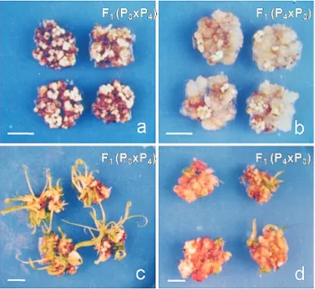
transluscent calli. In the hybrids involving these two par-ents, the reciprocal differences were significant for all of the fourin vitrocharacters studied. The quantity of embryo-genic and non-embryoembryo-genic portions of calli and the num-ber of regenerated plantlets in the reciprocal hybrids involving P3and P4are shown in Figure 2.
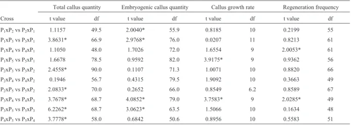
The Hayman (1954a) analysis for a 5x5 diallel set of pearl millet lines revealed items ‘c’ and ‘d’ to be significant for the quantity and frequency of regeneration of embryo-genic calli, whereas only item ‘d’ was significant for total quantity of calli and growth rate (Table 1). Among the ten hybrid combinations from a 5x5 diallel set, the reciprocal differences were significant for all of the fourin vitro char-acters in the cross involving P3 and P4 inbreds. In the
remaining hybrids, the reciprocal differences were signifi-cant for only one or two characters (Table 2). Conse-quently, the cross involving P3and P4 was chosen for a
detailed analysis of the reciprocal differences. One set of crosses, viz., P3xP4and its reciprocal P4xP3, was used to
produce the reciprocal F2s as well as reciprocal
of the fourin vitrocharacters of the 16 families were exam-ined.
Studentst-test, used to detect differences in the means of each of the fourin vitrocharacters, revealed significant differences between P3 and P4, between reciprocal F1s
(P3xP4and P4xP3), and between reciprocal backcrosses for
BC1 (callus growth rate) and BC2(total callus quantity)
(Table 3). A two-way ANOVA used to assess the differ-ences for each character revealed significant differdiffer-ences (p < 0.05) among the 16 families as expected, but no differ-ences between the replications (p > 0.05); a significant in-teraction factor (p < 0.01) was also observed (data not shown).
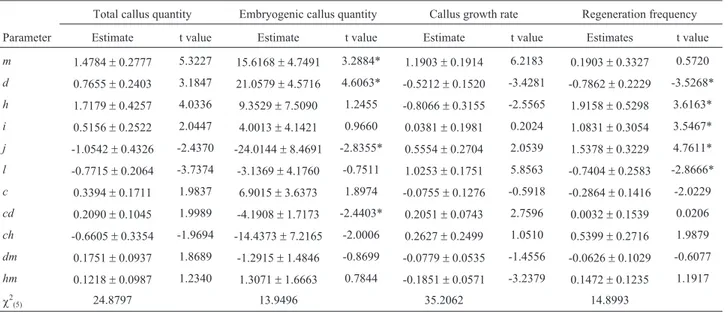
Embryogenic callus quantity and regeneration fre-quency showed a good fit to the genetic model, although the chi-square test of the goodness of fit was not significant (p > 0.01); the other two characters did not fit the model (p < 0.01) (Table 4). For embryogenic callus quantity, the pa-rametersd,jandcd, representing the additive effect, addi-tive x dominance interactions and the interaction between cytoplasmic and nuclear-determined effects, respectively, were significant. For the regeneration frequency, all of the parameters that provided estimates of the main effects and that represented the nuclear genetic contributions to the family means were found to be significant. The dominance increment of loci (h) for the nuclear genetic contribution was positive, while the ‘l’ increment for pairs of loci was
Figure 1- Various developmental stages during somatic embryogenesis in pearl millet. (A) A single ovoid proembryoid, (B) Stalked globular embryoids, (C) Scanning electron micrograph of a stalked globular embryoid, (D) Heart-shaped somatic embryo, (E) Scanning electron micrograph of a somatic em-bryo with collar-like scutellum (200x), and (F) Somatic emem-bryo with root and shoot primordia. Scale bar = 10mm.
Figure 2- Differences in the quantities of embryogenic (A) and non-embryogenic (B) portions of callus and in the number of regenerated plantlets in the corresponding hybrid F1(P3xP4) (C) and reciprocal hybrid
negative. The maternal effects included an additive genetic component (dm) and dominance (hm); the former affected total callus quantity, whereas the latter affected total callus quantity, embryogenic callus quantity and regeneration fre-quency.
Discussion
Cytoplasmic inheritance results in persistent recipro-cal differences and reflects an unequal contribution of cyto-plasmic determinants from male and female gametes to the zygote (Jinks, 1964). The maternal tissue effects are tran-sient and change with the genotype of the mother. Since the cytoplasmic component includes either the chloroplast or mitochondrial DNA, a distinction between maternal and cytoplasmic modes of inheritance becomes necessary. This distinction is also important in order to manipulate the par-ticular character, depending on its inheritance patterns. An in-depth study of the progenies other than the parents and F1s (that is F2s and backcrosses) is required when fitting
ge-netic models that allow estimation of cytoplasmic and ma-ternal effects and their adequacy in describing the observed reciprocal differences.
In a previous study, the ANOVA of genetic compo-nents from a 5x5 diallel cross revealed that GCA and SCA were significant (p < 0.05) for the fourin vitrocharacters studied, namely total callus quantity, embryogenic callus quantity, callus growth rate and frequency of regeneration (Satyavathi VV, 1998, PhD thesis, Andhra University, Visakhapatnam, India). GCA analysis indicated P4as the
best general combiner for total callus quantity and growth rate, P3for embryogenic callus quantity, and P1as well as
P3for regeneration frequency. SCA analysis revealed that
some of the crosses (P1xP3, P3xP4and P3xP5) were the best
specific combinations for one or more of thein vitro char-acters studied. In general, hybrids involving two poor gen-eral combiners showed better SCA effects. For callus growth rate and regeneration frequency, combinations of one better and one poor combiner produced better SCA ef-fects. Similar GCA and SCA effects have been reported in
Table 1- Hayman analyses of variance for the fourin vitrocharacters obtained from a 5x5 diallel analysis.
Mean squares
Item Degrees of freedom Total callus quantity Embryogenic callus quantity Callus growth rate Regeneration frequency
a 4 0.0209* 155.5555* 0.0394* 6.6107*
b 10 0.0078 61.6698* 0.0151* 4.6833*
b1 1 0.0006 1.5819 0.0505 3.0489*
b2 4 0.0185* 61.4082* 0.0214* 5.6812*
b3 5 0.0074 73.8966* 0.0029 4.2119*
c 4 0.0134 78.6379* 0.0023 0.4479*
d 6 0.0057* 13.0646* 0.0075* 0.7657*
a– additive genetic variation,b– dominance variation,b1– mean dominance deviation,i.e., the overall difference between the F1s compared to their
mid-parent values,b2– the variation in mean dominance deviations of the F1s from their mid-parent values within each array, over arrays,b3– dominance
deviations that are unique to individual F1s,c– average reciprocal differences,d– further reciprocal differences not accounted for byc. Each item was
tested against its own block interaction. *p < 0.05.
Table 2- Reciprocal differences for the fourin vitrocharacters in the different crosses (F1s).
Total callus quantity Embryogenic callus quantity Callus growth rate Regeneration frequency
Cross t value df t value df t value df t value df
P1xP2vs P2xP1 1.1157 49.5 2.0040* 55.9 0.8185 10 0.2199 55
P1xP3vs P3xP1 3.8631* 66.9 2.9768* 76.0 0.0207 11 0.8213 61
P1xP4vs P4xP1 1.1050 48.0 1.7026 72.0 1.6554 9 2.0053* 61
P1xP5vs P5xP1 1.6678 78.5 0.9592 82.0 3.9175* 9 0.9362 56
P2xP3vs P3xP2 2.4558* 90.0 0.1107 71.3 1.0071 10 0.8820 66
P2xP4vs P4xP2 0.1946 56.7 0.4315 79.5 1.9092 10 0.3663 49
P2xP5vs P5xP2 2.0833* 70.0 0.2652 66.0 0.8549 6.2 0.8589 67
P3xP4vs P4xP3 3.7678* 68.7 4.0852* 79.0 3.7583* 9 2.0285* 49
P3xP5vs P5xP3 6.2262* 68.7 3.0623* 63.5 1.5066 10 0.1634 48
P4xP5vs P5xP4 3.7778* 58.0 0.6842 50.6 0.8956 10 0.5583 51
rice (Abe and Futsuhara, 1991), maize (Petolino and Thompson, 1987), winter wheat (Ouet al., 1989),Allium ampeloprasum (Silverstand et al., 1995), Helianthus annuusL (Sarrafiet al., 1996), eggplant (Chakravarthiet al., 2010) and rapeseed (Etedalet al., 2012).
Based on Hayman’s analysis, significant additive ge-netic variation was observed for all of the fourin vitro char-acters. The most striking feature observed was the presence of highly significant average reciprocal effects (c) for embryogenic callus quantity and regeneration frequency. Thedcomponent was also significant for all of the charac-ters studied.
Our results revealed that embryogenic callus quantity and regeneration frequency showed a good fit to the genetic model of Mather and Jinks (1982), whereas for the other
two characters (total callus quantity and growth rate), this model was not a good fit, as indicated by the highly signifi-cantX2values (Table 4).
For embryogenic callus quantity, the estimates (addi-tive) and (additivex dominance) were significant among the nuclear parameters. The interaction between the cyto-plasm and nuclear additive component (‘cd’) was also sig-nificant. These findings suggested that the reciprocal differences for embryogenic callus quantity could be attrib-uted to the interaction between cytoplasmic and nuclear de-terminants. Narasimhuluet al. (1989) reported a similar observation for shoot morphogenesis inBrassicaspp. and suggested that both cytoplasm and nuclear genomes con-tain determinants important for temporal regulation and co-ordinated synthesis of factors that promote shoot
mor-Table 4- Estimates of the genetic parameters for nuclear and cytoplasmic components and chi-square values for the fourin vitrocharacters
Total callus quantity Embryogenic callus quantity Callus growth rate Regeneration frequency
Parameter Estimate t value Estimate t value Estimate t value Estimates t value
m 1.4784±0.2777 5.3227 15.6168±4.7491 3.2884* 1.1903±0.1914 6.2183 0.1903±0.3327 0.5720
d 0.7655±0.2403 3.1847 21.0579±4.5716 4.6063* -0.5212±0.1520 -3.4281 -0.7862±0.2229 -3.5268*
h 1.7179±0.4257 4.0336 9.3529±7.5090 1.2455 -0.8066±0.3155 -2.5565 1.9158±0.5298 3.6163*
i 0.5156±0.2522 2.0447 4.0013±4.1421 0.9660 0.0381±0.1981 0.2024 1.0831±0.3054 3.5467*
j -1.0542±0.4326 -2.4370 -24.0144±8.4691 -2.8355* 0.5554±0.2704 2.0539 1.5378±0.3229 4.7611*
l -0.7715±0.2064 -3.7374 -3.1369±4.1760 -0.7511 1.0253±0.1751 5.8563 -0.7404±0.2583 -2.8666*
c 0.3394±0.1711 1.9837 6.9015±3.6373 1.8974 -0.0755±0.1276 -0.5918 -0.2864±0.1416 -2.0229
cd 0.2090±0.1045 1.9989 -4.1908±1.7173 -2.4403* 0.2051±0.0743 2.7596 0.0032±0.1539 0.0206
ch -0.6605±0.3354 -1.9694 -14.4373±7.2165 -2.0006 0.2627±0.2499 1.0510 0.5399±0.2716 1.9879
dm 0.1751±0.0937 1.8689 -1.2915±1.4846 -0.8699 -0.0779±0.0535 -1.4556 -0.0626±0.1029 -0.6077
hm 0.1218±0.0987 1.2340 1.3071±1.6663 0.7844 -0.1851±0.0571 -3.2379 0.1472±0.1235 1.1917
c2(5) 24.8797 13.9496 35.2062 14.8993
cd– interaction between cytoplasm and the additive (d) nuclear-determined effect,ch– interaction between cytoplasm and the dominance (h) nu-clear-determined effect,d– additive genetic component,dm– maternal effects traceable to the additive genetic component,h– dominance,hm– domi-nance effects of the maternal genotype,i– additive by additive interaction,j– additive by dominance interaction,l– dominance by dominance interac-tion,m– population mean. Chi-square test for the goodness of fit was not significant (p > 0.01). *p < 0.01.
Table 3- Comparison of the average values of the two parents and corresponding reciprocal populations of F1s, F2s and backcrosses.
Total callus quantity Embryogenic callus quantity Callus growth rate Regeneration frequency
Population t value df t value df t value df t value df
Parents P3vs P4 7.1459* 10 9.4947* 10 9.9550* 10 8.6273* 10
F1(P3xP4) vs F1(P4xP3) 3.1727* 9 2.4650* 6.7 3.2509* 5.2 2.9635* 9
F2(P3xP4) x (P3xP4) vs (P4xP3) x (P4xP3) 1.1015 198 1.8707 198 1.2970 192 1.0463 197
F2(P3xP4) x (P4xP3) vs (P4xP3) x (P3xP4) 0.7636 187 0.9143 185 1.0214 146.3 0.5181 173
BC1(P3x(P3xP4) vs (P3xP4)xP3) 0.1773 83 0.3511 83 2.7090* 50.2 0.5573 81
BC1(P3xP4xP3) vs (P4xP3)xP3) 0.3147 96 0.6032 96 3.4493* 52.1 1.9679 93
BC2P4x(P3xP4) vs (P3xP4)xP4 2.5505* 72.8 1.3323 75 1.8129 64.7 0.5853 74
BC2P4x(P4xP3) vs (P4xP3)xP4 2.6741* 63.6 2.7113* 63.4 0.0010 65 1.5185 65
phogenesis. Janilaet al.(2013), based on genetic analyses of resistance to late leaf spot (LLS) in interspecific hybrids of groundnut, reported that a combination of nuclear and maternal gene effects was involved in the resistance factor.
For regeneration frequency in which the additive-dominance model was adequate, five out of the 11 esti-mates were significant, confirming the predominant role of nuclear gene control in the expression of this character. The positive (h) increment of loci and the negative (l) incre-ments for pairs of loci suggested the occurrence of non-allelic interaction of the duplicate kind in the expression of this character. This type of duplicate epistasis was consid-ered to be associated with characters expressed in direc-tional selection (Powell and Caligari, 1987). As inferred from the work of Satyavathiet al.(2006), regeneration fre-quency is associated with the d2 gene that controls the
dwarf nature of the plant. The suggestion by Powell and Caligari (1987) regarding the operation of directional selec-tion is consistent with the origin of P3(d2dwarf line)
devel-oped by Burton and Fortson (1966) and the association or linkage between thed2locus and loci controllingin vitro
re-generation frequency. The P3line was the result of breeders
selecting for homozygosis dwarf stature (d2d2),i.e.,
unidi-rectional selection for reduction in height. Since at least some of the loci controllingin vitroregeneration are linked to thed2locus, the selection for dwarfism might also have
indirectly affected the linked loci for regeneration. Both linkage and selection (direct and indirect) for the two characters (dwarf nature and regeneration) might have resulted in the characteristic and visually identifiable callus morphology noted in the dwarf parent. For Cyclamen persicum, Pueschelet al.(2003) postulated a hypothesis of two dominant epistatic genes controlling the capacity for regeneration and stated that the trait can easily be integrated into other genotypes of economic interest by crossings. Moreover, linked DNA-markers would enable early selec-tion of desired genotypes capable of regenerating somatic embryos and would spare the time and labor involved inin vitroscreening. Dodiget al.(2008) studied the correlation between tissue culture and agronomic traits in wheat. Agro-nomic traits with highly positive direct effects on tissue cul-ture traits were considered as suitable predictors of goodin vitro plant regeneration. These authors found productive tillering to have a significant (positive) direct effect on all tissue culture traits.
Our study revealed that for two characters, embryo-genic callus quantity and regeneration frequency, the for-mer showed a greater proportion of cytoplasmic nuclear interaction whereas the latter showed a greater role of nu-clear factors. Mathiaset al.(1986) reported the role of spe-cific interactions between the nuclear and cytoplasm in determining tissue culture responses in Chinese spring wheat. The possibility that regeneration from tissue culture is partly controlled by specific nuclear-cytoplasmic interac-tions has also been suggested by Henryet al.(1994).
Since the regeneration frequency is dependent upon embryogenic callus quantity, thed2cytoplasm might also
contribute (in addition to directional selection and linkage) to the characteristic response and morphological appear-ance of the callus in dwarf parents. This conclusion raises the possibility of manipulating these in vitro characters through the selection of appropriate lineages, despite their complex inheritance patterns.
Acknowledgments
The authors thank the University Grants Commis-sion, Government of India for the Junior and Senior Re-search Fellowship to VVS.
References
Abe T and Futsuhara Y (1991) Diallel analysis of callus growth and plant regeneration in rice seed-callus. Jpn J Genet 66:129-140.
Bebeli PJ (1995) Cytoplasmic effects on tissue culture response in wheat. J Genet Breed 49:201-208.
Ben-Amer IM and Boner A (1997) Effect of cytoplasm on imma-ture embryo culimma-ture in wheat (Triticum aestivumL.). Cereal Res Commun 25:135-140.
Burton GW and Fortson JC (1966) Inheritance and utilization of five dwarfs in pearl millet (Pennisetum typhoides) breeding. Crop Sci 6:69-72.
Cavalli LL (1952) An analysis of linkage in quantitative inheri-tance. In: Rieve ECR and Waddington CH (eds) Quantita-tive Inheritance, HMSO, London, pp 135-144.
Chakravarthi DVN,Rao YV,Rao MVS and Manga V (2010) Ge-netic analysis ofin vitrocallus and production of multiple shoots in eggplant. Plant Cell Tissue Organ Cult 102:87-97. Dodig D, Zoric M, Mitic N, Nikolic R, King SR, Lalevic B and
Surlan-Momirovic G (2008) Tissue culture and agronomic traits relationship in wheat. Plant Cell Tissue Organ Cult 95:107-114.
Ekiz A and Konzak CF (1991) Nuclear and cytoplasmic control of anther culture response in wheat: I. Analysis of alloplasmic lines. Crop Sci 31:1421-1427.
Etedal F, Khandan A, Motalebi-Azar A, Hasanzadeh Z, Sadeghan A, Pezeshki A, Pezeshki A and Kazemiani S (2012) Biomet-ric-genetic analysis ofin vitrocallus proliferation in rape-seed (Brassica napus). Afr J Agric Res 48:6474-6478. Foroughi-Wehr B, Friedt W and Wenzel G (1982) On the genetic
improvement of androgenetic haploid formation in
Hordeum vulgareL. Theor Appl Genet 62:233-239. Ge YX, Wei JL, Lu MY and Wei JK (1994) Studies on thein vitro
culture of immature embryos of homonuclear-heterocyto-plasmic maize lines. Acta Agric Univ Pekinensis 20:397-401.
Griffing B (1956) Concept of general and specific combining abil-ity in relation to diallel crossing systems.Aust J Biol Sci9:463-493.
Hayman BI (1954a) The analysis of variance of diallel tables. Biometrics 10:235-244.
Hayman BI (1958) The separation of epistatic from additive and dominance variation in generation mean. Heredity 12:371-390.
Henry Y, Vain P and de Buyser J (1994) Genetic analysis ofin vi-troplant tissue culture responses and regeneration capaci-ties. Euphytica 79:45-58.
Hou L, Ultrich SE and Kleinhofs A (1994) Inheritance of anther culture traits in barley. Crop Sci 34:1243-1247.
Ikeuchi M, Sugimoto K andIwase A (2013) Plant callus: mecha-nisms of induction and repression. Plant Cell 9:3159-3173. Janila P, Ramaiah V,Rathore A, Rupakula A, Reddy KR, Waliyar
F and Nigam SN (2013) Genetic analysis of resistance to late leaf spot in interspecific groundnuts. Euphytica 193:13-25. Jinks JL (1964) Extra Chromosomal Inheritance. Prentice Hall,
London, 177 p.
Keyes GJ, Collins GB and Taylor NL (1980) Genetic variation in tissue cultures of red clover. Theor Appl Genet 58:265-271. Krottje PA, Wofford DS and Quesenberry KH (1996) Heritability
estimates for callus growth and regeneration inDesmodium. Theor Appl Genet 93:568-573.
Li Z, Duan S, Kong J, Li S, Li Y and Zhu Y (2007) A single ge-netic locus in chromosome 1 controls conditional browing during the induction of calli from mature seeds ofOryza sativa ssp. Indica. Plant Cell Tissue Organ Cult 89:237-245. Mather K and Jinks JL (1982) Biometrical Genetics. 3rdedition.
Chapman and Hall, London, 396 p.
Mathias RJ, Fukui K and Law CN (1986) Cytoplasmic effects on the tissue culture response of wheat (Triticum aestivum) cal-lus. Theor Appl Genet 72:70-75.
Murashige T and Skoog F (1962) A revised medium for rapid growth and bioassays with tobacco tissue cultures. Physiol Plant 15:473-497.
Mythili PK, Satyavathi V, Pavan Kumar G, Rao MVS and Manga V (1997) Genetic analysis of short term callus culture and morphogenesis in pearl millet,Pennisetum glaucum. Plant Cell Tissue Organ Culture 50:139-142.
Narasimhulu SB, Chopra VL and Shyam Prakash (1989) The in-fluence of cytoplasmic differences on shoot morphogenesis inBrassica carinataA. Br. Euphytica 40:241-243. Ou G, Wang WC and Nguyan HT (1989) Inheritance of somatic
embryogenesis and organ regeneration from immature em-bryo cultures of winter wheat. Theor Appl Genet 78:137-142.
Petolino JF and Thompson SA (1987) Genetic analysis of anther culture response in maize. Theor Appl Genet 74:284-286. Powell W (1988) Diallel analysis of barley anther culture
re-sponse. Genome 30:152-157.
Powell W and Caligari PDS (1987) Thein vitrogenetics of barley (Hordeum vulgareL.): detection and analysis of reciprocal differences for culture response to 2,4-dichlorophenoxy-acetic acid. Heredity 59:293-299.
Pueschel AK, Schwenkel HG and Winkelmann T (2003) Inheri-tance of the ability for regeneration via somatic embryo-genesis inCyclamen persicum. Plant Cell Tissue Organ Cul-ture 72:43-51.
Sarrafi A, Roustan JP, Fallot J and Alibert G (1996) Genetic anal-ysis of organogenesis in the cotyledons of zygotic embryos of sunflower (Helianthus annuus L.). Theor Appl Genet 92:225-229.
Satyavathi VV, Subbarao MV, Manga V and Chittibabu M (2006) Genetics of some in vitro characters in pearl millet. Euphytica 148:243-249.
Silverstand CHJ, Jacobsen E and Van Harten AM (1995) Genetic variation and control of plant regeneration in leek (Allium ampeloprasumL.). Plant Breed 114:333-336.
Singsit C and Veilleux RE (1989) Intra and inter specific trans-mission of androgenetic competence in diploid potato spe-cies. Euphytica 43:105-112.
Taylor TE and Veilleux RE (1992) Inheritance of competences for leaf disc regeneration, anther culture and protoplast culture inSolanum phurejaand correlations among them. Plant Cell Tissue Organ Cult 31:95-103.
Tomes DT and Smith OS (1985) The effect of parental genotype on initiation of embryogenic callus from elite maize (Zea maysL.) germplasm. Theor Appl Genet 70:505-509. Walton PD and Brown DC (1988) ScreeningMedicagowild
spe-cies for callus formation and the genetics of somatic em-bryogenesis. J Genet 67:95-100.
Wan Y, Sorrensen Y and Liang GH (1988) Genetic control ofin vitroregeneration in alfalfa (Medicago sativaL.). Euphytica 39:3-9.
Wang X, Wu R, Lin X, Bai Y, Song C, Yu X, Xu C, Zhao N, Dong Y and Liu B (2013) Tissue culture-induced genetic and epigenetic alterations in rice pure-lines, F1hybrids and
poly-ploids. BMC Plant Biol 13:e77.
Willman MR, Schroll SM and Hodges TK (1989) Inheritance of somatic embryogenesis and plantlet regeneration from pri-mary (Type I) callus in maize. In Vitro Cell Dev Biol Plant 25:95-100.
Associate Editor: Everaldo Gonçalves de Barros