Diogo Rafael Coelho da Silva Araújo
dezembro de 2014
Functional characterization of the
grapevine aquaporin VvXIP1
UMinho|20
14
Diogo Rafael Coelho da Silva Ar
aújo
Functional characterization of t
he grape
vine aquaporin VvXIP1
Universidade do Minho
Diogo Rafael Coelho da Silva Araújo
dezembro de 2014
Dissertação de Mestrado
Mestrado em Biofísica e Bionanossistemas
Functional characterization of the
grapevine aquaporin VvXIP1
Universidade do Minho
Escola de Ciências
Trabalho efetuado sob a orientação do
Professor Doutor Hernâni Varanda Gerós
e do
iii
Acknowledgements
I would like to thank Professor Hernâni and Henrique for supervising me and supporting me doing my research work, as well as for their invaluable scientific advices which made me a better thinker and a better researcher.
Many thanks to the people in my laboratory, Rose Sousa, Vera Castro, Luís Loureiro, Marília Gonçalves, Luís Giraldo, Rupesh Rajput, Marslin Gregory, Walid Khouadja, António Freitas and Weina Wou, for their friendship and for making me feel at home from the start. I couldn’t have asked for better company. Special thanks to António Teixeira, Viviana Martins and Artur Conde for their patience and for helping me whenever I needed.
To Ana and Miguel for all the time you spent with me during this past year and for your solid friendship, I am deeply grateful.
To the friends I made during my Bachelor’s and master’s degrees that helped me becoming a better and a happier person, thank you!
To Sónia, not only for being involved in each detail of my life but also for helping me keep my head straight, a very special thank.
To my family, for their constant care and support thorough all the days of my life, specially during this year, and for their help to keep me positive in every situation, I am extremely grateful, and always will be.
This work was funded by FCT (Fundação para a Ciência e Tecnologia), Lisboa, Portugal, as a project grant, PTDC/AGR-ALI/100636/2008 and by the European project INNOVINE ref. 311775
iv
Functional characterization of the grapevine aquaporin VvXIP1
Abstract
The diffusion of water and small solutes through biological membranes is facilitated by aquaporins, members of the Major Intrinsic Proteins (MIPs) superfamily. As sessile organisms, plant species have been found to have more aquaporin genes than organisms from the other kingdoms of life. Among all the variety of MIPs, a subfamily called Uncharacterized Intrinsic Proteins (XIP) has been recently described in several fungi and eudicot plants. In the present study, the localization, expression and functional characterization of the grapevine VvXIP1 were performed. Phylogenetic analysis revealed that VvXIP1 is evolutionarily close to XIPs from cotton and peach. Co-localization studies with ZmPIP2;5-GFP and VvXIP1-RFP fusion proteins in transiently transformed Nicotiana bethamiana leaves after infection with Agrobacterium tumefaciens revealed that VvXIP1 is intracellular, and location prediction with the tool PredictProtein suggests that it locates in the tonoplast. Stopped flow spectrometry was used to evaluate water transport capacity of VvXIP1 in microsomal vesicles purified from the yeast strain YSK1172 transformed with the construct pVV214-VvXIP1. Results showed that VvXIP1 is unable to transport water but mediates glycerol transport. Growth experiments of the transformed yeast on solid media revealed that VvXIP1 is able to transport (besides glycerol) copper and H2O2.
Furthermore, when CM-H2DCFDA was used to measure H2O2 uptake by spectrofluorimetry, yeast
cells expressing VvXIP1 showed a much higher rate of fluorescence increase after H2O2 addition
than control cells transformed with the empty vector. Similarly, VvXIP1-transformed yeasts showed a 3.5 - fold higher rate of O2 production than the control, as measured with a Clark
electrode, when H2O2 was added to the incubation media. Both results strongly corroborated that
VvXIP1 is able to transport H2O2. Additional growth experiments in solid media with 2.1 M sorbitol
showed that the yeast transformed with the construct pVV214-VvXIP1 exhibited higher water stress sensitivity than the yeast transformed with the empty vector. To provide further insights into gene function, the expression of VvXIP1 was studied by qPCR in different organs and in response to different treatments/stressors. Results showed VvXIP1 steady-state transcript levels were much higher in leaves than in berries, canes or flowers from field grown cv. Vinhão grapevines. Moreover, VvXIP1 expression was downregulated in leaves from potted grapevines cv. Aragonez submitted to severe water deficit. In agreement, VvXIP1 was downregulated by ABA and salt stress in in vitro cultured cells. Based on the observation that VvXIP1 is able to transport
v
copper, the steady-state levels of the corresponding mRNA were measured in cv. Vinhão leaves from field-grown grapevines treated with the copper based fungicide Bordeux Mixture. The observed downregulation of the expression of VvXIP1 should have major importance in plant response to copper toxicity, contributing do the regulation of the intracellular levels of the heavy metal in coordination with the copper transporters VvCTrs recently studied in our group.
vi
Caraterização functional da aquaporina de Videira VvXIP1
Resumo
A difusão da água e de pequenos solutos através da membrana biológica é facilitada por aquaporinas, membros da superfamília Major Intrinsic Proteins (MIPs). Como organismos imóveis, as plantas possuem um número maior de genes de aquaporinas que os organismos de outros reinos. De entre todos os membros da superfamília MIP, uma subfamília, denominada Uncharacterized Intrinic Proteins (XIPs), foi recentemente descrita em fungos e plantas dicotiledóneas. No presente trabalho de tese foi estudada a localização e a função da proteína VvXIP1 de videira, bem como os níveis de expressão do mRNA correspondente em diferentes tecidos. A análise filogenética revelou que a proteína VvXIP1 é evolutivamente próxima de XIPs de algodão e pessegueiro. Os estudos de localização subcelular envolveram uma abordagem de transformação transiente de folhas de Nicotiana bethamiana por infiltração com Agrobacterium tumefaciens. Os resultados mostraram que a fluorescência verde da fusão ZmPIP2;5-GFP se localizava na membrana plasmática, ao contrário da fluorescência vermelha da fusão VvXIP1-RFP localizada provavelmente ao nível do tonoplasto, tal como previsto pela ferramenta bioinformática PredictProtein. A técnica “stopped flow” foi utilizada para avaliar a capacidade da aquaporina VvXIP1 transportar água em vesículas microssomais purificadas de células da estirpe de levedura YSK1172 transformada com o plasmídeo pVV214 - VvXIP1. Os resultados mostraram que a aquaporina é incapaz de transportar a água, mas é permeável ao glicerol. Estudos de crescimento em placa da levedura transformada mostraram que VvXIP1 é capaz de transportar cobre e H2O2 para além do glicerol. Quando se utilizou a sonda fluorescente
CM-H2DCFDApara medir por espectrofluorimetria a incorporação de H2O2, os resultados mostraram
que as células da levedura a expressar a proteína VvXIP1 proporcionaram um aumento maior de fluorescência do que o controlo quando se adicionou H2O2 ao meio de reação. Em paralelo,
estudos com o eléctrodo de Clark mostraram que a levedura a expressar a proteína VvXIP1 produzia mais O2 que a levedura transformada com o vector vazio após a adição de H2O2 ao meio
de reação. Ambas a evidências experimentais suportaram que a aquaporina VvXIP1 é capaz de transportar H2O2. A estirpe de levedura transformada com o plasmídeo pVV214 - VvXIP1 mostrou
ainda uma sensibilidade aumentada ao stresse hídrico que a levedura transformada com o plasmídeo vazio. Para obter uma melhor compreensão da função do gene VvXIP1, os níveis de expressão foram estudados por qPCR em diferentes órgãos e em resposta a diferentes tratamentos / stresses. Em videiras da casta Vinhão de uma vinha comercial o gene VvXIP1 foi
vii
mais expresso nas folhas do que em bagos, canas ou flores. Foi ainda observado que a expressão do gene VvXIP1 foi regulada negativamente em folhas de videiras envasadas da casta Aragonez submetidas a stresse hídrico severo. Em paralelo, em culturas celulares de videira a produção de mRNA foi regulada negativamente por 150 μM de ABA e 200 mM de NaCl. Por se ter descoberto que VvXIP1 é capaz de transportar cobre, foram medidos os níveis de mRNA em folhas de videiras da casta Vinhão tratadas com a Calda Bordalesa que é um fungicida à base de cobre. A diminuição observada dos níveis de transcritos de VvXIP1 pode constituir uma resposta da planta à toxicidade do cobre, contribuindo para a regulação dos níveis intracelulares do metal pesado em coordenação com os transportadores de cobre VvCTrs recentemente estudados no nosso grupo de investigação.
viii
Table of contents
Abstract ... iv
Resumo ... vi
Abbreviations and Acronyms... x
INTRODUCTION ... 12
Aquaporins are multifunctional water and solute channels widespread in living organisms ...13
Plant aquaporins ...15
Aquaporins in grapevine ...16
Materials and Methods ... 18
Plant material ...19
Growth assays ...20
Fluorescence assays ...20
Clark electrode assays ...21
RNA isolation from grapevine berries and leaves ...21
Co-localization studies ...21
Real-time PCR studies ...22
Yeast membrane Isolation ...22
Stopped flow spectroscopy ...23
Statistical analysis ...23
RESULTS ... 24
VvXIP1 sequence analysis ...25
Subcellular localization of VvXIP1 ...27
Transport Studies ...28
H2O2 transport ...28
Stopped flow experiments to study water transport ...30
Glycerol transport ...32
Copper transport ...33
Role of VvXIP1 in osmotic stress response ...35
VvXIP1 expression studies ...36
DISCUSSION ... 38
ix
VvXIP1 specificity is puzzling ...40
VvXIP1 is expressed in leaves and downregulated by water-deficit stress ...41
REFERENCES ... 42
APPENDICES ... 47
Appendix 1 - Sequence Accession Numbers ...48
x
Abbreviations and Acronyms
CM-H2DCFDA -5-(and-6)-chloromethyl-2’,7’-dichlorodihydrofluorescein diacetate acetyl ester
ABA - Abcisic acid Ea - Activation energy
AQP - Aquaporin
BLAST - Basic Alignment Search Tool CSB - Cabernet Sauvignon Berry
CTAB - Cetyltrimethyl ammonium bromide cv. - Cultivar
DTT - Dithiothreitol
EST - Expressed Sequence Tag Pgly - Glycerol permeability coeficient
GIP - GlypF-like Intrinsic Proteins GFP - Green Fluorescent Protein HIP - Hybrid Intrinsic Proteins MIP - Major Intrinsic Protein MS - Murashige and Skoog NIP - NOD26-like Intrinsic Protein OD - Optic density
Pf- - Permeability coefficient
PMSF - Phenylmethylsulfonyl fluoride PIP - Plasma membrane Intrinsic Protein PEG - Polyethylene glycol
PCR - Polymerase chain reaction PVP - Polyvinylpyrrolidone ROS - Reactive oxygen species RFP - Red Fluorescent Protein RT-PCR - Reverse transcriptase-PCR SA - Salicylic acid
xi
SIP - Small Basic Intrinsic Protein SC - Synthetic Complement TIP - Tonoplast Intrinsic Protein
TRIS - Tris(hydroxymethyl)aminomethane VvCTR1 - Vitis vinifera Copper Transporter 1
XIP - X Intrinsic protein / Uncharacterized intrinsic protein YNB - Yeast Nitrogen Base
12
13
Aquaporins are multifunctional water and solute channels widespread in living organisms
Transport of nutrients, ions, metabolic waste products and even water across cell membranes is of the utmost importance in all-living systems. Most transport processes across membranes are mediated by integral membrane proteins, sometimes functioning in association with extracytoplasmic receptor proteins and/or cytoplasmic energy-coupling and regulatory proteins. Transport proteins are often referred to as transporters, carriers, permeases, porters or channels. Important solutes like sugars, organic acids, minerals, protons and water cross the plasma membrane and cytosolic membranes through specific transport systems that play key roles in the cell metabolism. This is reflected in the genome of all organisms where up to 14% of transcribed genes code for transport proteins (reviewed by Conde et al., 2010).
The membrane that surrounds the cell and its compartments is known to be impermeable to charged and polar molecules. Classical studies have provided a solid basis for the lipid permeability of water and all physiologically relevant solutes, but they were unable to explain the high velocity of water and solute exchange or the low activation energy observed for such phenomena. More recently, it was shown that in addition to the intrinsic passive permeability of the bilayer to water, aquaporins can mediate the transport of water and small solutes (Gomes et al., 2009). In plants, it is known that the loss of aquaporin function is associated with growth impairment and reduced viability, but the role of these channels for survival in native environments is not fully understood (Ludwig and Dynowski, 2009).
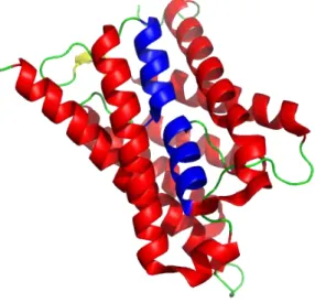
Aquaporins and aquaglyceroporins are the two clades of proteins dividing the Major Intrinsic Proteins (MIP) superfamily. MIPs are channels forming a hydrophilic pathway for small-uncharged molecules across the lipid bilayer of biological membranes that are virtually present in all living organisms. The division between aquaporins and aquaglyceroporins is based not only on sequence similarities but also on transport activities (Bienert et al., 2011). Despite the sometimes large sequence difference between MIPs from different clades, the overall structure is highly conserved. Structures from x-ray and electron crystallography of MIPs revealed a tetrameric quaternary structure in which each monomer consists of six membrane spanning helices (H1 to H6) connected by five loops (A-E). Loop B (cytoplasmic) and loop E (extracellular) form two half-membrane spanning helices (HB and HE) and interact with each other from opposing sides through two highly conserved aspargine-proline-alanine (NPA) boxes, forming a narrow region of the pore (Figure 1). A constriction region about 8 Å from the NPA boxes toward the periplasmic side, termed the aromatic/arginine (ar/R) region, is formed by two residues from
14
H2 and H5 and two residues from loop E. This region is a primary selection filter and is a major checkpoint for solute permeability (Danielson and Johanson, 2008).
Figure 1 – 3D structure of the mutant aquaporin from spinach SoPIP2;1. Adapted with pyMol (PyMOL Molecular Graphics System, Version 1.5.0.4 Schrödinger, LLC) from Nyblom et al. (2009).
Unlike aquaglyceroporins, AQPs are not widespread in bacteria and archea, in which water transport occurs through passive diffusion, unspecific pores, or through some aquaglyceroporins like the Lactococcus lactis GlaLlac, which is oddly able to transport water and glycerol (Gomes et al., 2009).
AQy1 and AQy2 were initially identified in S. cerevisiae, although, further research revealed that yeasts can have between one and four AQP genes (Soveral et al., 2012). In filamentous fungi a wide variety of AQPs have been described (Gomes et al., 2009).
Although invertebrates represent 97% of the animal species, their AQPs are still poorly described, with only few species having one or more aquaporins functionally characterized. Some are insects, including the two Drosophila species, having six homologues each; the mosquitoes Aedes aegypti, Anopholes gambiae and Culex pipiens with five homologues and the leafhopper Cicadella viridis. In the nematode Caenorhabditis elegans eleven MIP genes were identified, from which eight are aquaporins. In amphibians, over 17 MIPs have been identified and classified into clusters. Furthermore, the heterologous expression of aquaporins in Xenopus laevis oocytes is recurrently used as a powerful resource to study transport activities.
In mammals, 13 AQP isoform have been found which play major roles in the body homeostasis like re-absorbing water in the kidney and maintaining the cerebrospinal fluid balance in the brain (Bienert et al., 2011).
15
Plant aquaporins
Plants display a much larger diversity in MIPs than other organisms. While, as previously referred, 13 isoforms have been identified in mammals, 35 have been found in Arabidopsis, 55 in poplar, 71 in cotton and at least 36 in maize (Chaumont et al., 2001). Also, even the genome of the evolutionarily early land plant Physcomitrella patens encodes 23 different isoforms (Bienert et al., 2011). Plant MIPs perform key functions in transmembrane water conductance in a broad variety of physiological processes, such as uptake from the soil, transcellular and root-to-shoot transport, cellular water homeostasis and osmotic driven growth control. However, studies have revealed that some isoforms play important roles in many other processes, such as gas and nutrient uptake and translocation, metalloid homeostasis, nitrogen remobilization, signal transduction (Bienert et al., 2011).
The MIP superfamily in plants was initially divided into four subfamilies: the Plasma Membrane Intrinsic Proteins (PIPs), Tonoplast Intrinsic Proteins (TIPs), Nodulin-like Intrinsic Proteins (NIPs) and the Small Basic Intrinsic Proteins (SIPs). More recently, two new subfamilies were identified in the moss P. patens: the GlypF-like Intrinsic Proteins (GIPs) and the Hybrid Intrinsic Proteins (HIPs). Another subfamily called the Uncharacterized Intrinsic Proteins (XIPs) was identified, with members in a number of dicotyledonous plants, including tomato and grapevine (Danielson and Johanson, 2008; Hachez and Chaumont, 2010).
Interestingly, XIPs are present in a wide variety of eudicot plant species, but not in Brassicaceae (e.g. Arabidopsis). Although XIP genes have also been identified in the genomes of many fungi and a protozoan species, they remain undetected in monocots (Gupta et al., 2009). The most conserved feature of XIP protein sequences, which could be used as a signature for this subfamily, is the NPARC motif, with a cysteine residue located after the second NPA motif (Danielson and Johanson, 2008; Gupta et al., 2009). In addition, XIPs show considerable amino acid variation at both the first NPA motif and the ar/R filter. Based on the four amino acids defining the ar/R filters, XIPs from eudicots can be divided into four subclasses, two of which have an ar/R signature similar to that found in some plant NIPs, while the other two are even more hydrophobic (Bienert et al., 2011).
16
Aquaporins in grapevine
Grapes (Vitis spp.) are economically the most important fruit species in the world (Conde et al., 2007). Both development and maturation of grape berries have received considerable scientific interest because of both the uniqueness of such processes to plant biology and the importance of these fruits as a significant component of the human diet and wine industry. In the wine producers’ perspective, the most important attribute of Vitis vinifera is its capacity to store huge quantities of sugar, phenolics and other key compounds in the berries. From the plant point of view, the ripe phenotype is the summation of biochemical and physiological changes that occur during fruit development and make the organ edible and desirable to allow the dispersion of the seeds by animal activity (Conde et al., 2011). Therefore, understanding how and when various components, including water, accumulate in the berry, such as sugars, organic acids, phenolics, ions, is of critical importance to adjust grape growing practices and thus modify wine typology.
In grapevine, 23 MIPs were identified with genome sequence analysis in a single cultivar, but there are bound to be even more (Shelden et al., 2009; Tyerman et al., 2012). Several studies have linked aquaporin activity to the vine water status. Also, AQP expression varies according to environmental conditions and affects berry development. V. vinifera cultivars have different tolerance to water stress. Thus, different patterns of expression of VvPIP1;1 were observed in Chardonnay and Grenache. While Chardonnay exhibited a significant increase in VvPIP1;1 expression under water stress, Grenache did not show any alteration and this was shown to be related with differences in AQP regulation (Vandeleur et al., 2009). MIP expression also changes during maturation of grapes and this phenomenon is correlated with the increase in hydraulic resistance observed in the post-veraison stages (Tyerman et al., 2012).
In a recent study by our group (Noronha et al., 2014) the localization, expression, and functional characterization of a small basic intrinsic protein (SIP) from the grapevine were assessed. Results showed that VvSIP1 was expressed in leaves and berries from field-grown vines, and in leaves and stems from in vitro plantlets, but not in roots. When expressed in tobacco mesophyll cells and in S. cerevisiae, fluorescent-tagged VvSIP1 was localized at the endoplasmic reticulum (ER). This aquaporin was able to transport water but not glycerol, urea, sorbitol, glucose, or inositol. VvSIP1 expression in Xenopus oocytes failed to increase the water permeability of the plasma membrane. VvSIP1-His-tag was solubilized and purified to homogeneity from yeast ER membranes and the reconstitution of the purified protein in phosphatidylethanolamine liposomes confirmed its water channel activity.
17
In the present study we characterized two different XIP genes from grapevine - VvXIP1 and VvXIP2 -, and performed the localization, expression and functional characterization of VvXIP1. Co-localization studies with ZmPIP2;5-GFP and VvXIP1-RFP fusion proteins in transiently transformed Nicotiana bethamiana leaves revealed that VvXIP1 is intracellular. Heterologous expression in yeast revealed that VvXIP1 is able to transport copper, glycerol and H2O2.
Importantly, stopped flow spectrometry with microsomal vesicles purified from a transformed yeast strain showed that VvXIP1 is unable to transport water. VvXIP1 steady-state transcript levels were much higher in leaves than in berries, canes or flowers from field grown cv. Vinhão grapevines. VvXIP1 expression was low in leaves from potted grapevines cv. Aragonez submitted to severe water deficit. Accordingly, ABA and salt stress downregulated VvXIP1.
A manuscript based on this work is being prepared for publication in a peer reviewed journal.
18
19
In silico studies
Phylogenetic analysis was performed in silico using amino acid sequences from Vitis vinifera, Arabidopsis thaliana, Gossypium hirsutum, Populus trichocarpa, Prunus persica, Nicotiana tabacum, Lotus japonicas, Ricinus communis, Physcomitrella patens, Aspergillus terreus, Fusarium oxysporum, Penicillium marneffei, Hypocrea virens, Hypocrea jecorina and Selaginella moellendorffii obtained from the National Center of Biotechnology (NCBI), Uniprot and PlantGDB using the BLAST tool. The alignment of sequences was performed with PRANKSTER (Hasegawa et al., 1985; Tamura et al., 1993; Whelan et al., 2001; Loytynoja et al., 2005) and Genedoc (Nicholas et al., 1997). The trees were created using these alignments with PROTDIST, NEIGHBOR and RETREE from the PHYLIP software package (Felsenstein J. 1989). An alignment with VvXIP1 and several plant XIPs was performed and TOPCONS (Bernsel et al., 2009) was used to identify the transmembrane helix domains. The accession numbers of all the sequences used are listed in Appendix 1.
Plant material
Cv. Vinhão berries, canes, flowers and leaves were collected form a commercial vineyard near Guimarães. Potted cv. Aragonez plants were grown in a greenhouse at ITQB, subjected to different watering regimes during 4 weeks: full irrigation (FI – control), with plant watering every two days; and non-irrigation (NI), no watering. Leaves were collected when NI plants water potential was -1.3 < Ψpd < -0.9 MPa (Conde et al., 2014).
Cell suspensions of V. vinifera L. (Cabernet Sauvignon Berry - CSB) were freshly established from somatic callus that had been previously initiated from Cabernet Sauvignon berry pulp. They were maintained on modified Murashige and Skoog (MS) medium (Murashige and Skoog, 1962; Decendit et al., 1996), supplemented with 2% (w/v) sucrose in 250 ml flasks on a rotary shaker at 100 rpm in the dark, at 23 ºC according to Conde et al. (2014).
Cells were subcultured weekly by transferring 10 ml aliquots into 40 ml of fresh medium. In order to study the effect of different treatments on VvXIP1 expression, 5 ml aliquots were incubated overnight with 200 mM NaCl, polyethylene glycol (PEG) 2% (w/v), 150 µM abscisic acid (ABA), or 150 µM salicylic acid (SA) at 23 ºC. After each treatment, cells were immediately frozen in liquid nitrogen and stored at -80 ºC as performed by Noronha et al. (2014).
20
Growth assays
The following yeast strains were used: YSK1172, MPY2 and MPY3. YSK1172 is a deletion mutant for AQy aquaporins and MPY2 and MPY3 are double and triple mutants (MPY17 strain with ctr1Δctr3Δ, ctr1Δctr3Δctr2Δ), respectively, in copper transporters unable to grow with respiratory substrates such as glycerol and ethanol which requires high amounts of copper. AQy-null strain was transformed with pVV214-VvXIP and pVV214-Empty Vector. MPY2 and MPY3 strains were transformed with pVV214-VvXIP, pVV214-Empty Vector and with pVV214-VvCTR1. VvCTR1 is a copper transporter recently identified and functionally characterized (Martins et al., 2014a). The pVV214-VvXIP1 construct was prepared by Gateway (QIAGEN) and the primers can be found in appendix 1.
YSK yeast cells were grown in synthetic medium with 7 g/l YNB, 1.3 g/l SC, 2% Agar powder, 2% glucose, 300 mg/100 ml leucine and 250 mg/100 ml Triptophan, washed and resuspended in sterile water to a final OD600 nm = 1. Different dilutions (0.1, 0.01 and 0.001) were
made and 10 μl from each suspension were spotted in solid medium with the following conditions/stressors: i) 2.1M sorbitol, ii) 1% and 2% of ethanol and the same percentages of glycerol, iii) 30 and 60mM Boric acid (adjusted to pH 5.5 with Tris), iv) 5mM CuSO4, v) 0.35 and
0.7 mM H2O2. Appropriate control plates inoculated with non-complemented mutants were
prepared.
MPY yeast cells were grown in synthetic medium containing 7g/L YNB, 1.3g/L SC, 2% Agar powder, 2% glucose, 25 mg/100mL histidine, 30 mg/100ml Lysine and 25 mg/100mL Triptophan, washed and resuspended in sterile water to a final OD600 nm = 1; Different dilutions
(0.1, 0.01 and 0.001) dilutions were made and 10 μl from each suspension were spotted in solid medium containing 10 and 100 μM CuSO4. Yeast growth was observed during several days
and images were collected with Chemidox XRS (Bio-Rad). Fluorescence assays
Yeast cells from YSK1172 AQy-null strain were transformed with VvXIP1 and pVV214-Empty vector constructs and pre-cultured in YNB+SC solid medium. Liquid cultures were then grown in the dark overnight in presence of 1 μM 5-(and-6)-chloromethyl-2’,7’-dichlorodihydrofluorescein diacetate acetyl ester (CM-H2DCFDA, Molecular Probes), a fluorophore
21
1999; Bienert et al., 2007). In its acetylated form, the dye can freely diffuse into yeast cells but, once inside, the fluorochrome is deacetylated and unable to cross the membrane making the cells susceptible to oxidation by ROS. After incubation, cells were washed three times with MOPS buffer (pH 7) and resuspended in this buffer to a final OD600 nm =1.4. Fluorescence of 2 ml yeast
suspension was followed over time at 20°C in a spectrofluorometer at an excitation/emission of 492/527 nm (Perkin Elmer Luminecense Spectrometer LS 50).
Clark electrode assays
Yeast cells from YSK1172 AQy-null strain were transformed with VvXIP1 and pVV214-Empty vector constructs and pre-cultured in YNB+SC solid medium. Liquid cultures were then grown overnight. Cells were then washed three times and resuspended in water to a final final OD600 nm = 1. Hydrogen peroxide was then added to the cell suspension to a final concentration of
50 μM and the oxygen formation followed using a Clark electrode coupled to an YSI 5300 Biological Oxygen Monitor.
RNA isolation from grapevine berries and leaves
RNA from grape berries and leaves was isolated using QIAGEN RNeasy Plant Mini kit following the manufacturer’s instructions. The extraction buffer was changed to 2% cetyltrimethyl ammonium bromide (CTAB), 2% soluble polyvinylpyrrolidone (PVP) K-30, 300 mM TRIS-HCl (pH 8.0), 25 mM EDTA, 2.0M NaCl, and 2% (v/v) β-mercaptoethanol. After an in-column DNase treatment, the RNA integrity was checked in a 1% agarose gel, and the first-strand cDNA synthesis was performed with the LongRange 2Step RT-PCR (Quiagen), following manufacturer’s instructions.
Co-localization studies
The pH7RWG2-VvXIP1-RFP construct was prepared by Gateway (QIAGEN) and the primers can be found in appendix 1. Recombination sequences were introduced by PCR in the VvXIP1 cDNA without a stop codon and the fragment recombined into the entry vector pDONR221 using the BP clonase enzyme. VvXIP1 cDNA was then recombined into the pH7RWG2 vector by the LR clonase enzyme. pDE1001-ZmPIP2;5-GFP was kindly supplied by François Chaumont and was obtained as described by Chaumont et al. (2000). The two constructs were separately introduced in
22
Agrobacterium tumefaciens (GV3101) and transient transformation of Nicotiana benthamiana leaf epidermal cells was performed according to Sparkes et al. (2006). Bacterial cells were cultivated overnight in liquid LB medium up to the exponential-stationary phase and then diluted to OD600nm=0.1 with infiltration buffer (50 mM MES pH 5.6, 2 mM Na3PO4, 0.5% glucose, and 100
μM acetosyringone). Diluted cells were cultivated again until the culture reached an OD600nm=0.2.
Four-week-old N. benthamiana plants were infiltrated with the bacterial cultures and leaf discs were examined under the confocal microscope 2 days later.
Real-time PCR studies
Quantitative Real-time PCRs were performed with a QuantiTect SYBR Green PCR Kit (Qiagen) and in a CFX96 Real-Time detection System (Bio-Rad), at an annealing temperature of 55ºC. RNA and cDNA samples were obtained as described above. Experiments were carried out in biological triplicates with the software Bio-Rad CFX Manager (Bio-Rad), using VvGAPDH as an internal control. To verify the absence of unspecific and primer-dimer amplification melting curves were performed after each run. The primers used to study the expression of VvXIP1 and VvGAPDH can be found in appendix 2.
Yeast membrane Isolation
YSK1172 yeast cells transformed with pVV214-VvXIP1 were cultivated overnight in YNB medium with leucine and tryptophan. Cells were washed, resuspended in digestion buffer (1.35 M sorbitol, 10mM citric acid, 30 mM Na2HPO4, 1 mM EGTA, pH 7.4) with 30 mM dithiothreitol
(DTT), centrifuged and spheroplasts were obtained by digestion with zymolyase 20T (in digestion buffer). After complete digestion, monitored in phase contrast microscope, spheroplasts were pelleted and homogenized in a potter homogenizer after resuspended in HEPES-lysis buffer [20 mM HEPES, 50 mM potassium acetate, 100 mM sorbitol, 2 mM EDTA, 1 mM phenylmethylsulphonyl fluoride (PMSF), 1 mM DTT, pH 6.8]. The homogenate was centrifuged at 1000 g for 10 min and the supernatant saved. The membranes were then pelleted at 100 000 g for 30 min, resuspended in Resuspension buffer (100 mM mannitol, 10 mM Tris-HEPES, pH 7.5), centrifuged again at 100 000 g for 30 min, flash frozen and stored at -80 ºC These procedures were performed according Rodrigues et al. (2013) and Noronha et al. (2014).
23
Stopped flow spectroscopy
Membrane permeability of the microsomal fraction from transformed yeasts was studied with the stopped flow technique (HI-TECH Scientific PQ/SF-53) in the Laboratory headed by Prof. Graça Soveral at Faculdade de farmácia, Universidade de Lisboa (Lisbon). Five runs were usually stored and analysed in each experimental condition, as described before (Soveral et al., 1997; Noronha et al., 2014). To study water transport, vesicles resuspended in 100 mM mannitol, 10 mM TRIS-HEPES (pH 7.5) (0.1 ml, 0.4 mg protein ml-1) were mixed with a 340 mM mannitol, 10 mM
TRIS-HEPES (pH 7.5) solution at 23 ºC to produce an inwardly directed gradient of impermeant solute (osmotic gradient 240 mOsmol). The kinetics of vesicle shrinkage were measured from the time course of 90 º scattered light intensity at 400 nm until a stable light scatter signal was reached. Experiments were performed at 14-33 ºC to study activation energies.
To study glycerol transport, vesicles resuspended in 100 mM mannitol, 10 mM TRIS-HEPES (pH 7.5) (0.1 ml, 0.4 mg protein ml-1) were mixed with a solution containing 100 mM
mannitol, 240 mM glycerol and 10 mM TRIS-HEPES (pH 7.5) at 23 ºC to produce an inwardly directed gradient (osmotic gradient 240 mOsmol). The kinetics of vesicle shrinkage and swelling were measured from the time course of 90 º scattered light intensity at 400 nm until a stable light scatter signal was reached. The water permeability coefficient (Pf) and the glycerol
permeability coefficient (Pgly) were estimated by fitting the light scatter signal to a single
exponential curve with the equation Pf = k(V0/A) [1/Vw/(osmout)∞], where Vw is the molar volume of
water, V0/A is the initial volume to area ratio of the vesicle population and (osmout)∞ is the final
medium osmolarity after the application of the osmotic gradient. The osmolarity of each solution was determined from freezing point depression by a semi-micro-osmometer (Knauer GmbH, Germany). The activation energies (Ea) were obtained from the slope of an Arrhenius plot (ln Pf or
ln Pgly as a function of 1/T) multiplied by the gas constant R. Vesicle size (initial volume) was
determined by quasi-elastic light scattering (QELS) by a particle sizer (BI-90 Brookhaven Instruments) as described by Noronha et al. (2014).
Statistical analysis
The results obtained were statistically verified by analysis of variances tests (one-way and two-way ANOVA using Prism v. 6 (GraphPad Software, Inc.) Post-hoc multiple comparisons were performed using the HSD Tukey test.
24
25
VvXIP1 sequence analysis
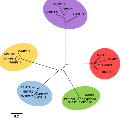
A phylogenetic tree constructed with amino acid sequences from SIPs, PIPs, TIPS, NIPs and XIPs from Vitis vinifera, Gossypium hirsutum, Populus trichocarpa, Arabidopsis thaliana and Nicotiana tabacum shows a clear clustering of the analyzed MIP sub-families in 5 separated groups (Figure 2). The tree depicted in Figure 3 was constructed only with XIP members from different plant and fungi species to make a close-up analysis of the phylogenetic relationships within XIP family. As can be seen, VvXIP1 is phylogenetically close to GhXIP1 from cotton and to PpXIP1 from peach.
Figure 2 – Phylogenetic analysis of several Plant MIPs. Sequences from PIPs, NIPS, SIPs, TIPs and XIPs
from Vitis vinifera (Vv), Arabidopsis thaliana (At), Gossypium hirsutum (Gh), Populus trichocarpa (Pt) and
Nicotiana tabacum (Nt)(NCBI and Uniprot access numbers in Appendix) were aligned using PRANKSTER (Hasegawa et al., 1985; Tamura et al., 1993; Whelan et al., 2001; Loytynoja et al., 2005). The phylogenetic tree was built using genedoc (Nicholas et al., 1997), Phylip (Felsenstein J, 1989) and MEGA4 (Tamura et al., 2007).
26
Figure 3 – Phylogenetic tree comparing XIP proteins from plants and fungi. Amino acid sequences from V.
vinifera (Vv), P. trichocarpa (Pt), Prunus. persica (Pp), G. hirsutum (Gh),Ipomoea nil (In), N. tabacum (Nt), Lotus japonicas (Lj), Ricinus communis (Rc), Physcomitrella patens (Ppat), Aspergillus terreus (Ate), Fusarium oxysporum (Fo), Penicillium marneffei (Pm), Hypocrea virens (Hv), Hypocrea jecorina (Hj)and
Selaginella moellendorffii (Sm) were aligned using PRANKSTER (Hasegawa et al., 1985; Tamura et al., 1993; Whelan et al., 2001; Loytynoja et al., 2005). The phylogenetic tree was built using genedoc (Nicholas et al., 1997), Phylip (Felsenstein J) and MEGA4 (Tamura et al., 2007).
Figure 4 shows the alignments of amino acid sequences from XIPs from V. vinifera (VvXIP1), P. trichocarpa (PtXIP1), P. persica (PpXIP), G. hirsutum (GhXIP1;1), I. nil (InXIP1;1), N. tabacum (NtXIP1;1), L. japonicas (LjXIP1), R. communis (RcXIP). As can be seen, all the proteins share the highly conserved NPARC motif, despite the slight variations in the first NPA (NPV in most of the aligned species). Using TOPCONS it was also possible to identify the regions corresponding to the six transmembrane helixes.
27
Figure 4 – Alignment of eight plant XIPs (VvXIP1, PtXIP1, PpXIP, GhXIP1;1, InXIP1;1 NtXIP1;1, LjXIP1 and RcXIP) showing six transmembrane helix domains (Black lines) and the conserved ‘NPV’ and ‘NPARC’ motifs (gray lines).
Subcellular localization of VvXIP1
As referred to in “Material and Methods”, the subcellular localization of VvXIP1-RFP was studied after Agrobacterium tumefaciens mediated transient expression in Nicotiana benthamiana leaf cells. Results are shown in Figure 5 when leaves were observed under the confocal microscope. As can be seen, the green fluorescence from ZmPIP2;5-GFP is clearly located at the cell periphery, at the plasma membrane, as shown before (Chaumont et al., 2000). However, the red fluorescence from RFP does not co-localize with the green fluorescence signal, showing a more diffuse pattern, probably between the vacuole - that occupies most of the intracellular space forcing the cytosol with its surrounding plasma membrane against the cell wall - and the plasma membrane (Figure 5). Thus VvXIP1 does not localize to the plasma membrane, in agreement with the prediction performed by the program “PredictProtein”.
28
Figure 5 – Subcellular localization of VvXIP1 in Nicotiana benthamiana. Laboratory grown plants were
infiltrated with Agrobacterium tumefaciens strains transformed with pH7RWG2-VvXIP1 and
pDE1001-ZmPIP-GFP plasmids. Images were acquired 2 days after infiltration in a confocal microscope.
Transport Studies H2O2 transport
To test whether VvXIP1 can mediate the transport of H2O2, the YSK1172 AQy-null yeast mutant
strain was transformed with the construct pVV214-VvXIP1 and H2O2 incorporation was studied
with three distinct approaches: i) rate of fluorescence variation of the fluorescent probe CM-H2DCFDA, ii) rate of O2 production resulting from intracellular breakdown of H2O2, and iii) yeast
growth inhibition in solid medium supplemented with H2O2.
When CM-H2DCFDA was used to measure H2O2 uptake by spectrofluorimetry, yeast cells
expressing VvXIP1 showed a much higher rate of fluorescence increase after H2O2 addition than
control cells transformed with the empty vector (Figure 6A). Similarly, VvXIP1-transformed yeast showed a 3.5 - fold higher rate of O2 production than the control, as measured with a Clark
29
Figure 6 – H2O2 transport by yeast cells expressing VvXIP1. Cultures of the yeast strain YSK 1172 AQynull
transformed with the empty vector pVV214 or pVV214 carrying VvXIP1 were incubated with CM-H2DCFDA
probe overnight and the fluorescence response after 600 μM H2O2 addition was monitored with a
spectrofluorimeter (A). O2 release monitored with a Clark electrode in response to 50 μM H2O2 (B).
Regarding the growth experiments in solid media, results showed that the growth of the yeast transformed with pVV214-VvXIP1 was severely inhibited when YNB media were supplemented with toxic levels of H2O2, 0.35 and 0.7 mM, when compared to controls (Figure 7).
30
Figure 7 – H2O2 sensitivity of yeast cells expressing VvXIP1. Cultures of YSK 1172 AQynull yeast cells
transformed with the pVV214-Empty vector and pVV214-VvXIP1 were spotted at an OD600 nm of 0.1 and
0.01 on medium containing the indicated concentrations of H2O2 and growth was recorded after 3 days at
30 ºC.
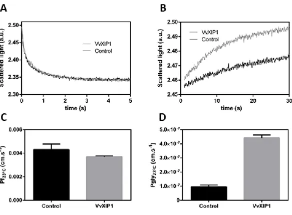
Stopped flow experiments to study water transport
Stopped flow light-scattering spectrophotometry was used to check if VvXIP1 is a water channel. As reported in “Material and Methods”, a microsomal fraction from YSK1172 yeast cells expressing VvXIP1 was used to study membrane permeability. Control microsomal vesicles were obtained from yeasts transformed with the empty vector. The vesicle size, measured by QELS analysis, was homogeneous in all batches with a mean hydrodynamic diameter of 375 ± 62 nm (n=8) (data not shown). Vesicles were challenged with a hypertonic mannitol solution to study water efflux. Results showed that the rate of water efflux (Figure 8A), and thus the osmotic permeability coefficient (Pf; Figure 8C), in yeast membrane vesicles with VvXIP1 were similar to
control vesicles. Also, the activation energies (Ea) were similar in both vesicle batches (Figure 9A
and C). Altogether, these clear-cut results strongly suggested that VvXIP1 is unable to transport water.
31
Figure 8 – Stopped flow experiment for evaluation of the water and glycerol permeability in yeast membrane vesicles. Normalized scattered light intensity obtained from stopped flow experiments with
membrane vesicles collected from yeast cells transformed with pVV214-VvXIP1 (grey) or the empty vector
(black), suddenly exposed to an osmotic gradient of 240 mOsm with a mannitol solution (A) and with a glycerol solution (B). The water and glycerol permeability coefficients are represented in C and D respectively.
32
Figure 9 – Stopped flow experiment to evaluate the energetics of water and glycerol permeability in yeast vesicles. Normalized scattered light intensity was obtained from stopped flow experiments performed according to a temperature gradient. Membrane vesicles collected from yeast cells transformed with
pVV214-VvXIP1 (grey) or the empty vector (black) were suddenly exposed to an osmotic gradient of 240 mOsm. The gradient was built with mannitol (A) to evaluate water transport and with glycerol (B) to evaluate glycerol transport. The activation energy for water (C) and glycerol (D) transport were calculated from the Arrhenius plots.
Glycerol transport
Taking advantage of the stopped flow approach, the capacity of VvXIP1 to transport glycerol was studied. As shown in Figure 8B the swelling rate of the membrane vesicles from VvXIP1-expressing yeast cells when challenged with a hypertonic glycerol solution was much higher than in control vesicles. The corresponding permeability coefficients are shown in Figure 8D. Channel mediated glycerol transport was confirmed when the Ea were measured. As can be seen in
Figures 9B and D, Ea value associated with glycerol transport in vesicles from VvXIP1-expressing
yeast cells was much lower than in vesicles from the yeast transformed with the empty vector, strongly suggesting protein-mediated diffusion.
33
To further demonstrate that VvXIP1 can transport glycerol, transformed yeast cells were grown in glycerol rich medium. Results showed that cells expressing VvXIP1 grew better than control cells (empty vector) in media supplemented with 1 and 2% ethanol and glycerol, confirming that glycerol is a substrate of VvXIP1 (Figure 10), and, therefore, validating the results from the stopped flow experiments
Figure 10 –Yeast growth assays in glycerol rich media by cells expressing VvXIP1. Cultures of YSK 1172
AQynull yeast cells transformed with pVV214-Empty vector and pVV214-VvXIP1 were spotted at OD600 nmof
0.1 and 0.01 on medium containing the indicated concentrations of ethanol and glycerol and growth was recorded after 3 days at 30 ºC.
Copper transport
As referred to in “Material Methods”, the yeast strains MPY2, MPY3 and YSK1172 were used to study if VvXIP1 can transport Cu. MPY2 and MPY3 are double and triple mutants in copper transporters, respectively, and cannot survive when cultivated with respiratory substrates due to the lack of means to uptake high amounts of Cu required for respiration. These yeast strains were transformed with pVV214-VvXIP1 construct to evaluate if respiration capacity was recovered. Furthermore, MPY strains were also transformed with pVV214-VvCTR1, a copper transporter recently identified and characterized in our group (Martins et al., 2014a). Results showed that VvXIP1 and VvCTR1 were able to restore yeast growth when both mutant strains were plated onto 100 μM CuSO4 (Figure 11A). Also, the transformation of YSK yeast strain with
34
VvCTR1 increased yeast sensitivity to the toxic effects of Cu, and thus reduced the growth in YNB media supplemented with 5 mM CuSO4 (Figure 11B), suggesting that in addition to H2O2 and
glycerol VvXIP1 also transports Cu.
Figure 11 – Cu effect on the growth of yeast cells expressing VvXIP1. Cultures of MPY2 (A), MPY3 (A) and YSK 1172 (B) yeast cells transformed with pVV214-Empty vector, pVV214-VvCTR1 (only used in MPY
strains) and pVV214-VvXIP1 were spotted at OD600 nm of 0.1 and 0.01 on medium containing the indicated
35
Role of VvXIP1 in osmotic stress response
It has been reported that aquaporin expression in yeast may lead to an osmotic stress oversensitivity (Leitão et al., 2012). In the present study YSK 1172 AQy-null yeast cells were transformed with pVV214-VvXIP1 and incubated in YNB media supplemented with 2.1 M sorbitol. As shown in Figure 12, transformed cells with VvXIP1 grew less than control cells transformed with the empty vector suggesting that they are more sensitive to osmotic stress.
Figure 12 – Sorbitol induced osmotic stress sensitivity of yeast cells expressing VvXIP1. Cultures of YSK
1172 AQynull yeast cells transformed with the pVV214-Empty vector and pVV214-VvXIP1 were spotted at
OD600 nm of 0.1 0.01 on medium containing the indicated concentration of sorbitol and growth was
36
VvXIP1 expression studies
The expression of VvXIP1 was studied in different organs in field grown cv. Vinhão grapevines. Total RNA was isolated from leaves, berries, canes and flowers. As shown in Figure 13, steady-state transcript levels of VvXIP1 were much higher in leaves than in the remaining organs.
Figure 13 – VvXIP1 expression in several organs from grapevine determined by quantitative real-time PCR.
Transcript levels in berries, canes, flowers and leaves from cv. Vinhão.
To study the effect of water stress on the expression of VvXIP1, transcript levels were measured in leaves from potted cv. Aragonez grapevines cultivated in a greenhouse at ITQB (Oeiras – Portugal) in the scope of a collaboration in progress with the group headed by Prof. Manuela Chaves. As can be seen in Figure 14A, VvXIP1 steady-state transcript levels were lower in leaves from non-irrigated plants than in leaves from plants watered every two days, suggesting that water deficit downregulates VvXIP1.
Following the results from heterologous expression experiments which suggested that VvXIP1 is able to mediate Cu transport, we decided to study whether the application of Bordeaux mixture in field conditions could affect the expression of VvXIP1. This task took advantage of the work developed in the scope of the PhD project of Viviana Martins (Martins et al., 2014a; Martins et al., 2014b). As shown in Figure 14B, in leaves from cv. Vinhão vines treated with Bordeaux mixture (+ Cu) a downregulation of VvXIP1 expression was observed when compared to plants treated with a conventional triazole-based fungicide (- Cu).
Despite the necessary cautions needed to extrapolate the results to a multicellular level, suspension-cultured cells have provided a convenient experimental system to study several important aspects of plant cell physiology, including gene expression and functional studies
37
(Conde et al., 2006; Noronha et al., 2014; Conde et al., 2014). Additionally, in cell suspensions, the plasma membrane is readily amenable for challenging with exogenous stressors/elicitators. In the present study cultured CSB cells were used to study VvXIP1 expression after an overnight incubation with 100 mM NaCl, 150 µM ABA, 150 µM SA and 2% (w/v) PEG. VvXIP1 transcript levels were downregulated by ABA and NaCl but not affected by SA and PEG (Figure 14C).
Figure 14 - VvXIP1 expression in response to potential stressors/elicitators determined by q PCR. VvXIP1
steady-state transcript levels in leaves from field grown cv. Vinhão grapevines in response to copper treatments (Bordeaux mixture) (A), in potted grapevines (cv. Aragonez) under drought stress (B) and in CSB cell suspensions treated with salt, ABA, SA and PEG (C).
38
39
VvXIP1 is a grapevine intracellular solute channel
Despite the overall structure similarities of aquaporins, they are highly divergent among different subfamilies (Gomes et al., 2009; Lopez et al., 2012). The differences in amino acid sequences are even more relevant in plants because as sessile organisms they have much more MIP genes than animals, bacteria or fungi (Bienert et al., 2007; Gomes et al., 2009). As reported to before, XIPs - Uncharacterized Intrinsic Proteins - are present in some plant species and fungi, but their nature and true physiological role are still no clear. As shown in Figure 2, when aligned with several plant TIPs, PIPs, SIPs and NIPs, plant XIPs cluster in a clearly distinct monophyletic group, completely separated from the other MIP subfamilies, suggesting specific transport characteristics. Moreover, VvXIP1 identified in the present study is highly similar to other XIPs from cotton and peach and show strong evolutionary divergence from fungi XIPs.
The divergence in the composition of the NPA motives has been proposed to reflect differences in the substrate specificity, as previously observed in NIPs (Bienert et al., 2011). In the present study, results showed that the composition of the first NPA motive might change considerably within XIP sequences from different species. As shown in “Results”, the alanine of VvXIP1 is replaced by valine in XIPs from P. persica, G. hirsutum and N. tabacum, by isoleucine in Ipomoea nil and P. trichocarpa, and by threonine in R. communis. The specificity of the aquaporin VvXIP1 revealed rather striking, as discussed below.
After their discovery at the plasma membrane of erythrocytes, researchers found that, similarly to other transporters and channels, aquaporins could also localize at intracellular membranes and transport other substrates than water (Wudick et al., 2009). Actually, it is known that TIPs have a key role in water transport at the tonoplast level and are tightly regulated by water deficit conditions (Tyerman et al., 2012). In addition, several aquaporins can be located to the membrane of other organelles, including the endoplasmatic reticulum. This is the case of VvSIP1 that specifically transports water across the ER of grapevine cells (Noronha et al., 2014). In a previous work it was shown that the XIP aquaporin from tobacco NtXIP1;1 is located at the plasma membrane (Bienert et al., 2011), but in the present study clear-cut results showed that VvXIP1 is indeed not targeted to the plasma membrane. After a transient transformation of tobacco leaves, the red fluorescent protein-tagged VvXIP1 did not co-localize with the green fluorescent protein-tagged ZmPIP2;5, which is a known plasma membrane protein. To ascertain the precise localization of VvXIP1, future work will involve the utilization of intracellular membrane
40
markers, including the endoplasmatic reticulum marker HDEL–YFP, the Golgi apparatus marker ST-YFP, and the tonoplast marker ZmTIP2;1-YFP .
VvXIP1 specificity is puzzling
What should the physiological role be of an intracellular aquaporin which mediates the incorporation H2O2, Cu, glycerol, but not water? Why substrates structurally and functionally so
different could share the same channel? Is VvXIP1 involved in detoxifying damaging molecules by trapping them in the vacuole? Is there any physiological redundancy between VvXIP1 and VvNIPs? The addressing of these key questions in future work will expand our knowledge on the role of intracellular aquaporins and will shed light into their structure-function relationships.
H2O2 is an important molecule with dual functions, because it is an oxidative stress
inducer but also a signaling molecule in plants (Bolwell, 1999; Foyer et al., 1999; Neill et al., 2001). In the present study we demonstrated with distinct approaches that VvXIP1 indeed transports H2O2 after its heterologous expression in yeast. But, intriguingly, this aquaporin is
unable to transport water, which is indeed a very peculiar finding since H2O and H2O2 display high
structural and electrostatic similarity, and MIPs from other subfamilies that are able to transport H2O2 are also water channels (Bienert et al., 2011).
H2O transport capacity of VvXIP1 was studied by stopped flow scattered-light
spectrophotometry in yeast membrane vesicles, a technique recently used in our group to confirm water transport capacity of the intracellular aquaporin VvSIP1 (Noronha et al., 2014). Clear-cut results showed that the permeability to water of membrane vesicles isolated from the yeast strain YSK1172 overexpressing VvXIP1 was similar to control. Also, no changes were observed in Ea of water transport across the membrane of the vesicles. This behavior is similar to
XIPs from Solanaceae (Bienert et al., 2011), but PtXIP2;1 and PtXIP3;3 from P. trichocarpa are water channels, although their water permeability is low when compared to members of the PIP subfamily (Lopez et al., 2012).
Stopped flow experiments also showed that glycerol permeates VvXIP1. Glycerol is a common substrate for non-water conducting MIPs, particularly from the NIP and XIP subfamilies (Bienert et al., 2011).
The role of VvXIP1 in Cu transport was unequivocally demonstrated in transformed yeasts. Cu is an important plant micronutrient, but highly toxic at high concentrations, leading to a reduction in photosynthetic activity, damage to lipids, proteins and DNA and, eventually, cell
41
death. Recent results obtained in our group showed that the treatment of the grapevine with the Cu-based fungicide Bordeaux mixture caused a transcriptional reprogramming of the expression of the VvCTrs (V. vinifera copper transporters) and a significant shift in grape berry composition and wine quality (Martins et al., 2012; Martins et al, 2014a; Martins et al., 2014b). Following the observation that VvXIP1 may transport Cu, we found that VvXIP1 transcript levels in leaves are downregulated after Bordeaux mixture application in the vineyard. These results open a new avenue of research on how VvXIP1 cooperates with the Cu transporters VvCTrs in the regulation of Cu compartmentation/detoxification in grape cells.
VvXIP1 is expressed in leaves and downregulated by water-deficit stress
Results suggested that VvXIP1 should play an important role in grapevine leaves because VvXIP1 steady-state transcript levels where very high in leaf tissues. Also, VvXIP1 is likely regulated by drought stress, because the levels of mRNAs were reduced by water deficit in leaves from cv. Aragonez. In agreement, in CSB cultured cells, VvXIP1 transcripts were downregulated by ABA and salt. The role of ABA on plant response to abiotic stresses, including water deficit and salt stress is well-known (Tyerman et al., 2012). Thus, the treatment of maize roots with ABA results over 1–2 h in a transient increase in hydraulic conductivity of the whole organ and of cortical cells and also rapidly enhances the expression of some PIP isoforms (Maurel et al., 2008). In the present study we also showed that VvXIP1 greatly increased yeast cell sensitivity to water-deficit stress imposed by sorbitol, clearly confirming that VvXIP1 has an important role in osmotic regulation. Considering that this protein was proven to be unable to transport water, this effect could be due to its role on the homeostasis of osmotically active solutes. The involvement of aquaporins in osmotic stress has been extensively studied (Tyerman et al., 2012), but the specific role of VvXIP1 deserves further investigation.
In summary, the functional analysis of VvXIP1 reveled an aquaporin with rather atypical features, being unable to transport water but facilitating the transport of heavy metals like Cu. The complete understanding of the puzzling role of this protein may still need intense investigation in the coming years, but the way to get there will be exiting.
42
REFERENCES
Bernsel A, Viklund H, Hennerdal A, Elofsson A. (2009) TOPCONS: consensus prediction of membrane protein topology. Nucleic Acids Research 37: 465-468
Bienert G, Bienert M, Jahn T, Boutry M, Chaumont F. (2011) Solanaceae XIPs are plasma membrane aquaporins that facilitate the transport of many uncharged substrates. The Plant Journal 66: 306–31
Bienert G, Møller A, Kristiansen K, Schulz A, Møller I, Schjoerring J, Jahn T. (2007) Specific Aquaporins Facilitate the Diffusion of Hydrogen Peroxide across Membranes. Journal of Biological Chemistry 282: 1183-1192
Bolwell G. (1999) Role of active oxygen species and NO in plant defence. Current Opinion in Plant Biology 2: 287–294
Chaumont F, Barrieu F, Jung R, Chrispeels MJ. (2000) Plasma Membrane Intrinsic Proteins from Maize Cluster in Two Sequence Subgroups with Differential Aquaporin Activity. Plant Physiology 122: 1025-1034
Chaumont F, Barrieu F, Wojcik E, Chrispeels MJ, Jung R. (2001) Aquaporins constitute a large and highly divergent protein family in maize. Plant Physiology 125: 1206–1215.
Conde A, Diallinas G, Chaumont F, Chaves M, Gerós H. (2010) Transporters, channels or simple diffusion? Dogmas, atypical roles and complexity in transport systems. International Journal of Biochemistry & Cell Biology 42: 857-868
Conde A, Martins M, Noronha H, Conde C, Fontes N, Gerós H. (2011) Solute transport across plant cell membranes. Canal BQ 8: 20-34
Conde A, Regalado A, Rodrigues D, Costa M, Blumwald E, Chaves M, Gerós H. (2014) Polyols in grape berry – transport and metabolic adjustments as a physiological strategy for water-deficit stress tolerance in grapevine. Journal of Experimental Botany. In press, DOI: 10.1093/jxb/eru446
Conde C, Agasse A, Glissant D, Tavares R, Gerós H, Delrot S. (2006) Pathways of Glucose Regulation of Monosaccharide Transport in Grape Cells. Plant Physiology 141:1563–1577
43
Conde C, Silva P, Fontes N, Dias A, Tavares R, Sousa MJ, Agasse A, Delrot S, Gerós H. (2007) Biochemical Changes throughout Grape Berry Development and Fruit and Wine Quality. Food 1(1): 1-22
D. Gomes D, Agasse A, Thiébaud P, Delrot S, Gerós H, Chaumont F. (2009) Aquaporins are multifunctional water and solute transporters highly divergent in living organisms. Biochimica et Biophysica Acta 1788: 1213–1228
D. Lopez D, Bronner G, Brunel N, Auguin D, Bourgerie S, Brignolas F, Carpin S, Tournaire-Roux C, Maurel C, Fumanal B, Martin F, Sakr S, Label P, Julien J, Gousset-Dupont A, Venisse J. (2012) Insights into Populus XIP aquaporins: evolutionary expansion, protein functionality, and environmentalregulation. Journal of Experimental Botany 63: 2217-30
Danielson J and Johanson U. (2008) Unexpected complexity of the Aquaporin gene family in the moss Physcomitrella patens. BMC Plant Biology 8-45
Decendit A, Ramawat KG, Waffo P, Deffieux G, Badoc A, Mérillon JM. (1996) Anthocyanins, catechins, condensed tannins and piceid production in Vitis vinifera cell bioreactor cultures. Biotechnology Letters 18: 659–662.
Felsenstein, J. (1989) PHYLIP - Phylogeny Inference Package (Version 3.2). Cladistics 5: 164-166.
Foyer CH, Lopez-Delgado H, Dat J, Scott M. (1997) Hydrogen peroxide and glutathione-associated mechanisms of acclimatory stress tolerance and signaling. Physiologia Plantarum 100: 241–254
Gupta AB and Sankararamakrishnan R. (2009) Genome-wide analysis of major intrinsic proteins in the tree plant Populus trichocarpa:characterization of XIP subfamily of aquaporins from evolutionary perspective. BMC Plant Biology 9: 134
Hachez C and Chaumont F. (2010) Aquaporins: A Family of Highly Regulated Multifunctional Channels. In: MIPs and Their Role in the Exchange of Metalloids, Bienert G, Jahn T (eds) Landes Bioscience pp 1-18
Halliwell B, and Gutteridge J. (1999) Free Radicals in Biology and Medicine. 3rd Ed., Oxford University Press pp. 105–245
44
Hasegawa M, Kishino H, Yano T. (1985) Dating of the human-ape splitting by a molecular clock of mitochondrial DNA. JME 2:160-174.
Johanson U, Karlsson M, Johansson I, Gustavsson S, Sjovall S, Fraysse L, Weig AR, Kjellbom P. (2001) The complete set of genes encoding major intrinsic proteins in Arabidopsis provides a framework for a new nomenclature for major intrinsic proteins in plants. Plant Physiology 126: 1358–1369
Leitão L, Prista C, Moura T, Loureiro-Dias M, Soveral G. (2012) Grapevine Aquaporins: Gating of a Tonoplast Intrinsic Protein (TIP2;1) by Cytosolic pH. Plos One 7: 1-10
Loytynoja A, Goldman N. (2005) An algorithm for progressive multiple alignment of sequences with insertions. Proceedings of the National Academy of Sciences of the United States of America 102: 10557-10562.
Ludewig U and Dynowski M. (2009) Plant aquaporin selectivity: where transport assays, computer simulations and physiology meet. Cellular and Molecular Life Sciences 66:3161–3175 Martins V, Bassil E, Hanana M, Blumwald E, Gerós H (2014a) Copper homeostasis in grapevine: Functional characterization of the Vitis vinifera copper transporter 1. Planta 240: 91-101
Martins V, Teixeira A, Bassil E, Blumwald E, Gerós H. (2014b) Metabolic changes of Vitis vinifera berries and leaves exposed to Bordeaux mixture. Plant Physiology and Biochemistry 82: 270-278 Maurel C, Santoni V, Luu DT, Wudick MM, Verdoucq L. (2009) The cellular dynamics of plant aquaporin expression and functions. Current Opinion in Plant Biology 12: 690-98.
Maurel C, Verdoucq L, Luu D, Santoni V. (2008) Plant aquaporins: membrane channels with multiple integrated functions. Annual Review of Plant Biology 59: 595– 624
Murashige T, Skoog F. (1962) A revised medium for rapid growth and bioassays with tobacco tissue cultures. Physiologia Plantarum 15: 473–497
Neill S, Desikan R, Clarke A, Hurst R, Hancock J. (2002) Hydrogen peroxide and nitric oxide as signaling molecules in plants. Journal of Experimental Botany 53: 1237-1247
Nicholas KB, Nicholas HB Jr, Deerfield DW. (1997) GeneDoc: Analysis and Visualization of Genetic Variation. EMBNEW.NEWS 4:14
45
Noronha H, Agasse A, Martins AP, Berny M, Gomes D, Zarrouk O, Thiebaud P, Delrot S, Soveral G, Chaumont F, Gerós H. (2014) The grape aquaporin VvSIP transports water across the ER membrane. Journal of Experimental Botany 65: 981-993
Nyblom, M., Frick, A., Wang, Y., Ekvall, M., Hallgren, K., Hedfalk, K., Neutze, R., Tajkhorshid E, Tornroth-Horsefield S. (2009) Structural and functional analysis of SoPIP2.1 mutants adds insight into plant aquaporin gating. Journal of Molecular Biology 387: 653-668
Rodrigues J, Silva RD, Noronha H, Pedras A, Gerós H, Côrte-Real M. (2013) Flow cytometry as a novel tool for structural and functional characterization of isolated yeast vacuoles. Microbiology 159: 848-856
Rost B, Yachdav G, Liu J. (2004) The PredictProtein Server. Nucleic Acids Research 32: 321-326 Shelden MC, Howitt SM, Kaiser BN, Tyerman D. (2009) Identification and functional characterisation of aquaporins in the grapevine, Vitis vinifera. Functional Plant Biology 36(12): 1065-1078
Soveral G, Macey RI, Moura TF. (1997) Water permeability of brush border membrane vesicles from kidney proximal tubule. Journal of Membrane Biology 158: 219-228
Soveral G, Prista C, Moura T, Loureiro-Dias M. (2011) Yeast water channels: an overviewof orthodox aquaporins. Biology of the cell 103: 35–54
Sparkes IA, Runions J, Kearns A, Hawes C. (2006) Rapid transient expression of fluorescent fusion proteins in tobacco plants and generation of stably transformed plants. Nature Protocols 1: 2019-2025
Tamura K, Dudley J, Nei M, Kumar S. (2007) MEGA4: Molecular Evolutionary Genetics Analysis (MEGA) software version 4.0. Molecular Biology and Evolution 24: 1596-1599.
Tamura K, Nei M. (1993) Estimation of the number of nucleotide substitutions in the control region of mitochondrial DNA in humans and chimpanzees. Molecular Biology and Evolution 10:512-526.
46
Tyerman SD, Chaves M, Barrieu F. (2012) Water Relations of the Grape Berry and Aquaporins. In: The Biochemistry of the Grape Berry, Gerós H, Chaves M, Delrot S (eds.) Bentham Science pp. 3-22
Vandeleur RK, Mayo G, Shelden MC, Gilliham M, KaiserBN, Tyerman SD. (2009) The role of plasma membrane intrinsic protein aquaporins in water transport through roots: diurnal and drought stress responses reveal different strategies between isohydric and anisohydric cultivars of grapevine. Plant Physiology 149: 445-60.
Whelan S, Goldman N. (2001) A General Empirical Model of Protein Evolution Derived from Multiple Protein Families Using a Maximum-Likelihood Approach. Molecular Biology and Evolution 18:691-699
47
48
Appendix 1 - Sequence Accession Numbers
Table I – Accession numbers used to perform phylogenetic analysis between different plant MIPs (Figure 2).
Aquaporin subfamily Description Accession Number
PIPs
Gossypium hirsutum PIP1;6 DAA33860 Populus trichocarpa PIP1;1 EEE92649 Nicotiana tabacum PIP1 AAL33585 Arabidopsis thaliana PIP1;4 NP_567178
Vitis vinifera PIP1;2 ABN14348
NIPs
Gossypium hirsutum NIP6;1 DAA33875 Populus trichocarpa NIP6;1 XP_002297797 Arabidopsis thaliana NIP6;1 NP_178191
Vitis vinifera NIP6;1 XP_002272988
SIPs
Populus trichocarpa SIP1;2 XP_002325395 Gossypium hirsutum SIP1;2 DAA33876
Arabidopsis thaliana SIP1;2 NP_187059 Vitis vinifera SIP1 NP_001267877
TIPs
Gossypium hirsutum TIP2 ACP28878 Populus trichocarpa TIP2 XP_006381378 Arabidopsis thaliana TIP2 AAM65100
Nicotiana tabacum TIP1 BAF95576 Vitis vinifera TIP1;2 NP_001267926
XIPs
Gossypium hirsutum XIP1;1 D9DBX6 Populus trichocarpa XIP1 EEE86940 Nicotiana tabacum XIP1;1 ADO66667
49
Table II – Accession numbers used to build a phylogenetic tree comparing XIP proteins from plants and fungi (Figure 3). The sequences from VvXIP1, PtXIP1, PpXIP, GhXIP1;1, InXIP1;1 NtXIP1;1, LjXIP1 and RcXIP were also used in the alignment displayed in Figure 4 and Appendix 3
Description Accession number
Vitis vinifera XIP1 F6I152
Gossypium hirsutum XIP1;1 D9DBX6 Populus trichocarpa XIP1 EEE86940 Nicotiana tabacum XIP1;1 ADO66667
Prunus persica XIP1 M5VMI6
Ipomoea nil XIP1;1 E3UMZ5
Ricinus communis XIP1 B9T717
Penicillium marneffei XIP1 B6QIR3
Fusarium oxysporum XIP1 N4U8H1
Selaginella moellendorffii
XIP1 XP_002971714
Physcomitrella patens XIP1 XP_001758094 Aspergillus terreus XIP1 Q0CWK8
Hypocrea virens XIP1 G9N6L5
Hypocrea jecorina XIP1 G0RFI6
Appendix 2 - Primer Sequences
Table III – Primer sequences used to perform quantitative real-time PCR and to insert VvXIP1 in expression vectors
Primer forward Primer reverse
qPCR GAPDH CACGGTCAGTGGAAGCATCATGA CCTTGTCAGTGAACACACCAGTTGACTC qPCR VvXIP1 ATCATGTCGGTTGTTGTTGC CAGCGCGTGAGAAAGAGATA
GATEWAY VvXIP1 GGGGACAAGTTTGTACAAAAAAG CAGGCTTCCAAATGGGTTCACAC AATGGGGTTG GGGGACCACTTTGTACAAGAAAGCTGGG TCTATCAATG AATGCCTCAATATATTC