RESUMO.- [Infecção simultânea de Neospora caninum
e Herpesvirus bovino tipo 5 em casos espontâneos de aborto bovino.] Não está demonstrado até ao momento, que o Herpesvírus bovino tipo 5 (BoHV-5) seja um agente causal de aborto bovino. Uma vez que as lesões cerebrais tanto de Neospora caninum como de Herpesvírus bovino tipo 1(BoHV-1) têm características similares, é necessária uma avaliação microscópica cuidadosa, bem como exames laboratoriais adicionais, para obter um diagnóstico final preciso. O objetivo do presente trabalho foi investigar a presença de infeções por BoHV-1, BoHV-5 e N. caninum em
Concomitant infection of
Neospora caninum
and Bovine
Herpesvirus type 5 in spontaneous bovine abortions
1Maia S. Marin2, Eleonora L. Morrell3, Sandra E. Pérez2, María R. Leunda3,
Dadín P. Moore2, Leandro R. Jones4, Carlos M. Campero3 and Anselmo C. Odeón3*
ABSTRACT.- Marin M.S., Morrell E.L., Pérez S.E., Leunda M.R., Moore D.P., Jones L.R., Cam-pero C.M. & Odeón A.C. 2013. Concomitant infection of Neospora caninum and Bovine Herpesvirus type 5 in spontaneous bovine abortions. Pesquisa Veterinária Brasileira 33(11):1291-1295. Área de Investigación en Producción y Sanidad Animal, Instituto Nacional de Tecnología Agropecuaria (INTA), Estación Experimental Agropecuaria Balcarce, Ruta 226 Km 73.5 (7620), Balcarce, Buenos Aires, Argentina. E-mail: aodeon@balcarce.inta.gov.ar
Bovine Herpesvirus type 5 (BoHV-5) has not been conclusively demonstrated to cau-se bovine abortion. Brain lesions produced by Neospora caninum and Bovine Herpesvirus type 1 (BoHV-1) exhibit common features. Therefore, careful microscopic evaluation and additional diagnostic procedures are required to achieve an accurate final etiological diag -nosis. The aim of the present work was to investigate the occurrence of infections due to BoHV-1, BoHV-5 and N. caninum in 68 cases of spontaneous bovine abortions which sho-wed microscopic lesions in the fetal central nervous system. This study allosho-wed the identi-fication of 4 (5.9%) fetuses with dual infection by BoHV-5 and N. caninum and 33 (48.5%) cases in which N. caninum was the sole pathogen identified. All cases were negative to BoHV-1. The results of this study provide evidence that dual infection by BoHV-5 and N. caninum occur during pregnancy in cattle; however, the role of BoHV-5 as a primary cause of bovine abortion needs further research. Molecular diagnosis of BoHV-5 and N. caninum confirmed the importance of applying complementary assays to improve the sensitivity of diagnosing bovine abortion.
INDEX TERMS: Abortion, Bovine Herpesvirus type 5, cattle, encephalitis, Neospora caninum.
1 Received on September 13, 2012
Accepted for publication on April 12, 2013.
2 Consejo Nacional de Investigaciones Científicas y Técnicas (CONICET),
Rivadavia 1917, C1033AAJ, Buenos Aires, Argentina.
3 Instituto Nacional de Tecnología Agropecuaria (INTA), Estación
Experi-mental Agropecuaria Balcarce, Ruta 226 Km 73.5 (7620), Balcarce, Buenos Aires, Argentina. *Corresponding author: aodeon@balcarce.inta.gov.ar
4 División de Biología Molecular, Estación de Fotobiología Playa Unión,
CC 15 (9103), Playa Unión, Rawson, Chubut, Argentina
68 casos de aborto espontâneo, nos quais se verificaram lesões microscópicas no sistema nervoso central. Foram encontrados 4 (5,9%) fetos com infeção simultânea de BoHV-5 e N. caninum e 33 (48,5%) casos com infeção exclusiva de N. caninum. Todos os casos foram negativos a BoHV-1. Os resultados deste estudo demonstram que a infeção dual por BoHV-5 y N. caninum está presente durante a gestçao dos bovinos. Apesar disso, o papel de BoHV-5 como agente primário causal de aborto, carece de mais investigaçao. O diagnóstico molecular de BoHV-5 e N. caninum confirmou a importância de se aplicar ensaios complementares para melhorar a sensibilidade do diagnóstico de aborto bovino.
TERMOS DE INDEXAÇÃO: Aborto, Herpesvírus bovino tipo 5, bo-vinos, encefalite, Neospora caninum.
INTRODUCTION
cat-tle. These viruses belong to the family Herpesviridae, subfa-mily Alphaherpesvirinae, genus Varicellovirus (Roizman & Pellett 2001). BoHV-1 is an important pathogen of cattle, causing significant economic losses to the cattle industry worldwide (Takiuchi et al. 2005). It is responsible for a va-riety of clinical syndromes, including respiratory disease, conjunctivitis, abortion and genital infections. Sporadically, BoHV-1 is neuroinvasive and can cause encephalitis. On the other hand, BoHV-5 is a primary etiological agent of non--suppurative meningoencephalitis in calves (Pérez et al. 2002) and has occasionally been isolated from aborted bo-vine fetuses (Schudel et al. 1986). Both viruses have been implicated as causes of bovine abortion (Smith 1997). Ho-wever,the role of BoHV-5 as an etiologic agent has not been conclusively demonstrated.
Neospora caninum is one of the most important agents causing abortion in cattle worldwide. Cows of any age may abort, from 3 months of gestation to term, with most abor-tions occurring at 5-6 months of gestation. Fetuses may die in the uterus, be reabsorbed, mummified, autolyzed, stillborn, born alive with clinical signs, or born clinically normal but persistently infected. This protozoan produces distinctive fetal lesions, particularly in the central nervous system (CNS), which consist of focal areas of necrotizing multifocal encephalitis and the presence of sporadic cysts and/or tachyzoites (Collantes-Fernández et al. 2006). On the other hand, considering that latent infection in the CNS may also occur even in calves without clinical signs, the fin -ding of microscopic lesions are relevant for the diagnosis of Neospora-related abortion (Collantes-Fernández et al. 2006, Moore et al. 2008).
Brain lesions produced by Neospora and BoHV-1 exhibit common features (Brower et al. 2008). Therefore, careful microscopic evaluation and additional diagnostic procedu-res are necessary to achieve an accurate conclusive etiolo-gical diagnosis. The aim of the present work was to inves-tigate the occurrence of infections due to BoHV-1, BoHV-5 and N. caninum in 68 cases of spontaneous bovine abor-tions which showed microscopic lesions in the fetal CNS.
MATERIALS AND METHODS
Specimens from 383 aborted fetuses submitted for routine labo-ratory diagnosis of abortion from 2004 to 2010 were analyzed in this retrospective study. The fetuses were from beef and dairy herds of the “Humid Pampas” of Argentina. Diagnosis of bovine abortion was performed at the Animal Health Group, Veterinary Diagnosis Laboratories, National Institute of Agricultural Techno-logy (INTA), Balcarce, Argentina. The routine laboratory metho-dology for the diagnosis of bovine abortion has been previously described by Campero et al. (2003).
For this study,68/383 bovine fetuses were selected based on the histopathological lesions observed in the fetal CNS
(non-su-ppurative meningitis, perivascular cuffings, hemorrhages, diffuse
gliosis, focal gliosis, non-suppurative encephalitis and necrotizing encephalitis). For each fetus submitted for diagnosis, information on cattle production system (beef or dairy), age, sex and degree of autolysis was recorded. Tissues (lung, spleen, liver, kidney, lymph nodes) and abomasal content from all fetuses were collected for
microbiological diagnosis by culture and/or a direct fluorescent
antibody test (DFAT) for common reproductive pathogens (
Cam-pylobacter fetus, Tritrichomonas foetus, Brucella abortus, Leptospi-ra spp.). Homogenates of spleen and lymph nodes were processed for viral isolation on Madin-Darby Bovine Kidney (MDBK) cells. After four blind passages of 48-72 h each, cultures were tested for bovine viral diarrhea virus (BVDV) and BoHV antigens by DFAT with a commercially available polyclonal antibody (American Bio-Research, Sevierville, TN, USA). Fetal tissues collected at ne-cropsy were stored at -80°C for further analysis. For this study, DNA extracted from fetal CNS was analyzed by molecular biology
procedures. Fetal fluids (thoracic and abdominal) collected at ne -cropsy were tested by a serum neutralization test to determine
the presence of specific antibodies to BoHV and BVDV (Odeón et
al. 2001). Infection by N. caninum was evaluated by indirect fluo
-rescent antibody test (IFAT) using dilution of fetal fluids at a ra
-tio of 1:25 (Moore et al. 2002). Fetal tissues were fixed in 10% neutral buffered formalin, embedded in paraffin, and stained with
hematoxylin and eosin (HE) for routine histological examination. CNS was also processed by immunohistochemistry (IHC) for de-tection of Neospora caninum (Campero et al. 2003) and BoHV-5
antigens (Brower et al. 2008). For this purpose, a BoHV-5-specific
monoclonal antibody (L6G, VMRD, Pullman, WA, USA) (Chung et al. 1994) was used. IHC to BoHV-5 was only performed in fetuses which were BoHV-5 positive by polymerase chain reaction (PCR).
DNA was extracted from frozen CNS using a commercial kit (Dneasy Blood and Tissue Kit, Qiagen Inc., Valencia, CA, USA). A nested-PCR technique according to Campos et al. (2009) and a multiplex-PCR technique according to Claus et al. (2005) were used to detect BoHV-1 and BoHV-5 glycoprotein C (gC) gene. DNA from reference strains Los Angeles 38 (LA38) (BoHV-1.1), Coo-per (BoHV-1.1), N569 (BoHV-5a) and A663 (BoHV-5b) were used in this study as positive controls. Nested-PCR for N. caninum was performed according to Buxton et al. (1998). DNA from tachyzo-ites of the strain NC-1 of N. caninum was used as positive
con-trol. Aliquots of ultrapure sterile water were included as negative controls in all PCR procedures. All BoHV-5 positive samples were
confirmed by sequencing of the PCR products, obtained according
to Campos et al. (2009), using the Sanger method in capillary
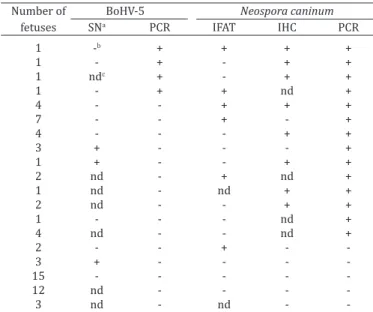
se-Table 1. Summary of the results obtained by serum neutralization test, IFAT, IHC and PCR for BoHV-5 and
Neospolra caninum in 68 spontaneously aborted bovine
fetuses with microscopic lesions in the CNS
Number of BoHV-5 Neospora caninum
fetuses SNa PCR IFAT IHC PCR
1 -b + + + +
1 - + - + +
1 ndc + - + +
1 - + + nd +
4 - - + + +
7 - - + - +
4 - - - + +
3 + - - - +
1 + - - + +
2 nd - + nd +
1 nd - nd + +
2 nd - - + +
1 - - - nd +
4 nd - - nd +
2 - - +
3 + -
15 - -
12 nd -
3 nd - nd
-a Serum neutralizing antibodies to BoHV, b – Negative, + Positive, c nd =
quencers (Institute of Biotechnology, INTA, Castelar, Argentina). Corresponding nucleotide sequences were deposited in GenBank database at the website of the National Institutes of Health (NIH) (NCBI–National Center for Biotechnology Information, NIH, USA). Nucleotide sequences were aligned and analyzed with MEGA 5.1
(Center for Evolutionary Medicine & Informatics Biodesign
Insti-tute and School of Life Sciences Arizona State University, Tempe, USA) (Tamura et al. 2011) and the phylogenetic tree was cons-tructed based on the neighbor-joining clustering method.
RESULTS
Macroscopic evaluation of the fetuses showed that all had a moderate to severe degree of autolysis. Viral isolation and microbiological cultures for the sixty-eight bovine aborted fetuses analyzed in this study were negative. Furthermore, BoHV-1 DNA was not amplified in any case. Thirty-three of 68 fetuses (48.5%) were positive to Neospora caninum by PCR, and 15 were also positive to N. caninum by IHC (Table 1).
BoHV-5 and N. caninum DNA was detected by PCR in the CNS of 4 fetuses (Table 1). BoHV-5 genome was detec-ted in fetal CNS by using two differential PCR methods for BoHV-1 and BoHV-5, according to Claus et al. (2005) and Campos et al. (2009), as described in Materials and Metho-ds. Nucleotide sequences of the amplified BoHV-5 products were deposited in the GenBank database (accession num-bers KC412233 to KC412236). These sequences from the BoHV gC gene were used to study the phylogenetic rela-tionships of the viral strains identified in the fetal CNS with the reference strains of BoHV-5 (A663 [accession number DQ173719.1], N569 [accession number DQ173726.1]) and
BoHV-1 (LA38 [accession number KC412237] and Cooper [accession number KC412238]) and with reference strains of other related viruses (Suid Herpesvirus type 1 [acces-sion number EU719641.1] and Caprine Herpesvirus type 1 [accession number JX993260.1]). This analysis revealed that genomes detected in the four aborted fetuses are clo-sely related to the BoHV-5 Argentinean reference strain (A663) (Fig.1). Three of the fetuses with dual infection be-longed to beef herds; information on the procedence of the remaining fetus is lacking. Gestational ages of the BoHV-5 positive fetuses were 4, 6 and, 7 months (2 cases). Specific antibodies to N. caninum in fetal fluids were detected in 2 out of 4 fetuses with dual infection (Table 1). N. caninum was also identified by IHC in the CNS of 3 out of 4 aborted fetuses (Table 1). Due to the severe degree of autolysis, IHC could not be performed on one fetus. BoHV-5 was not de-tected by IHC in the CNS of dually infected fetuses. However, viral antigens were detected when IHC was performed on BoHV-5-infected MDBK cells (data not shown), demonstra-ting that other causes, likely related to tissue preservation, were involved in the lack of antigen detection.
Comparison of microscopic lesions from fetuses with dual infection and fetuses with solely N. caninum-infection were compared (Table 2). For this comparison, only the 4 fetuses which were consistently positive by IFAT, IHC and PCR were selected. Predominant microscopic lesions in fe-tal CNS are shown in Figure 2, 3 and 4. Focal necrotizing encephalitis was consistently observed in the fetuses in which only N. caninum infection was detected. Perivascular cuffings, gliosis, and multifocal non-suppurative encepha -litis were also observed in the 3 fetuses with concomitant BoHV-5 and N. caninum infection. It was not possible to describe the CNS lesions in one fetus which had a high de-gree of autolysis.
DISCUSSION
Previous studies have demonstrated that, under optimal la-boratory conditions, a final diagnosis of the cause of bovine Table 2. Histophatological lesions in the CNS of aborted fetuses
dually infected with Neospora caninum and BoHV-5 and in aborted fetuses in which only N. caninum was detected
Lesion Fetus
Neospora caninum and N. caninum
BoHV-5 (dual infection) (single infection) No. 1 No. 2 No. 3 No. 4 No. 1 No. 2 No. 3 No. 4
NSM nd 1 nd nd nd 3 1 2
PVC 0 2 0 nd 0 0 1 0
HEM 1 1 0 nd 0 2 0 1
DG 1 0 1 nd 0 0 0 0
FG 2 2 1 nd 0 0 0 1
NSE 3 2 3 nd 1 1 2 3
NE 1 1 2 nd 3 0 2 3
NSM = non-suppurative meningitis, PVC = perivascular cuffing, HEM = hemorrhage, DG = diffuse gliosis, FG = focal gliosis, NSE = multifocal non-suppurative encephalitis, NE = focal necrotizing encephalitis, nd = not determined (absence of meninges or severely autolytic tissue), 0 = absence of lesion, 1 = mild lesion (less than 25% of the tissue affected), 2 = moderate lesion (between 25 and 50% of the tissue affected), 3 = severe lesion (more than 50% of the tissue affected).
Fig.1. Neighbor-joining phylogenetic tree of BoHV isolates ba-sed on the analysis of the gC fragment. Cooper and LA38 are BoHV-1 reference strains and A663 and N569 are BoHV-5 reference strains. SuHV-1 corresponds to a Suid Herpesvirus type 1 reference strain (Bartha) and CapHV-1 corresponds to a Caprine Herpesvirus type 1 reference strain (Biser). 07/415, 07/447, 07/487 and 08/523 are the cases that were PCR po-sitive for BoHV-5 in fetal CNS. The tree is drawn to scale, with branch lengths in the same units as those of the evolutionary distances used to infer the phylogenetic tree. The evolutiona-ry distances were computed using the Maximum Composite Likelihood method and are in the units of the number of base substitutions per site. There were a total of 285 positions in
the final dataset. Overall average evolutionary divergence
abortion is only achieved in 25 to 45% of cases (Campero et al. 2006). In this work, the presence of microscopic lesions in the CNS of fetuses was suggestive of an infectious cause of abortion. Molecular diagnosis techniques, such as PCR, are important ancillary tests because they improve the sen-sitivity of the diagnosis, particularly when fetal autolysis is present.
Viral isolation from samples of abortions (fetuses and placenta) represents the “gold standard” for a conclusive diagnosis of BoHV infection. However, isolation is difficult, time-consuming and requires samples to be in optimal con-ditions to achieve the most favorable result. In fact, conta-mination, inadequate transportation conditions, autolysis, and other factors may all adversely affect viral isolation (Takiuchi et al. 2005). Routine diagnosis of Neospora cani-num is based on histopathological changes in fetal tissues and identification of parasites by IHC. However, confirma -tion of N. caninum infection using IHC has low sensitivity (Baszler et al. 1999). In the present work, BoHV was not isolated, and N. caninum was identified by IHC only in 3 out of 4 aborted fetuses that presented a concomitant infec-tion with BoHV-5, as detected by PCR. Moreover, in order to prove that BoHV-5 infection was the cause of abortion, viral antigen detection by IHC was also attempted. Howe-ver, as expected, BoHV-5 was not detected in CNS sections. Tissue conditions were not optimal since fetal specimens had been long-term stored in formaldehyde, which is de-trimental for antigen detection by IHC (Beckstead 1994). It is also likely that the use of a monoclonal antibody to detect BoHV-5, which is highly specific but not sensitive enough, had also influenced the final outcome (Haines & Chelack 1991). Furthermore, fetal autolysis might also be responsible for the lack of antigen detection by this techni-que (Kirkbride 1986). Therefore, in these cases, the use of
PCR is a more rapid and sensitive approach for diagnosis than virus isolation or IHC. Indeed, PCR should be conside-red a useful complementary tool for the diagnosis of both pathogens in fetuses with histopathological lesions in the CNS.
The prevalence of N. caninum found in this study (48.5%) is in agreement to that previously reported by Moore et al. (2008) by using PCR. Even though these data indicate the relevance of this parasite as a cause of bovine abortion, the presence of other pathogens cannot be excluded (Campe-ro et al. 1998). Moreover, the presence of N. caninum DNA does not imply the parasite as the primary cause of abor-tion and microscopic observaabor-tion of fetal tissues should be performed for the final diagnosis of Neospora-related abor-tion (Moore et al. 2008). On the other hand, pathological synergism by dual infections, like in these cases, has been previously hypothesized (Campero et al. 2003).
In the present work, BoHV-5 DNA was detected in fetu-ses with microscopic lesions in the CNS which were indi-cative of necrotizing meningoencephalitis and which were also in agreement with histopathological lesions described for fetal N. caninum-infections. The presence of BoHV-5 was confirmed by sequencing of PCR products and the cor -responding phylogenetic analysis. The nucleotide sequence of the PCR-amplified gC gene fragment of 4 BoHV-5 cases was compared with the sequences of BoHV-1 and BoHV-5 reference strains. This analysis showed that viral genomes identified in the four aborted fetuses are closely related to Fig.2. Focal non-suppurative encephalitis from a bovine aborted
fetus. This type of lesion was a common finding in fetuses in
which BoHV-5 was detected. HE, Bar = 100µm.
Fig.3. Vasculitis and non-suppurative encephalitis from a bovine aborted fetus. Lesions commonly observed in fetuses where BoHV-5 DNA was detected. HE, Bar = 100µm.
Fig. 4. Focal necrotizing encephalitis from a bovine aborted fetus. This type of lesion is frequently observed in cases in which only N. caninum was detected. HE, Bar = 100µm.
2 3
the BoHV-5b Argentinean reference strain (A663). Interes-tingly, Maidana et al. (2011) demonstrated that circulation of this subtype “b” prototype strain in Argentina was tran-sient, when compared to the commonly isolated subtype “a” strains. In a previous report by Schudel et al. (1986), BoHV-5 was isolated from an aborted fetus. However, no reference was made to fetal CNS lesions. No other cases of BoHV-5 have been associated with bovine abortion in Ar-gentina prior to this study. As it is described in cases of bo-vine abortion in which BoHV-1 infection is involved, small amounts of viral antigens may be commonly found in the CNS. However, microscopic lesions due to herpetic infec-tion may be rare or difficult to recognize due to autolysis of the aborted fetuses. Nevertheless, a recent study detected the presence of encephalitis in aborted fetuses with diag-nosis of BoHV-1 infection (Brower et al. 2008).
The results of this retrospective study are relevant since this is the first identification of BoHV-5 genome in the CNS of aborted fetuses which also showed neurological lesions and concomitant infection with N. caninum. These results suggest that infection by alpha-herpesviruses should be considered in the diagnosis of cases in which fetal ence-phalitis is present. Despite the findings of the present stu -dy, it cannot be ruled out the possibility that the presence of these pathogens and the lesions observed in fetal CNS are simply due to an incidental infection not related to the cause of abortion, particularly when virus isolation failed to demonstrate the presence of BoHV-5.
Because the CNS has a limited range of morphologic responses to injury, causes of abortion related to different infections can result in almost similar lesions. The results of this study demonstrate that, in some cases, brain micros-copic lesions may be indistinguishable between BoHV and N. caninum, mainly when typical parasite cysts are not pre-sent in the CNS of aborted bovine fetuses. The possibility of identifying BoHV-5 using PCR, as described here, is impor-tant because it can improve the sensitivity of the diagno-sis of bovine abortion. Furthermore, it also contributes to the differentiation of the type of BoHV involved. Molecular diagnosis of BoHV-5 and N. caninum confirmed the useful -ness of PCR in the identification of causative agents of bovi -ne abortions, especially when autolysis is present.
Acknowledgements.- We would like to thank to Dr Andrea Verna for her
contribution and support to the work. Continuous support from INTA is also highly acknowledged. We thank the members of the Specialized Vete-rinary Diagnostic Service of INTA Balcarce for attending the abortion ca-ses and collecting the samples used in this study. This work was financially supported by INTA, Specific Projects AESA-203981 and AESA-201711.
REFERENCES
Baszler T.V., Gay L.J., Long M.T. & Mathison B.A. 1999. Detection by PCR of
Neospora caninum in fetal tissues from spontaneous bovine abortions. J. Clin. Microbiol. 37(12):4059-4064.
Beckstead J.H. 1994. A simple technique for preservation of fixation-sen -sitive antigens in paraffin-embedded tissues. J. Histochem. Cytochem. 42(8):1127-1134.
Brower A., Homb K.M., Bochsler P., Porter R., Woods K., Ubl S., Krueger D., Cigel F. & Toohey-Kurth K. 2008. Encephalitis in aborted fetuses associated with Bovine Herpesvirus 1 infection. J. Vet. Diagn. Invest. 20(3):297-303.
Buxton D., Maley S., Wright S., Thomson K.M., Rae A.G. & Innes E.A. 1998. The pathogenesis of experimental neosporosis in pregnant sheep. J. Comp. Pathol. 118(4):267-279.
Campero C.M., Anderson M.L., Conosciuto G., Odriozola E., Bretschneider G. & Poso M.A. 1998. Neospora caninum-associated abortion in a dairy herd in Argentina. Vet. Rec. 143(8):228-229.
Campero C.M., Moore D.P., Odeón A.C., Cipolla A.L. & Odriozola E. 2003. Aeti-ology of bovine abortion in Argentina. Vet. Res. Commun. 27(5):359-369. Campos F.S., Franco A.C., Hübner S.O., Oliveira M.T., Silva A.D., Esteves P.A., Roehe P.M. & Rijsewijk F.A. 2009. High prevalence of co-infections with bovine herpesvirus 1 and 5 found in cattle in southern Brazil. Vet. Mi-crobiol. 139(1/2):67-73.
Chung C.S., Pearson L.D., Ayers V.K. & Collins J.K. 1994. Monoclonal anti-bodies that distinguish between encephalitogenic bovine herpesvirus type 1.3 and respiratory bovine herpesvirus type 1.1. Clin. Diagn. Lab. Immunol. 1(1):83-88.
Claus M., Alfieri A., Folgueras-Flatschart A., Wosiacki S., Médici K. & Alfieri A. 2005. Rapid detection and differentiation of bovine herpesvirus 1 and 5 glycoprotein C gene in clinical specimens by multiplex-PCR. J. Virol. Methods. 128(1/2): 183-188.
Collantes-Fernández E., Rodríguez-Bertos A., Arnáiz-Seco I., Moreno B., Aduriz G. & Ortega-Mora L.M. 2006. Influence of the stage of pregnancy on Neospora caninum distribution, parasite loads and lesions in aborted bovine foetuses. Theriogenology 65(3):629-641.
Haines D.M. & Chelack B.J. 1991. Technical considerations for developing enzyme immunohistochemical staining procedures on formalin-fixed, paraffin-embedded tisues for diagnostic pathology. J. Vet. Diagn. Invest. 3(1):101-112.
Kirkbride C.A. 1986. Examination of bovine and ovine fetuses. Vet. Clin. North Am. Food Anim. Pract. 2(1):61-83.
Maidana S.S., Ladelfa M.F., Pérez S.E., Lomónaco P.M., Del Médico Zajac M.P., Odeón A.C., Blanco Viera J., Combessies G., Fondevila N., Palacios M., Thiry J., Muylkens B., Thiry E. & Romera S.A. 2011. Characterization of BoHV-5 field strains circulation and report of transient specific subtype of bovine herpesvirus 5 in Argentina. BMC Vet. Res. 7:8.
Moore D.P., Campero C.M., Odeón A.C., Posso M.A., Cano D., Leunda M.R., Basso W., Venturini M.C. & Späth E. 2002. Seroepidemiology of beef and dairy herds and fetal study of Neospora caninum in Argentina. Vet. Para-sitol. 107(4):303-316.
Moore D.P., Regidor-Cerrillo J., Morrell E., Poso M.A., Cano D.B., Leunda M.R., Linschinky L., Odeón A.C., Odriozola E., Ortega-Mora L.M. & Campero C.M. 2008. The role of Neospora caninum and Toxoplasma gondii in spontane-ous bovine abortion in Argentina. Vet. Parasitol. 156(3/4):163-167. Odeón A.C., Späth E.J.A., Paloma E.J., Leunda M.R., Fernández Sainz I.J.,
Pérez S.E., Kaiser G.C., Draghi M.G., Cetra B.M. & Cano A. 2001. Prevalen-cia de anticuerpos al virus de diarrea viral bovina, herpesvirus bovino y virus sincicial respiratorio bovino en Argentina. Revista de Medicina Veterinaria. 82:216-220.
Pérez S.E., Bretschneider G., Leunda M.R., Osorio E.A., Flores E.F. & Odeón A.C. 2002. Primary infection, latency and reactivation of Bovine Herpes-virus Type 5 in the bovine nervous system. Vet. Pathol. 39(4):437-444. Roizman B. & Pellett P. 2001. The Family Herpesviridae: a brief introduc-tion, p.1929-1939. In: Knipe D. & Howley P. (Eds), Fields Virology. 4th ed.
Lippincott Williams and Wilkins, Philadelphia.
Schudel A.A., Carrillo B.J., Wyler R. & Metzler A.E. 1986. Infections of calves with antigenic variants of bovine herpesvirus 1 (BHV-1) and neurologi-cal disease. J. Vet. Med. B 33(4):303-310.
Smith K.C. 1997. Herpesviral abortion in domestic animals. Vet. J. 153(3):253-268.
Takiuchi E., Médici K.C., Alfieri A.F. & Alfieri A.A. 2005. Bovine herpesvi -rus type 1 abortions detected by a semi-nested PCR in Brazilian cattle herds. Res. Vet. Sci. 79(1):85-88.