C A S O C L Í N I C O
A N K Y L O S I N G S P O N D Y L I T I S
A N D S P I N A L C O R D I N J U R Y
Pedro Machado,
*Jan Gawronski,
*Angela Gall
*Palavras-chave: Espondilite Anquilosante;
Fractu-ra VertebFractu-ral; Lesão Medular; Osteoporose.
Introduction
Ankylosing spondylitis (AS) is a chronic inflamma-tory disease affecting mainly the axial skeleton and characterized by ossification of the spinal discs, jo-ints, and ligaments, leading to progressive rigidity and altered biomechanical properties of the spi-ne.1It can be associated with impaired balance, os-teoporosis, and a reduced ability to take protective measures during a fall.2, 3As a consequence of the-se changes this population is at an increathe-sed risk of vertebral fractures and spinal cord injury (SCI). The initial skeletal vertebral trauma can often be mis-sed predisposing the patient to a secondary neuro-logical injury. Inadequate awareness of these inju-ries and inappropriate management, both in the pre-hospital and in the hospital setting can have devastating consequences. Given the higher inci-dence of SCI and the increased morbidity and mor-tality rates in patients with AS who suffer a SCI (fre-quently due to respiratory impairment), it is criti-cal that prevention strategies be developed to avoid this devastating complication of the disease.4, 5
Case report
A 72-year-old white male with a long history of AS accidentally fell at home from an upright standing position, landing on his back. On admission to the local hospital, he complained of back pain only. There was no head injury. Spinal and pelvic radio-graphs revealed typical features of advanced AS with bilateral total hip replacement (Figure 1) wi-thout features of an injury or fracture to the verte-bral column. Recent right sided rib fracture was found on chest radiograph.
The patient was admitted to hospital for analge-sic control of severe spinal pain and mobilisation. *Spinal Cord Injury Centre, Royal National Orthopaedic Hospital,
Stanmore, London, United Kingdom
Abstract
Ankylosing spondylitis patients are reported to be at greater risk for vertebral fractures and spinal cord injury. We describe the case of a 72-year-old male with a long history of ankylosing spondylitis who sustained a vertebral fracture after minor trauma. The fracture was initially missed on conventional radiographs but was later diagnosed by magnetic resonance imaging, after the development of new neurological symptoms. With this case report the authors outline the factors that increase the inci-dence of vertebral fractures and spinal cord injury in ankylosing spondylitis patients and discuss pre-vention strategies to avoid this devastating compli-cation of the disease.
Keywords: Ankylosing Spondylitis; Vertebral
Frac-ture; Spinal Cord Injury; Osteoporosis.
Resumo
Os doentes com espondilite anquilosante apresen-tam um risco aumentado de fracturas vertebrais e de lesão medular. Os autores descrevem o caso de um doente do sexo masculino, com 72 anos de ida-de, com espondilite anquilosante avançada, que sofreu uma fractura vertebral após um pequeno traumatismo. A fractura não foi inicialmente diag-nosticada nas radiografias convencionais, mas foi mais tarde identificada por ressonância magnéti-ca, após o aparecimento de novos sinais neuroló-gicos. Com este caso clínico, os autores salientam os factores que aumentam o risco de fracturas ver-tebrais e de lesão medular nos doentes com espon-dilite anquilosante e discutem estratégias de pre-venção que permitam evitar esta dramática com-plicação da doença.
A N K Y L O S I N G S P O N D Y L I T I S A N D S P I N A L C O R D I N J U R Y
Five days after the initial insult, the patient deve-loped loss of sensation from the abdominal wall below, with loss of power, reflexes and tone in both lower limbs and loss of anal sphincter (no volun-tary anal contraction and no anal sensation) and bladder function.
Urgent magnetic resonance imaging (MRI) re-vealed a T10-T11 fracture dislocation with sugges-tion of cord transacsugges-tion at that level (Figure 2). Whilst an inpatient at the local hospital, he was treated for right sided haemothorax.
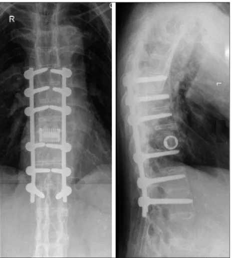
The patient was then transferred to our hospi-tal, a regional centre for traumatic spinal cord in-jury surgery and rehabilitation. Spinal cord inin-jury ASIA classification6on admission was T6 paraple-gia ASIA A. He underwent T7 to L1 posterior fusi-on for T10-T11 fracture dislocatifusi-on (Figure 3) and had a tracheostomy tube inserted as for manage-ment of respiratory impairmanage-ment and an anticipa-ted period of ventilatory care. The tracheostomy tube was later removed. He was subsequently transferred from intensive care to the Spinal Cord Injury Centre for a period of intensive interdisci-plinary rehabilitation to optimize function and re-integration and facilitate a safe discharge home. Due to suboptimal spinal stability at the T10-T11 level, 20 weeks after the first surgery the patient
un-derwent a T10-T11 cage insertion (Figure 4 and 5). Treatment included analgesics, low-molecular--weight heparin & compression stockings as pro-phylactic treatment for venous thrombo-embo-lism, proton-pump-inhibitor, laxatives and ade-quate nutrition (in the early stages of admission re-quired gastric feeding) and skin care. Dual-energy x-ray absorptiometry (DEXA) testing preformed at the lumbar spine and forearm revealed osteoporo-tic values according to the World Health Organiza-tion thresholds, and calcium, cholecalciferol and bisphosphonates were also prescribed.
Physiotherapy in the acute stages focussed on maintenance of joint range of movement and tis-sue extensibility whilst in the intensive care unit and maintenance of patent airways whilst intuba-ted (e.g. positioning, suction and shaking). He also required bilateral chest drains for post-operative recurrence of bilateral pleural effusions.
Three weeks after the first surgery the patient was cleared for mobilisation in a thoracic-lumbar--sacral orthosis (TLSO) and hoisted into a wheel-chair. On assessment of his musculoskeletal sys-tem he had features consistent with chronic AS e.g limited range of movement of his cervical spine, thoracic hyperkyphosis and limited shoulder and hip range of movement bilaterally. Limited range of Figure 1. Radiographs of the pelvis and thoracic and lumbar spine showing advanced features of ankylosing spondylitis (li-gamentous ossification, squaring of the vertebral bodies, syndesmophytosis and grade IV bilateral sacroiliitis) and bilateral total hip replacement.
P E D R O M A C H A D O E C O L.
hip flexion caused restriction of sitting position and required the use of a seating system fixed in ex-tension. Sitting tolerance was gradually increased and focus was made on an upper limb exercise programme, function improvement, standing pro-gramme, and identification of a suitable wheel-chair, cushion, backrest and home equipment to increase functional independence.
ASIA classification6on discharge was T6 para-plegia ASIA A. Urodynamics revealed a well sup-pressed bladder and long term oxybutynin was re-commended. He was able to undertake self-inter-mittent clean catheterisation. Due to physical res-triction imposed by fracture, AS and TLSO, the patient was not able to reliably perform his own bowel care. Skin was intact. There were no docu-mented episodes of autonomic dysreflexia, al-though given his level of injury he is at risk and therefore has received full education and was ad-vised to be supplied at all times with nifedipine capsules which can be pierced and expressed un-der the tongue in an emergency. He was taking nu-trition orally and there were no swallowing or com-munication issues. Minimal spasticity was control-led with baclofen. Lung function tests reveacontrol-led a mild restrictive ventilatory dysfunction not
com-promising his respiratory status. A six week surgi-cal and a three month medisurgi-cal and urology follow-up were scheduled.
Discussion
We describe the case of a 72-year-old man with a long history of AS who developed a complete SCI after a fall. There is data suggesting that patients with AS have an increased rate of SCI.4,7,8Alaranta
et al4recently determined that the incidence of SCI
in patients with AS was 11.4 times higher than in the population at large. The proportion of complete SCI also appears to be higher.4,5,8,9Although our pa-tient had a thoracic lesion, a higher incidence of cervical SCI has been noted in the population with AS.4Patients with AS who sustain a SCI are also ol-der than the general spinal cord-injured popula-tion.4,7,8,10,11
The higher incidence of SCI in AS is directly cor-related with the increased incidence of vertebral fractures in patients with AS. Cooper et al2found an odds ratio of 7.7 for clinically significant vertebral fractures, as compared with the general popula-tion, and noted that the cumulative incidence of Figure 2. T2 (left) and T1-weighted (right) sagittal MR images of thoracolumbar spine revealing a T10-T11 fracture dislocation with suggestion of cord transaction at that level.
A N K Y L O S I N G S P O N D Y L I T I S A N D S P I N A L C O R D I N J U R Y
coupling of the bone formation and bone resorpti-on processes. Therefore, although ectopic bresorpti-one formation occurs within the inflamed vertebral en-thesis, bone resorption, through increased osteo-clast activity, also occurs at an unregulated rate wi-thin the vertebra and promotes weakening of the spinal column.12The altered spinal biomechanics combined with the brittle quality of the osteopo-rotic bone in patients with AS greatly increase sus-ceptibility to vertebral column factures, even after minor, often trivial, trauma.2,4,7,17The case we pre-sent clearly illustrates the consequences of these mechanisms.
Regarding falls, they are the most frequent cau-se of SCI in patients with AS.1, 3The aetiology of the injury in our patient was also a fall. The propensity for fall is increased by poor posture, impaired ba-lance and impaired mobility. The poor posture is a result of changed biomechanics in the enclosed spine that leads to loss of normal curvatures and a shifting of the centre of gravity. The rigid, kyphotic spinal deformities as well as a variable degree of pe-ripheral joint arthritis are factors that exacerbate gait unsteadiness and thus increase susceptibility to falls and reduces the ability to take protective measures during a fall.1The spinal deformity can vertebral fractures appears to peak at 17% in the
third decade after diagnosis. Geusens et al12 re-cently reviewed the risk factors associated with ver-tebral fractures in the population with AS: sex (men more than women), age, low body mass index, os-teoporosis, disease duration, degree of syndesmo-phytosis, peripheral joint involvement, increased modified Stoke AS Spine Score, increased restric-tion of spinal movement and increased occiput-to-wall distance. These data fit well in the case we des-cribe.
Fracture risk is associated with bone properties and fall-related risks. Regarding bone properties, the two central features of AS that promote the pa-thological remodelling of the vertebral column are inflammation and new bone formation. As the spi-ne in the patient with AS fuses through ligamen-tous ossification and syndesmophytosis, a rigid hyperkyphotic deformity develops. Biomechani-cally, the fused spine acts as a rigid lever that is in-capable of appropriately dissipating the energy of a traumatic event.13-15Although new bone forma-tion is central to the pathogenesis of AS, this pa-thological entity is also associated with osteoporo-sis and low bone mineral density.12,16This see-mingly paradoxical finding is attributed to an
un-Figure 3. Thoracolumbar spine radiograph after T7 to L1 posterior instrumented fusion for T10-11 fracture. Screws were placed into T7 to T9 bilaterally and then into T11 to L1.Two rods were used.
tability is directly related to the AS disease process. AS promotes the ossification of spinal ligaments, which then also fracture as part of the injury pat-tern, further decreasing the structural support available to the spinal column. Any fracture that re-sults usually involves all 3 columns resulting in ins-tability, as in the case we report (Figure 2). Moreo-ver, the greater instability significantly increases the risk of iatrogenic SCI during patient transpor-tation and manoeuvres aimed at reducing fractu-re dislocation. Accordingly, gfractu-reat cafractu-re should be used whenever patients with AS and spinal fractu-res are moved, and reduction manoeuvfractu-res, if ne-cessary, should be performed only by experienced individuals.
The incidence of SCI is also increased in the set-ting of AS because of the greater incidence of spi-nal epidural hematoma.7, 19, 20Where an interval oc-curs between trauma and the onset of neurologi-cal signs or worsening of the neurologineurologi-cal picture the formation of an epidural haematoma should be suspected and excluded by means of an MRI scan. The management of SCI in the population with AS is further complicated by an advanced patient age, decreased chest wall expansion and the pre-sence of multiple possible medical comorbidities, namely the extra-articular manifestations of the disease. Physiotherapy can be particularly challen-ging given the special musculoskeletal features of P E D R O M A C H A D O E C O L.
also compromise the vertical field of vision mak-ing it difficult for the individual to see what is in front of him/her.3
Other factors contribute to the increased inci-dence of SCI in patients with AS. As it happened in the case we report, vertebral fractures are often ini-tially missed in patients with AS. This is likely to be related to the usual background of trivial trauma (yielding a low index of suspicion), overlooked pain complaints by attributing axial pain to normal di-sease activity and also because fractures can be difficult to identify on conventional radiographs due to the distortion of normal anatomy by the os-sified spinous ligaments. Moreover, as it also hap-pened in our patient, the neurological findings can be subtle or non-existent on initial presentation with neurological deterioration occurring later. This has been identified by several authors.3, 18 The-refore, a thorough history and careful physical exa-mination are required to avoid underestimating the severity of the injury and more refined ima-ging (e.g CT, MRI) should be considered whenever a patient with AS presents with symptoms of new neck or back pain, no matter how minor or trivial the reported mechanism of injury.
Even when fractures are appropriately diagno-sed, spinal column fractures in patients with AS are notoriously unstable and this correlates with an increased incidence of SCI. This high level of
the AS patient.
The patient that we describe demonstrates many of the pitfalls that are present all along the treatment pathway. The unique nature of spinal column injuries in AS makes it imperative that it is treated with required caution from the start by the initial response team, by the accident and emer-gency staff, and by the treating orthopaedic or spi-nal injury team until definitive management is completed. This would be facilitated if the parame-dical personnel and meparame-dical staff are aware of the diagnosis. Medical alert bracelets or cards have been proposed as a mean to identifying patients with AS so that appropriate precautions can be ins-tituted by emergency services.3Other primary pre-vention strategies have also been proposed as me-ans of avoiding SCI in this susceptible popula-tion4,5: encouragement to install activity aids such as handrails beside all staircases and within
bathrooms, use of night lights in bedrooms and bathrooms, avoidance of loose area rugs that pre-sent a tripping risk, avoidance of excessive use of alcohol, avoidance of all contact sports or other high-impact physical activities, wearing seat belts at all times while driving and liberal use of car seat headrests. Moreover, DEXA scanning should be considered early in the disease stage, ideally befo-re fractubefo-res, and drug tbefo-reatment for osteoporosis should be considered in any patient with a T-sco-re lower than -2.5.12 Finally, an early and reliable diagnosis and treatment of AS is another very im-portant and relevant issue. There is a need for an early diagnosis in all patients with AS or undiffe-rentiated spondylarthropathies with axial involve-ment in order to prevent the pathological changes that increase the susceptibility to fractures and SCI. This is especially relevant given that more effecti-ve treatment options are now available.
A N K Y L O S I N G S P O N D Y L I T I S A N D S P I N A L C O R D I N J U R Y
Figure 5. Thoracolumbar spine radiograph after osteotomy and re-alignment of the spine with fixation rods from T7-L1 and cage insertion at the level of T10-T11.
Correspondence to:
Pedro Machado
Serviço de Reumatologia dos Hospitais da Universidade de Coimbra
Praceta Mota Pinto 3000-075 Coimbra, Portugal
E-mail: [email protected]
References:
1. Bot SD, Caspers M, Van Royen BJ, Toussaint HM, Kingma I. Biomechanical analysis of posture in pati-ents with spinal kyphosis due to ankylosing spondylitis: a pilot study. Rheumatology (Oxford) 1999;38:441-443.
2. Cooper C, Carbone L, Michet CJ, Atkinson EJ, O’Fal-lon WM, Melton LJ, 3rd. Fracture risk in patients with ankylosing spondylitis: a population based study. J Rheumatol 1994;21:1877-1882.
3. Thumbikat P, Hariharan RP, Ravichandran G, McClel-land MR, Mathew KM. Spinal cord injury in patients with ankylosing spondylitis: a 10-year review. Spine 2007;32:2989-2995.
4. Alaranta H, Luoto S, Konttinen YT. Traumatic spinal cord injury as a complication to ankylosing spondyli-tis. An extended report. Clin Exp Rheumatol 2002;20:66-68.
5. Jacobs WB, Fehlings MG. Ankylosing spondylitis and spinal cord injury: origin, incidence, management, and avoidance. Neurosurg Focus 2008;24:12. 6. Maynard FM, Jr, Bracken MB, Creasey G et al.
Inter-national Standards for Neurological and Functional Classification of Spinal Cord Injury. American Spinal Injury Association. Spinal Cord 1997;35:266-274. 7. Rowed DW. Management of cervical spinal cord
in-jury in ankylosing spondylitis: the intervertebral disc as a cause of cord compression. J Neurosurg 1992;77:241-246.
8. Tico N, Ramon S, Garcia-Ortun F et al. Traumatic spi-nal cord injury complicating ankylosing spondylitis. Spinal Cord 1998;36:349-352.
9. Murray GC, Persellin RH. Cervical fracture complica-ting ankylosing spondylitis: a report of eight cases and review of the literature. Am J Med 1981;70:1033-1041.
10. Apple DF, Jr., Anson C. Spinal cord injury occurring in patients with ankylosing spondylitis: a multicenter study. Orthopedics 1995;18:1005-1011.
11. Jackson AB, Dijkers M, Devivo MJ, Poczatek RB. A de-mographic profile of new traumatic spinal cord inju-ries: change and stability over 30 years. Arch Phys Med Rehabil 2004;85:1740-1748.
12. Geusens P, Vosse D, van der Linden S. Osteoporosis and vertebral fractures in ankylosing spondylitis. Curr Opin Rheumatol 2007;19:335-339.
13. Aufdermaur M. Pathogenesis of square bodies in ankylosing spondylitis. Ann Rheum Dis 1989;48:628--631.
14. de Vlam K, Lories RJ, Luyten FP. Mechanisms of pa-thologic new bone formation. Curr Rheumatol Rep 2006;8:332-337.
15. Chavassieux P, Seeman E, Delmas PD. Insights into material and structural basis of bone fragility from diseases associated with fractures: how determinants of the biomechanical properties of bone are compro-mised by disease. Endocr Rev 2007;28:151-164. 16. Karberg K, Zochling J, Sieper J, Felsenberg D, Braun J.
Bone loss is detected more frequently in patients with ankylosing spondylitis with syndesmophytes. J Rheumatol 2005;32:1290-1298.
17. Olerud C, Frost A, Bring J. Spinal fractures in patients with ankylosing spondylitis. Eur Spine J 1996;5:51-55. 18. Finkelstein JA, Chapman JR, Mirza S. Occult vertebral fractures in ankylosing spondylitis. Spinal Cord 1999;37:444-447.
19. Foo D, Sarkarati M, Marcelino V. Cervical spinal cord injury complicating ankylosing spondylitis. Paraple-gia 1985;23:358-363.
20. Garza-Mercado R. Traumatic extradural hematoma of the cervical spine. Neurosurgery 1989;24:410-414. P E D R O M A C H A D O E C O L.