UNIV ERSID ADE DE
LISBO A
F
ACULD ADE DEC
I ÊNCI ASDE P ART AM E NTO DE
BIOLOGI A V
EGE T ALDissertação
Characterization of peripheral blood B cell subpopulations in
Rheumatoid Arthritis
Cláudia Cristina Valente Quaresma
Mestrado em Biologia Molecular e Genética
U
NIV ERSID ADE DE LISBO AF
ACULD ADE DEC
I ÊNCI ASDE P ART AM E NTO DE
BIOLOGI A V
EGE T ALCharacterization of peripheral blood B cell subpopulations in
Rheumatoid Arthritis
Cláudia Cristina Valente Quaresma
Mestrado em Biologia Molecular e Genética
Dissertação orientada por:
Doutora Rita Alexandra Pedra Aguiar de Moura
Unidade de Investigação em Reumatologia – Instituto de Medicina Molecular (IMM)
Professora Doutora Maria Margarida BlasquesTelhada
Faculdade de Ciências da Universidade de Lisboa
Todas as afirmações efetuadas no presente documento são de exclusiva responsabilidade do seu autor, não cabendo qualquer responsabilidade à Faculdade de Ciências da Universidade de Lisboa pelos conteúdos nele apresentados.
i
R
ESUMOA artrite reumatoide (AR) é uma doença autoimune crónica, sistémica e de etiologia desconhecida, que afeta cerca de 0,5-1% da população mundial. A AR é caracterizada por uma poliartrite simétrica associada a dor e tumefação de múltiplas articulações, em particular das pequenas articulações das mãos. Embora não exista ainda cura para a AR, sabe-se que um tratamento adequado, assim como um diagnóstico precoce são de extrema importância para o doente e para a sociedade em geral. Se não for corretamente tratada, a AR conduz a destruição óssea e articular. Deste modo, é crucial que a terapêutica seja a mais adequada possível ao estado clínico do doente e implementada na fase inicial do curso da doença.
O tratamento da AR tem como principais objetivos a preservação da função e qualidade de vida do doente, minimizando a dor e sinais inflamatórios, a remissão e controlo das manifestações sistémicas. Os primeiros tratamentos administrados aos doentes com AR são drogas anti-inflamatórias não esteróides e corticóides, úteis no alívio dos sintomas clínicos e na diminuição da inflamação. Contudo, torna-se primordial o uso de drogas antirreumáticas modificadoras da doença como, por exemplo, o metotrexato (MTX), eficaz no controlo da atividade e progressão da doença. Todavia, a introdução das terapias biológicas como as drogas anti-fator de necrose tumoral (anti-TNF) ou o anticorpo monoclonal bloqueador do recetor da interleucina-6 (IL-6R) tocilizumab (TCZ), possibilitou atingir uma melhoria considerável na inibição da progressão da doença.
Várias doenças autoimunes são dependentes de células B, principalmente através da produção de autoanticorpos, tal como a AR. Estudos anteriores revelaram a importância das células B na patogénese da AR através de diversos mecanismos. As células B produzem autoanticorpos, como o fator reumatoide (FR) e anticorpos anti-proteínas citrulinadas (ACPA), que formam complexos imunes que se depositam nas articulações, causando inflamação. Além disso, as células B podem funcionar como células apresentadoras de antigénio e ativar células T, sendo também capazes de produzir citocinas uma vez ativadas e participar na organização de estruturas linfóides secundárias. A descoberta da eficácia da terapêutica de depleção de células B com rituximab (RTX) trouxe não só melhorias na qualidade de vida dos doentes, como também veio reforçar a posição-chave destas células nesta doença autoimune.
ii
O principal objetivo deste trabalho foi a caracterização das subpopulações de células B no sangue periférico de doentes com AR em fase inicial, sem terapêutica e em doentes com AR estabelecida. Adicionalmente, de forma a analisar as possíveis influências do tratamento no desenvolvimento dos processos imunológicos mediados pelas células B na evolução da AR, foi também comparado o efeito dos diversos tratamentos (MTX, anti-TNF e TCZ) no fenótipo das células B e na produção de citocinas e quimiciocinas diretamente relacionadas com a ativação de células B na AR. Para tal, foram incluídos neste estudo, um grupo de doentes com poliartrite não tratada (n=13) com menos de 1 ano de duração da doença, os quais foram classificados como AR iniciais (ERA). Foram também analisados doentes com AR estabelecida sob terapêutica com MTX (MTX, n=17) e MTX pré-biológico (MTX pre-bio, n=29). Dentro do grupo MTX pre-bio, foi realizada uma segunda colheita de sangue em doentes com AR oito meses após terem iniciado tratamento com anti-TNF (n=10) ou TCZ (n=7).
A análise da frequência das subpopulações das células B no sangue periférico foi efetuada usando o sistema de classificação IgD/ CD27 que permite a identificação das principais quatro subpopulações de células B (CD19+): naïve (IgD+CD27-), pre-switch-memory (IgD+CD27+), post-switch memory (IgD-CD27+) e double-negative (DN, IgD-CD27-). De forma a poder identificar e caracterizar a subpopulação de plasmócitos no sangue periférico, foi usado o sistema de classificação IgD/ CD38, que identifica os plasmócitos como IgD-CD38++, dentro da região das células B CD19+.
Neste estudo não foram observadas diferenças nas células B totais (CD19+) nos doentes com AR em comparação com os controlos saudáveis. Contudo, quando analisadas as subpopulações de células B, foram observadas alterações nas células B de memória em circulação, nomeadamente um aumento da subpopulação de células B de memória DN (IgD-CD27-) nos doentes com AR estabelecida tratados com MTX e MTX pré-biológico, em relação aos controlos saudáveis.
De modo a caracterizar o fenótipo das células B na fase inicial e estabelecida da AR, foram estudados vários marcadores celulares diretamente relacionados com a ativação e sobrevivência de células B (BAFF-R, TACI e BCMA), ativação celular (HLA-DR, CD69 e CD86), ativação mediada pelo recetor de células B (IgM), diferenciação (CD5), quimiotaxia das células B (CXCR5), apoptose mediada pelo complexo Fas (CD95) e ativação mediada pelo Toll-like receptor 9 (TLR9), através da análise da intensidade média da fluorescência (MFI). Adicionalmente, foram quantificados no soro de todos os grupos analisados os níveis
iii
de CXCL13, uma quimiocina importante para as células B, e o CD23 solúvel (sCD23), envolvido na maturação das células B.
O BAFF, uma citocina fundamental na sobrevivência das células B, tem sido sugerida como potencial alvo terapêutico para doenças autoimunes dependentes de células B, como a AR. Esta citocina apresenta 3 recetores: BAFF-R, TACI e BCMA. Neste estudo observou-se um aumento da MFI do TACI em doentes com AR estabelecida sob terapêutica com anti-TNF, em particular na subpopulação de células B post-switch memory. Nos restantes recetores do BAFF não foram encontradas diferenças significativas em comparação com os controlos saudáveis. O recetor TACI tem uma função ambígua, pois pode funcionar como recetor de ativação e/ ou de inibição das células B. É possível que o tratamento com anti-TNF induza um aumento da expressão de TACI nas células B de modo a inibir a sua ativação e o desenvolvimento da autoimunidade.
Estudos anteriores têm evidenciado que as células B, uma vez ativadas, aumentam a expressão de marcadores de ativação como o HLA-DR, CD69 e CD86. Neste trabalho, embora não tenham sido observadas diferenças na MFI do CD69 e CD86 nas subpopulações de células B analisadas nos grupos de doentes em comparação com os controlos saudáveis, foi encontrado um aumento significativo na MFI do HLA-DR em doentes com AR estabelecida sob terapêutica com anti-TNF e TCZ. Estudos anteriores demonstraram que as terapêuticas com anti-TNF e TCZ diminuem a infiltração celular observada na membrana sinovial dos doentes com AR. É possível que, devido ao efeito da terapêutica, as células B ativadas que infiltram as articulações regressem ao sangue periférico, justificando deste modo o aumento da MFI do HLA-DR. Foi também observada uma diminuição da MFI do CD86 nos doentes com AR estabelecida tratados com anti-TNF e TCZ em comparação com as colheitas baseline, o que sugere uma inibição da ativação das células B induzida pelo tratamento com imunosupressores.
A apoptose por meio do recetor de morte celular Fas (CD95) desempenha um papel central na manutenção da auto-tolerância imunológica periférica. De facto, alterações nesta via de apoptose foram demonstradas na patogénese de doenças autoimunes como a AR. Neste estudo, valores aumentados de MFI do CD95 foram observados nas células B de memória
post-switch em doentes com AR estabelecida após o tratamento com anti-TNF e TCZ
quando comparados com controlos saudáveis, o que poderá ser uma consequência direta do tratamento como forma de neutralizar a ativação de células B autoreativas.
iv
Estudos efetuados na AR e noutras doenças autoimunes têm revelado alterações nas células B CD5+. Estas células estão associadas à reabsorção óssea através da produção de IL-6, uma citocina que estimula a diferenciação de osteoclastos, as células responsáveis pela erosão óssea. Este marcador também tem sido sugerido como um regulador negativo da ativação de células B. Neste trabalho foi encontrada uma diminuição significativa da frequência de células B CD5+ em todos os grupos de doentes com AR estudados, em comparação com controlos saudáveis. A diminuição da frequência destas células em circulação em todos os grupos de doentes poderá ser resultante do recrutamento de células B CD5+ para a membrana sinovial, onde ocorre o principal processo inflamatório na AR, o que poderá contribuir para a erosão óssea. Além disso, foram observados aumentos dos valores de MFI do CD5 em doentes com AR estabelecida sob terapêutica com anti-TNF e TCZ, que poderão estar relacionados com a função inibitória deste marcador na ativação das células B.
A estimulação de células B através de TLRs também pode ser um mecanismo diretamente relacionado com autoimunidade. O TLR9, em particular, tem sido associado ao desenvolvimento da AR. Sabe-se que a associação do BCR e TLR9 pode ativar as células B autoreativas que reconhecem o DNA CpG endógeno libertado a partir de células apoptóticas e o DNA-CpG IgG dos complexos imunes nas articulações com AR, e induzir a transformação das células B em plasmócitos produtores do FR. Neste estudo, a expressão aumentada de TLR9 foi observada em todas as subpopulações de células B nos doentes com AR estabelecida após o tratamento com TCZ quando comparados com os controlos saudáveis. Além disso, foram observados aumentos nos valores de MFI do TLR9 nas células B post-switch memory e células DN em doentes tratados com MTX em comparação com os controlos saudáveis. Estas observações sugerem que as células B podem ter mecanismos alternativos para perpetuar a autoimunidade, através da ativação mediada pelo TLR9.
O CXCR5 é um recetor de quimiocina expresso nas células B de recirculação. Este recetor de quimiocina e o seu ligando, CXCL13, estão envolvidos na quimiotaxia de células B, estando regulados positivamente no líquido sinovial dos doentes com AR. Neste estudo, foi observado um aumento dos níveis de expressão (MFI) do CXCR5 nas células B e um aumento dos níveis séricos de CXCL13 em doentes com AR quando comparados com controlos saudáveis, o que pode indicar uma regulação positiva da quimiotaxia de células B na AR e o seu recrutamento para os locais de inflamação, como as articulações.
v
Durante a ativação de células B, o CD23 membranar (mCD23) é clivado por uma protease associada à célula e libertado como CD23 solúvel (sCD23). Níveis aumentados de sCD23 foram encontrados em doentes com AR, os quais estão diretamente relacionados com a erosão das articulações. Neste estudo, o aumento dos níveis séricos de sCD23 detetados em doentes ERA não tratados em comparação com os controlos saudáveis, poderá indicar uma ativação precoce das células B desde a fase inicial da doença.
Em conclusão, os resultados desta tese suportam a existência de alterações nas células B de memória em circulação na fase estabelecida da AR. Além disso, os níveis de expressão de marcadores celulares podem ser afetados pelos tratamentos com anti-TNF e TCZ, nomeadamente marcadores de ativação (HLA-DR, CD86). Doentes na fase inicial da doença, sem terapêutica apresentam diminuições significativas nas frequências de células B CD5+, expressão elevada de CXCR5 e níveis elevados de CXCL13 e sCD23 no soro em comparação com os controlos saudáveis, o que suporta a hipótese da ativação das células B e o seu envolvimento na patogénese e desenvolvimento da AR desde a fase inicial da doença.
Palavras-chave: Artrite Reumatoide, Células B, Marcadores celulares, Metotrexato, Anti-TNF, Tocilizumab
vi
A
BSTRACTRheumatoid arthritis (RA) is a systemic inflammatory autoimmune disease characterized by chronic pain and progressive joint damage. The etiology of RA is unknown and the disease prevalence in the adult population is 0.5-1% worldwide. If left untreated, RA leads to joint destruction, functional disability, comorbidity and reduced life expectancy.
Different effector pathways and cells are involved in the cascade of events leading to the progression and persistence of the disease. B cells play critical roles in RA physiopathology through diverse mechanisms. B cells produce autoantibodies such as rheumatoid factor (RF) and anti-citrullinated protein antibodies (ACPA), which form immune complexes that deposit in the joints, causing inflammation. Additionally, B cells can function as antigen presenting cells and activate T cells; release cytokines once activated and participate in ectopic lymphoid organogenesis.
The main goal of this work was to characterize B cell subpopulations in the peripheral blood from untreated early rheumatoid arthritis (ERA) patients and established treated RA patients. A full characterization of peripheral blood B cell subpopulations, serum cytokine and chemokine environment was performed.
It was found that established RA patients have disturbances in the frequencies of memory B cell subpopulations in circulation, namely a significant increase in double negative IgD-CD27- B cells. Also, the expression levels of cellular markers can be affected by TNF inhibitors and TCZ treatment, particularly activation markers (HLA-DR, CD86). Furthermore, untreated ERA patients have significantly decreased frequencies of CD5+ B cells, elevated CXCR5 expression and higher serum CXCL13 and sCD23 levels in comparison with controls, which support an early B cell activation in RA pathogenesis.
In conclusion, alterations in the mechanisms associated with B cells’ physiopathology are observed since early RA that might be modulated by treatment with TNF inhibitors and TCZ at later stages of the disease.
Keywords: Rheumatoid Arthritis, B cells, Cellular markers, Methotrexate, TNF-inhibitors, Tocilizumab
vii
L
IST OFA
BBREVIATIONSACPAs Anti-citrullinated protein antibodies AID Activation-induced cytidine deaminase Anti-CCP Anti-cyclic citrullinated peptide
APCs Antigen-presenting cells APRIL A proliferation-inducing ligand BAFF B cell activating factor
BAFF-R BAFF receptor
BCMA B cell maturation antigen
CDR Complementary determining region
CSR Class switch recombination
BCR B cell receptor
CRP C-reactive protein
CXCL13 Chemokine (C-X-C motif) ligand 13 CXCR5 Chemokine (C-X-C motif) receptor 5 DAS 28 Disease activity score of 28 joints
DCs Dendritic cells
DMARDs Disease modifying anti-rheumatic drugs
EA Early arthritis
EBV Epstein-Barr virus
ELISA Enzyme linked immunosorbent assay ERA Early rheumatoid arthritis
EULAR European League Against Rheumatism FDC Follicular dendritic cell
GC Germinal center
HLA Human leukocyte antigen
ICOS Inducible Costimulator Molecule
IFN-γ Interferon-gamma
Ig Immunoglobulin
IL Interleukin
MFI Mean of intensity fluorescence MHC Major histocompatibility complex
MTX Methotrexate
NC Normal controls
viii
OA Osteoarthritis
PAD Peptidylarginine Deiminase
PBMC Peripheral blood mononuclear cells
RA Rheumatoid arthritis
RF Rheumatoid factor
RTX Rituximab
SE Shared epitope
SF Synovial fluid
SLE Systemic lupus erythematous
SMZ Splenic marginal zone
TACI Transmembrane activator and calcium modulator cyclophilin ligand interactor
TCR T cell receptor
TdT Terminal deoxynucleotidyl transferase
Th T helper
TLRs Toll-like receptors
TNF Tumor necrosis factor
TNFR TNF receptor
ix
A
GRADECIMENTOSCom enorme satisfação e felicidade, escrevo neste momento estas palavras, que delimitam o final de uma fase muito importante na minha vida. Uma etapa que consegui alcançar após muito esforço pessoal, dedicação, esperança mas também com a preciosa ajuda de algumas pessoas que contribuíram de forma decisiva para a sua concretização.
O espaço limitado desta secção de agradecimentos, seguramente, não me permite agradecer, como devia, a todas as pessoas que, ao longo do meu Mestrado em Biologia Molecular e Genética me ajudaram, direta ou indiretamente, a cumprir os meus objetivos e a realizar mais esta etapa da minha formação académica.
Desta forma, deixo apenas algumas palavras, poucas, mas com um profundo e sincero sentimento de reconhecimento e gratidão.
Em primeiro lugar gostaria de agradecer ao Prof. João Eurico Fonseca pela oportunidade que me concedeu de integrar o grupo de Investigação em Reumatologia, um laboratório de tão elevada qualidade exigência, que muito contribuiu para a minha formação académica e profissional.
À Rita Moura expresso o meu profundo agradecimento pela orientação e apoio incondicionais que muito elevaram os meus conhecimentos científicos e estimularam o meu desejo e vontade constante de querer sempre dar o meu melhor. O facto de ter sempre acreditado em mim, pois não é fácil supervisionar o trabalho de quem entra no laboratório para iniciar um protocolo ao final do dia ou mesmo já de noite, após várias horas de trabalho. É certo que o mais fácil seria duvidar e contestar, mas naqueles momentos em que “o pânico dominava a minha cabeça”, a Rita sempre soube motivar-me e mostrar-me como conseguiríamos juntas “dar a volta por cima”.
“Do not stress!” by Rita Moura
À Professora Margarida Telhada, deixo uma enorme gratidão pela disponibilidade em aceitar a orientação da minha tese.
À Rita Vieira quero aqui demonstrar todo o meu carinho por ter sido uma verdadeira
PARTNER, não só como colega de laboratório, mas por todo o encorajamento e
determinação que me passava.
E porque “o copo estar meio cheio ou meio vazio é apenas uma questão de perspetiva…a Vieira mostrou-me, sem duvida o “copo meio cheio” que eu precisava de ver! Sem ela, a realização deste estudo jamais poderia ter sido conseguido!
x
A toda a Unidade de Investigação em Reumatologia quero também expressar todo o meu carinho e gratidão pelo apoio a nível de trabalho laboratorial, pela disponibilidade sempre demonstrada em esclarecer duvidas, estimular o raciocínio lógico-científico e sugestões científicas. É a todos vós que se deve todo o reconhecimento científico e a qualidade de investigação que é atribuída à UIR!
Aos Meus Pais, um enorme obrigada por acreditarem sempre em mim e naquilo que faço, por me deixarem sempre seguir os meus sonhos!
Ao Pedro, deixo um agradecimento muito especial, pois foi sempre quem esteve ao meu lado, nos bons e maus momentos, mostrando sempre um apoio incondicional a todos os níveis mesmo quando “a frustração se apoderava de mim e me descompensava
completamente”. Muito obrigada por estares AQUI!
Aos colegas do IPO, quero também deixar o meu carinho, pelo apoio que demonstraram na flexibilidade em trocas de turnos, e em especial aos “colegas que deram o bracinho à
causa”. Porque eram vocês que me “aturavam às 8:00 da manhã com aquele ar de quem tinha sido atropelada por um camião TIR”!
xi
I
NDEXResumo ... i
Abstract ...vi
List of Abbreviations ...vii
Agradecimentos ...ix
Index xi
List of figures ... xiv
List of tables ...xv
I. Introduction ... 1
1. Rheumatoid Arthritis ... 1
1.1. Definition ... 1
1.2. Etiology and pathophysiology ... 1
1.3. Genetic predisposition and environmental risk factors ... 3
1.4. Prognosis and treatment ... 3
2. B cells ... 5
2.1 Origin, development and function ... 5
2.2. Antibodies ... 7
3. B cells and rheumatoid arthritis ... 7
II. AIMS ... 9
III. MATERIALS AND METHODS ...10
Patients...10
Isolation of peripheral blood mononuclear cells ...10
Flow Cytometry ...10
ELISA ...11
xii
IV.
R
ESULTS ...121. Clinical characterization of patients ...12
2. Classification of B cell subpopulations ...13
2.1. Established RA patients have alterations in the frequency of memory B cell subpopulations in peripheral blood ...13
3. B cell markers and effect of treatment in comparison with healthy controls ...14
3.1. TACI expression increases on memory B cell subpopulations after treatment with TNF inhibitors, but no changes occur in BAFF-R and BCMA MFI values in all B cell subsets ...15
3.2. TNF inhibitors and TCZ treatment increase the expression of the activation marker HLA-DR on B cells, but have no effect on CD69 and CD86 ...16
3.3. CXCR5 expression is increased since early RA ...18
3.4. CD95 expression increases on memory B cell subpopulations after treatment with TNF inhibitors and TCZ ...18
3.5. IgM expression and the frequency of IgM+ B cells in circulation in ERA and established RA patients is similar to controls ...19
3.6. ERA and established RA patients have alterations in the expression levels of CD5 and in the frequency of circulating CD5+ B cells ...20
3.7. TLR9 expression increases after TCZ treatment, but not after MTX or TNF inhibitors ...20
4. B cell markers and effect of TNF inhibitors and TCZ treatment in comparison with baseline ...21
4.1. TNF inhibitors and TCZ treatment affect B cell expression levels of HLA-DR, CD86 and CD95 ...21
5. Quantification of B cell cytokines and chemokines ...23
5.1. ERA patients, but not established RA, have higher sCD23 levels in circulation ...23
5.2. Serum CXCL13 levels are increased in ERA and established RA patients ...24
V.DISCUSSION AND CONCLUSIONS ...25
xiii
VII.BIBLIOGRAPHY ...30
APPENDIX 1 ...43
1. 2010 ACR/EULAR classification criteria for rheumatoid arthritis ...43
APPENDIX 2 ...45
xiv
L
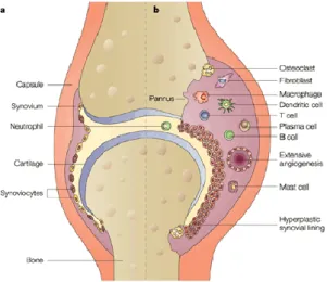
IST OF FIGURESFigure 1. Representative scheme illustrating the differences between healthy (a) and rheumatoid joints (b) ... 2
Figure 2. Summary of biologic treatments used in rheumatoid arthritis ... 5
Figure 3: B cells development ... 6
Figure 4. Frequency of B cell subpopulations in ERA and established RA patients treated with MTX, MTX pre-bio, TNF inhibitors and TCZ in comparison with healthy controls ...14
Figure 5. sCD23 serum levels are increased in ERA patients, but not in established RA ...23
Figure 6. CXCL13 serum levels are increased in ERA and established RA patients treated with MTX, MTX pre-bio and TNF inhibitors ...24
xv
L
IST OF TABLESTable 2. Clinical information of untreated ERA and established RA patients treated with
MTX, MTX pre-bio, TNF inhibitors or TCZ ...12
Table 2. MFI values of BAFF-R on B cell subpopulations ...15
Table 3: MFI values of BCMA on B cell subpopulations ...15
Table 4. MFI values of TACI on B cell subpopulations ...16
Table 5. MFI values of CD69 on B cell subpopulations ...16
Table 6. MFI values of CD86 on B cell subpopulations ...17
Table 7. MFI values of HLA-DR on B cell subpopulations ...17
Table 8. MFI values of CXCR5 on B cell subpopulations ...18
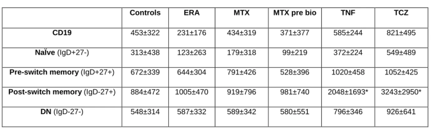
Table 9: MFI values of CD95 on B cell subpopulations ...18
Table 10. MFI values of IgM on B cell subpopulations ...19
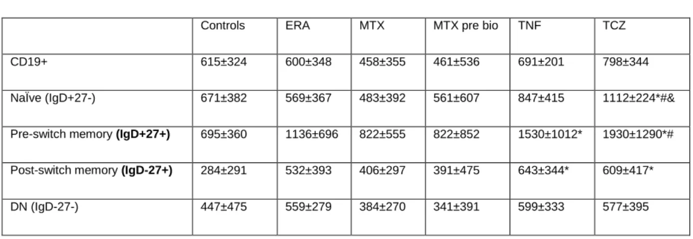
Table 11. MFI values of CD5 on B cell subpopulations ...20
Table 12. MFI values of TLR9 on B cell subpopulations ...20
Table 13: MFI values of HLA-DR on B cell subpopulations before and after TNF inhibitors ...21
Table 14. MFI values of HLA-DR on B cell subpopulations before and after TCZ ...22
Table 15. MFI values of CD95 on B cell subpopulations before and after TNF inhibitors ...22
Table 16. MFI values of CD86 on B cell subpopulations before and after TNF inhibitors ...22
1
I.
I
NTRODUCTION1. Rheumatoid Arthritis
1.1. Definition
Rheumatoid arthritis (RA) is a systemic inflammatory autoimmune disease characterized by chronic pain and progressive joint damage. The disease prevalence in the adult population is 0,5-1% worldwide. RA onset usually occurs between 30-50 years of age and it is more frequent in women, suggesting a specific hormonal component association. RA is associated with a high risk of functional disability, a diminished quality of life and increased mortality when compared to healthy individuals [1, 2]. Risk factors such as smoking, infectious agents (e.g., Epstein-Barr virus, cytomegalovirus) and their products, environment, obesity can contribute to a worse prognosis [2, 3].
In general, RA is clinically recognized as an inflammatory process affecting joints, but the presence of extra-articular manifestations makes it a systemic disease with different clinical patterns. RA affects mainly the small joints such as those of the hands, wrists and feet and, if not properly treated, leads to bone and cartilage destruction with complete loss of joint integrity [1, 2]. RA is responsible for high levels of functional impairments: 20 to 30% of people with RA become permanently work-disabled within three years of diagnosis and 50% after ten years [4-6].
For years, RA diagnosis was established according to the 1987 American College of Rheumatology (ACR) criteria [7] and at least four of the criteria had to be present for a positive classification as RA. Morning stiffness lasting at least 45 minutes, fatigue and symmetrical inflammation of large and small joints are the most common initial symptoms of RA. Recently, RA diagnosis is defined according to the 2010 ACR/ European League Against Rheumatism (EULAR) classification criteria (appendix 1) [8, 9].
1.2. Etiology and pathophysiology
The etiology of RA is unknown. Different effector pathways and cells are involved in the cascade of events leading to the progression and persistence of the disease [1, 10, 11].
The inflammatory process in RA synovium (Figure 1) is initiated when an unknown antigenic trigger induces the development of an inflammatory autoreactive response. Antigen-presenting cells (APCs) exhibit antigenic processed peptides to T cells, interacting through
2
the major histocompatibility complex (MHC) – T-cell receptor (TCR) and costimulatory signals via CD28-B7 family receptor CD80/86 [12, 13].
Figure 3. Representative scheme illustrating the differences between healthy (a) and rheumatoid joints (b). Adapted from[14]
B cells can function both as antibody producing cells and as efficient APCs[15]. In RA, autoreactive B cells produce autoantibodies such as rheumatoid factor (RF) and anti-cyclic citrullinated peptides (anti-CCP)[16], which are able to form immune complexes that deposit in the joints[16, 17]. These immune complexes activate monocytes through the low-affinity IgG receptor FcγRIIIa, leading to the production and release in synovial tissue of several proinflammatory cytokines such as tumor necrosis factor (TNF)[17], interleukin (IL)-1β and IL-6[18]. Activated synovial fibroblasts promote the expression of cell-adhesion molecules such as VCAM1 that contribute to an increase of cellular infiltration in the joints[19]. Additionally, the local production of metalloproteinases (MMP) such as MMP3 or MMP9, as well as the production of receptor activator for nuclear factor κB ligand (RANKL) increases cartilage and bone destruction [20, 21]. The inflamed synovial membrane begins to grow irregularly, forming invasive “pannus” tissue, which consequently invades and destroys cartilage and bone. RA pannus is mainly composed by mononuclear cells and fibroblasts. The inflammatory process is mediated by the activation of intracellular signaling pathways that stimulate the production and the release of multiple cytokines, chemokines, grow factors, proteinases and adhesion molecules that further increase joint erosion. In late-stages of RA, the pannus becomes fibrotic, minimally vascularized, with collagen fibres overlying articular cartilage [21-24].
3
1.3. Genetic predisposition and environmental risk factors
Rheumatoid Arthritis involves a complex interplay between environmental and genetic risks factors [25]. Heritability studies suggest that 60% of predispositions to RA are explained by genetic factors, leaving the remaining 40% to environmental and stochastic factors [25-27].
The genetic association in RA is due to the histocompatibility leucocyte antigen (HLA) DR genes, which reside in the MHC and participate in antigen presentation [28-30]. The risk for developing RA is related to the presence of specific alleles of the class II gene HLA-DRB1 [31] that encode a conserved sequence of aminoacids, the shared epitope (SE), in the third hypervariable region (HVR3) of the class II DRβ1 chain [30, 31]. This sequence is found in multiple RA-associated DR genes, including DR1, DR4, and DR14. The location of SE on MHC molecules suggests that it might have a role on the ability of HLA-DR to bind and present specific arthritogenic peptides, which might cause RA. In inflamed joints, MHC class II-dependent T cell activation by APCs (B cells, monocytes, dendritic cells) is considered a major driver of the disease, which reinforces the relevance of adaptive immunity in RA pathogenesis [28-31].
The environmental risk factors in RA include female gender, age, a previous family history of RA, smoking, obesity and some infectious agents [3, 32-35].
The best established environmental factor for RA is smoking. A study that included 13 monozygotic twin pairs discordant for RA and smoking, revealed that the smoker was the one who developed the disease in 12 out of 13 pairs [36]. Furthermore, additional studies have also indicated an association between smoking and autoantibody production, namely with anti-citrullinated protein antibodies (ACPA). The observation that RA associated autoantibodies RF and ACPA may be present for more than 10 years before disease onset suggests that risk factors are active several years prior to the development of the disease [26, 33, 36, 37].
Infectious agents such as Epstein-Barr virus [3], cytomegalovirus [38], Proteus species [39],
Mycobacterium tuberculosis [40], parvovirus B19 [41], Escherichia coli and their products
(e.g. heat-shock proteins (HSP)) [42] can also be considered risk factors. These associations have been supported by increased antibody titers against the infectious organism present in RA patients and the possibility of molecular mimicry [3, 38-42].
1.4. Prognosis and treatment
In early stages of RA, predictors such as high functional impairment, early involvement of several joints, high erythrocyte sedimentation rate or C-reactive protein levels at disease
4
onset, positivity for autoantibodies (RF, ACPA) [37, 43] and early radiographic changes can be related to a worse disease prognosis. The presence of specific alleles that encode the susceptibility epitope on MHC molecules might also influence the severity of the disease, since the risk of extra-articular and erosive disease is greater if the patients are homozygous [28, 44]. An adequate RA treatment is most important for the patient and the society. Uncontrolled active RA causes disability, decreased quality of life, and increases comorbidity, which results in loss of employment, high medical and social costs, and substantial morbidity and mortality. This impact of RA ultimately justifies expensive treatment [5, 25]. Although no cure has been found for RA, it is clear that establishing a diagnosis as early as possible and immediate treatment are the basis for a successful management of these patients [25].
The main goals of RA treatment are the preservation of function and quality of life, minimizing pain and inflammation, remission of symptoms and control of systemic manifestations [24, 45-48]
Treatment options are divided in three main classes: 1) nonsteroidal anti-inflammatory drugs (NSAIDs); 2) corticosteroids; and 3) disease modifying anti-rheumatic drugs (DMARDS) (synthetic and biological) [49, 50].
NSAIDs are efficient in relieving RA symptoms, but ineffective in the disease course. Corticosteroids are suppressors of the inflammatory response and previous studies have confirmed that are able to decrease the progression of the RA [51-54].
DMARDs have demonstrated efficiency in managing disease activity by the suppression of the inflammation [55, 56]. Synthetic DMARDs such as methotrexate (MTX) [57], leflunomide, sulfasalazine and hidroxychloroquine are widely used in RA and have proven to be highly beneficial in decreasing inflammation and joint damage [58]. Combinations of DMARDs have also proven efficiency [59-61]. Nevertheless, despite being treated some patients still have persistent disease activity and often present side effects such as gastrointestinal and pulmonary toxicity, headaches, fatigue and less commonly bone marrow suppression [58]. In these cases, when patients do not respond favorably to synthetic DMARDs, treatment with biologic DMARDs must be initiated (Figure 2).
TNF-inhibitory agents are the first line biologic treatment. Infliximab [62, 63], certolizumab [64], adalimumab and golimumab are monoclonal antibodies that target and neutralize circulating and synovial TNF [60]. Etanercept is a fusion protein that functions as a decoy receptor that binds to TNF and lymphotoxin (LT) family members)[50, 65]. Studies have shown that monotherapy with these drugs is effective [60]. Other biological agents include
5
abatacept, a fusion protein composed of the Fc region of IgG1 fused to the extracellular domain of cytotoxic T-lymphocyte antigen (CTLA)-4 that binds to the CD80/ CD86 molecule and [66, 67] inhibits T cell activation; tocilizumab, an IL-6 receptor antibody antagonist [18, 68]; and anakinra, an IL-1 receptor antagonist that has only a moderate therapeutic effect [69].
Additionally, RA treatment can also be achieved with rituximab (RTX), a monoclonal antibody that selectively depletes B cells. RTX is chimeric monoclonal IgG1 anti-CD20 antibody whose efficiency in RA treatment has brought a new interest to the role of B cells in RA pathogenesis [70-73].
Figure 2. Summary of biologic treatments used in rheumatoid arthritis. Adapted from [74]
2. B cells
2.1 Origin, development and function
B lymphocytes are white blood cells (6-10µm) whit a dense nucleus and little cytoplasm that express a membrane-bound Ig and originate and mature in the bone marrow. In humans, B cells correspond to 5-15% of total lymphocytes (2X10²). These cells are important mediators of humoral immune response, by producing and secreting proteins called immunoglobulins (Ig) or antibodies. B cells can also function as APCs and secrete cytokines that can further activate T cells and contribute to the development of an effective immune response [75, 76].
6
In humans, during fetal development, B cells are generated in the fetal liver and after birth, bone marrow assumes this function and continues throughout life. B cells arise from a lymphoid stem cell in the bone marrow and precede through several maturation stages, during which they express different cell surface markers. The B cell progenitor (pro-B cell) is the earliest distinctive B-lineage maturation stage. Pro-B cells proliferate in the bone marrow and differentiate into precursor B cells (pre-B cells). Pre-B cell stage is followed by an immature B cell stage that is not fully functional. B cells leave the bone marrow as naïve B cells when maturation is achieved by co-expression of IgM and IgD on their surface [75, 77-81]. The B cell differentiation stages in the bone marrow correspond to the antigen-independent phase of B cell development [81, 82]. Once they leave the bone marrow, naïve B cells, which have never encountered an antigen, circulate in the blood and lymphatic systems and are carried to secondary lymphoid organs (lymph nodes, spleen, Peyer’s Patches), where they further differentiate. After encountering an antigen, naïve B cells transform into large B-blasts and may follow two different pathways. Some cells proliferate and differentiate into short-living IgM producing plasma cells and a minority of B-blasts will differentiate to form germinal centers (GC) [82]. In GC reactions, B cells differentiate either into memory B cells or plasma cells (Figure 3).
Figure 3: B cells development adapted from [83]
Since an antigen is required for B cell activation and differentiation in the periphery, this stage comprises the antigen-dependent phase of B cell development. In the absence of antigen-induced activation, naïve B cells in the periphery become apoptotic [84].
7
2.2. Antibodies
Antibodies, also called immunoglobulins, are the antigen-binding proteins secreted by plasma cells, present on the B cell membrane or in circulation. Antibodies can eliminate pathogens by several mechanisms such as opsonization, complement activation and direct lysis of bacteria and/or neutralization of virus and toxins to prevent their entry into host cells [77, 84]. An antibody molecule has a common structure of four peptide chains, two identical light (L) chains and two identical heavy (H) chains and has five major classes based on the diversity of their heavy chain, namely IgM, (µ), IgG (γ), IgA (α), IgD (δ) and IgE (ɛ), each one with different structural and functional properties [77, 85, 86]. Antibody diversity is achieved by V(D)J recombination (V-variable, D-diversity and J-joining), somatic hypermutation (SHM) and class or isotype-switching [84, 86].
The V(D)J recombination is an ordered site-specific DNA rearrangement process that occurs in developing lymphocytes in the BM, in which V(D)J gene segments are randomly combined at recombination signal sequences (RSS) [84, 87]. RSS are recognized by an endonuclease coded by recombination activating gene (RAG)-1 and RAG-2, responsible for DNA double-stranded breaks following the 12-23 bp rule [84, 86]. The diversity generated by this process is further increased by terminal deoxynucleotidyl transferase (TdT) that adds nucleotides to gene segments junctions [84, 86]. SHM is a mechanism that occurs within GC in secondary lymphoid organs and involves the introduction of point mutations, mainly nucleotide substitutions, as well as occasional deletions and duplications at a very high rate into the DNA of heavy and light chain variable region genes, at CDR [86]. This differential selection is due to an increase in antibody affinity for an antigen, a process known as affinity maturation. CSR is an isotype switching deletional DNA recombination process that occurs in mature B cells and consists of replacing an expressed heavy-chain constant-region gene, usually IgM, with another one of a different biological function – IgG, IgA or IgE [86]. During this process, only 10 the effector functions of the antibody are changed. SHM and CSR are mediated by activation-induced cytidine deaminase (AID) that promotes the recombination event [88-90].
3. B cells and rheumatoid arthritis
For decades, RA was considered a T-cell driven disease. In the rheumatoid synovium, accumulation of CD4+ T lymphocytes suggested that macrophages could be activated by T cells, specifically T helper (Th)-1, to produce proinflammatory cytokines such as TNF, IL-1β and IFNγ [91-93].
In patients with RA, the increase of IL-17 levels in synovial fluid [94] suggests the involvement of Th17 cells and this association is directly proportional to the severity of joint
8
destruction [23, 95]. In fact, the percentage of Th17 cells is increased in RA synovial fluid [96]. These facts are supported by studies performed in animal models of arthritis that demonstrate that a local expression of Il-17 in mouse joints results in acute inflammation, while the incidence and severity of arthritis is markedly attenuated in IL-17-deficient mice [97, 98]. Contrarily to Th1, Th2, and Th17 cells, regulatory T cells (Tregs) CD4+CD25+ are characterized by low proliferative capacity upon triggering the T cell receptor (TCR) and by their ability to suppress CD4+ and CD8+ T-cell immune responses [99]. Several studies have proposed that Tregs are severely impaired in autoimmune rheumatic diseases, suggesting that in fact a breakdown of Treg-mediaded peripheral tolerance may have occurred [99-101]. Despite all the observations that support a role for T cells in RA development, the disappointing results obtained with anti-CD4 therapy in humans reinforced the notion that perhaps other parts of the inflammatory process of RA should be better studied and understood [50, 102, 103].
Although the cause that triggers RA autoimmune process remains unknown, it has been demonstrated that several types of cells from both innate and adaptative immune system actively participate and form complex networks of cell-cell interactions that contribute to the development and chronicity of synovitis and inflamation on RA [104, 105]. Several studies have pointed to B cell function as a critical factor in the development of RA [85, 106-108]. In RA, B cells are responsible for the production of autoantibodies [109], which include antibodies against cartilage components (anti-type II collagen or anti-CII; anti-human cartilage glycoprotein 39 or gp39); enzymes (glucose-6-fosfato-isomerase or anti-GPI, anti-enolase); nuclear proteins (anti-RA33) and stress proteins. RF and ACPAs are, however, the autoantibodies most specifically associated with this disease. RF are autoantibodies that directly bind to the Fc portion of normal human IgG and ACPAs are autoantibodies that recognize peptides or proteins containing citrulline, a non-standard aminoacid generated by the post-translational modification of arginine by peptidylarginine deiminase (PAD) enzymes, in a process known as citrullination. RF and ACPAs can form immune complexes that deposit in the joints, activate complement and cause inflammation [110]. The clinical usefulness of RF and ACPAs has been acknowledged due to their good diagnostic sensitivity and prognostic value, but the clinical significance and pathogenic roles of the other autoantibodies are unknown. Furthermore, the association between high titer of RF and worse prognosis indicate that RF may have an important role in the pathogenesis of RA, although there are RA patients seronegative for RF who also manifest the disease. Moreover, ACPAs-positive RA patients have a higher cellular infiltration in RA synovium and these autoantibodies are also helpful to predict the outcome of patients with undifferentiated arthritis [37, 109, 111].
9
B cells are also very efficient APCs that activate T cells through the expression of costimulatory molecules [112, 113]. A B cell is considered autoreactive when its B cell receptor (BCR) targets a self-antigen. In RA, B cells have the ability to present processed self-antigens to T cells, thus allowing the development of an autoreactive immune response. Indeed, RF+B cells play an important role in antigen presentation and in the induction of T cell activation [12, 114, 115].
B cells can also function as cytokine and chemokine-producing cells that promote leukocyte infiltration in the joints and the formation of ectopic lymphoid structures [116, 117], thus aggravating angiogenesis and synovial hyperplasia [15, 118].
The efficacy of B cell depletion therapy with RTX reinforced the relevance of B cells in RA pathogenesis [119].
II.
AIMS
The main goals of the present work were:
To characterize B cell subpopulations in the peripheral blood from untreated early rheumatoid arthritis (ERA) patients and established treated RA patients.
To analyze the cytokine and chemokine environment directly related with B cell activation since early RA.
To compare the effect of treatment options (methotrexate-MTX, TNF-inhibitors and tocilizumab-TCZ) on B cell phenotype and cytokine production in ERA and established RA patients.
10
III.
MATERIALS
AND
METHODS
Patients
Blood samples were collected from 13 consecutive patients with untreated polyarthritis (Rheumatology Department, Hospital de Santa Maria, Lisbon) of < 1 year disease duration. After a minimum follow-up of 3 months, the patients fulfilled the 2010 ACR/ EULAR criteria for RA and were classified as early RA (ERA). In addition, blood samples from 17 patients with established RA treated with methotrexate (MTX); 29 patients with established RA treated with MTX before starting biologic DMARD therapy (MTX pre-biologic, MTX pre-bio); 10 patients under treatment with TNF inhibitors and 7 patients under tocilizumab (TCZ) treatment were also collected for comparison (Rheumatology Department, Hospital de Santa Maria, Lisbon). Furthermore, blood samples from 15 healthy donors were also collected and processed for comparison.
This study was approved by the local ethics committee (Comissão de Ética do Hospital de Santa Maria), and all patients and healthy donors signed an informed consent form. Patient care was conducted in accordance with standard clinic practice, and the study was performed in accordance with the Declaration of Helsinki (2008).
Isolation of peripheral blood mononuclear cells
Peripheral blood mononuclear cells (PBMC) were isolated from 40 ml heparinized whole blood following density gradient centrifugation with Ficoll-Paque Plus (GE Healthcare, Sweden).Cells were washed twice in 1X phosphate buffered saline (PBS) and cellular viability was estimated with 0.4% Trypan blue (Sigma, USA).
Flow Cytometry
Immunophenotyping of B cells was performed in PBMC samples (1X10⁶ cells/ sample) using matched combinations of anti-human murine monoclonal antibodies (mAbs) conjugated to FITC, phycoerytrin (PE), peridinin chlorophyll protein (PerCP)-Cy5.5, allophycocyanin (APC), PE-Cy7, eFlour 450 and APC-eFluor780.Combinations of anti-CD19 conjugated to PerCP-Cy5.5 or APC, anti-IgD conjugated to PE-Cy7 or FITC, anti-CD27 conjugated to eFluor450 or FITC, anti-CD38 conjugated to APC-eFluor780, anti-BAFF-R conjugated to PE, anti-TACI conjugated to APC, anti-CD86 conjugated to PE, anti-CD69 conjugated to PerCP or APC, anti-IgM conjugated to PE, anti-CD5 conjugated to APC, anti-CXCR5 conjugated to
11
PE, anti-HLA-DR conjugated to APC, anti-CD95 conjugated to APC, anti-BCMA conjugated to PE and anti-TLR9 conjugated to APC were used. All antibodies were purchased from BD Pharmingen (USA), eBioscience (USA) and R&D Systems (United Kingdom). For cell surface stainings, PBMC were incubated with antibodies during 30 minutes, in the dark, at 4ºC. For TLR9 intracellular staining, PBMC were fixed during 20 minutes at room temperature with IC Fixation Buffer (eBioscience, USA), permeabilized with 1X Permeabilization Buffer (eBioscience, USA) and stained according to eBioscience intracellular antigen staining protocol. A total of 50.000 cells/ sample gated in CD19+ B cells were acquired with LSR Fortessa (BD). Data were analyzed with FlowJo (TreeStar, Stanford University, California, USA). All samples were acquired on the same day of the staining protocol.
ELISA
The B-lymphocyte chemoattractant (BLC) also known as C-X-C motif chemokine 13 (CXCL13) and the soluble form of CD23 (sCD23) were quantified in serum samples from all groups by enzyme-linked immunosorbent assay (ELISA) (R&D systems, United Kingdom), according to the manufacturer’s instructions. Samples were analyzed using plate reader Infinite M200 (Tecan, Switzerland).
Statistical analysis
Statistical differences were determined with GraphPad Prism (GraphPad, San Diego, USA). For populations that did not follow a Gaussian distribution, non-parametric tests were used. The Mann-Whitney test was used for comparisons between 2 independent groups. For comparisons between 3 or more groups, the Kruskal-Wallis and Dunn’s multiple comparison tests were used. The Wilcoxon matched pairs test was used for comparisons between 2 paired groups. Correlation analyses were performed using Spearman’s test. Differences were considered statistically significant for p < 0.05.
12
IV.
R
ESULTS1. Clinical characterization of patients
A group of untreated polyarthritis patients (n=13) with less than 1 year of disease duration and classified as early RA (ERA) according to the 2010 ACR/ EULAR criteria after a minimum follow-up of 3 months was included in this study. ERA patients had a mean ± standard deviation age of 58±14 years old, 85% were female, 69% were RF positive, 62% were anti-CCP positive and DAS28 score was 4.1±2.0. A group of established RA patients under MTX treatment (n=17) with a mean age of 55±14 years old, 81% female and a DAS28 of 2.5±1.2 was also included. Furthermore, a group of established RA patients under MTX treatment pre-biological therapy (MTX pre-bio, n=29) with a mean age of 57±11 years old, 83% female and a DAS28 of 5.1±1.3 was also analyzed. From the MTX pre-bio group, a second blood collection was performed to RA patients that had either initiated treatment with TNF-inhibitors (n=10) or with tocilizumab (TCZ) (n=7), after an average follow-up of 8 months of treatment. In addition, blood samples were collected from age and sex-matched healthy donors (n=15). The clinical information and data from all patients and healthy controls included in this study is indicated in Table 1.
Table 1. Clinical information of untreated ERA and establi shed RA patients treated with MTX, MTX pre-bio, TNF inhibitors or TCZ. Controls (n=15) ERA (n=13) MTX (n=17) MTX pre-bio (n=29) TNF (n=10) TCZ (n=7) Age (years) 52±7 58±14 55±14 57±11 56±15 61±10 Sex (% female) 73 85 81 83 80 83
Disease duration (years) NA ≤ 1 7±6 13±12 14±13 8±4
CRP (mg/dl) ND 1.4±2.1 0.5±0.6 1.1±1.2 1.9±2.5 0.1±0.1**#& ESR (mm/1st hour) ND 44±26 23±22 32±22 47±39 6±5**#& VAS NA 40±33 38±34# 69±21 62±21 63±13 DAS28 NA 4.1±2.0 2.5±1.2# 5.1±1.3 4.2±1.5 2.3±0.7# Swollen joints NA 4±4 1±2# 7±6 2±3 1±1 Tender joints NA 5±5 2±3# 10±8 6±5 1±1# RF (+) % ND 69 77 74 78 50 Anti-CCP (+) % ND 62 63 68 38 67
ERA - Early Rheumatoid Arthritis; RA - Rheumatoid Arthritis; CRP - C-reactive protein; ESR - Erythrocyte Sedimentation Rate;
VAS – Visual Analogue Scale; DAS28 – Disease Activity Score of 28 joints; RF – Rheumatoid Factor; Anti-CCP – anti-cyclic
citrullinated peptide; NA – not applicable; ND – not determined. Values are represented as mean ± standard deviation.
13
2. Classification of B cell subpopulations
To analyze the frequency of B cell subpopulations in the periphery, B cells were classified using the IgD/ CD27 classification system that allows the identification of four main B cell subsets (gated in CD19): naïve B cells (IgD+CD27-), pre-switch-memory (IgD+CD27+), post-switch memory (IgD-CD27+) and double-negative (DN, IgD-CD27-) B cells (appendix 2). A second classification system based on IgD/ CD38 (gated in CD19) was also used to identify circulating plasmablasts, defined as IgD-CD38++. The IgD/ CD38 classification system allows the identification of six B cell subpopulations: transitional (IgD+CD38++), naïve B cells (IgD+38+), IgD+ memory (IgD+CD38-), resting memory CD38-), post-GC memory (IgD-CD38+) B cells and plasmablasts (IgD-CD38++) (appendix 2). Although an analysis with both classification systems has been performed in the present work to define B cell subpopulations, the results presented in this thesis are based on IgD/ CD27 classification system (except plasmablasts) in order to compare with previous results obtained by our group in which IgD/ CD27 classification was used [120].
2.1. Established RA patients have alterations in the frequency of memory B cell subpopulations in peripheral blood
The analysis of the frequency of total CD19+ B cells has revealed that no significant differences were found in ERA and established RA patients treated with MTX, MTX pre-bio, TNF inhibitors or TCZ when compared to healthy controls. However, when analyzing B cell subpopulations, it was observed that established RA patients treated with MTX and MTX pre-bio had significantly increased frequencies of DN (IgD-CD27-) B cells in comparison to controls (p < 0.05). Furthermore, no significant differences were observed in the remaining B cell subpopulations analyzed (naïve, pre-switch memory and post-switch memory B cells) in all groups (Figure 4). Importantly, no significant correlations were found between the frequencies of all B cell subpopulations with age or with disease activity (DAS28) in all groups studied (data not shown).
14
Figure 5. Frequency of B cell subpopulations in ERA and established RA patients treated with MTX, MTX pre-bio, TNF
inhibitors and TCZ in comparison with healthy controls. Lines represent median percentage values. * p < 0.05 in comparison with Controls.
3. B cell markers and effect of treatment in comparison with healthy controls
In this study the expression of several cellular markers was analyzed by mean fluorescence intensity (MFI) to characterize B cell phenotype in circulation in untreated early and
15
established treated RA patients, according to their function. Thus, BAFF-R, TACI and BCMA were studied to analyze the expression of BAFF receptors on B cells; CD69, CD86 and HLA-DR were studied to analyze B cell activation; CXCR5 was studied to analyze B cell chemotaxis; CD95, also known as Fas receptor (FasR), was studied to analyze Fas-mediated apoptosis; IgM was analyzed as a component of the BCR; CD5 was studied to analyze B cell differentiation and TLR9 was studied to analyze the main TLR expressed by B cells. The MFI values obtained for all cellular markers were considered in all B cell subpopulations, except in plasmablasts (IgD-CD38++), due to the low number of events obtained during data acquisition and analysis.
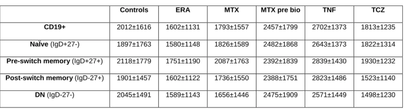
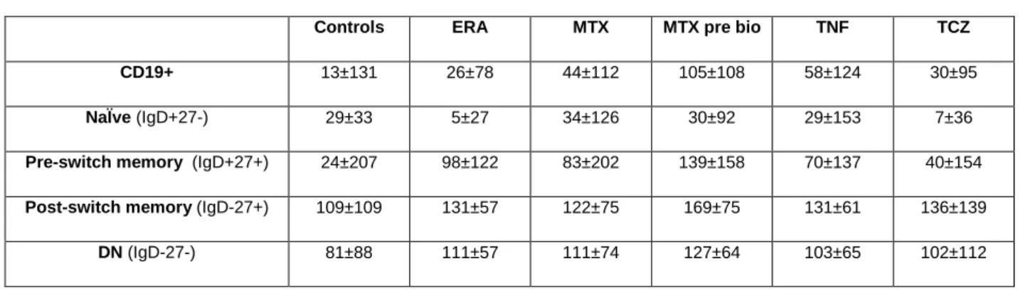
3.1. TACI expression increases on memory B cell subpopulations after treatment with TNF inhibitors, but no changes occur in BAFF-R and BCMA MFI values in all B cell subsets
B cell activating factor (BAFF) is an important cytokine for B cell activation and survival [108, 121, 122] that binds to three receptors: BAFF-R, TACI and BCMA. While no significant differences were found in BAFF-R (Table 2) and BCMA (Table 3) MFI levels in all groups analyzed in all B cell subpopulations, TACI MFI was significantly increased in total CD19+ B cells in established RA patients after treatment with TNF inhibitors in comparison with controls, namely in post-switch memory (p = 0.0170) and double negative B cells (p = 0.0084) (Table 4). Furthermore, TACI MFI levels were also significantly increased in double negative B cells of ERA patients when compared to controls (p = 0.0138).. Moreover, no significant differences were observed in the frequencies of BAFF-R+ B cells in all B cell subpopulations in all groups analyzed (data not shown).
Table 2. MFI values of BAFF-R on B cell subpopulations. Represented values are mean ± standard deviation (SD).
Controls ERA MTX MTX pre bio TNF TCZ CD19+ 2012±1616 1602±1131 1793±1557 2457±1799 2702±1373 1813±1235
NaÏve (IgD+27-) 1897±1763 1580±1148 1826±1589 2482±1868 2643±1373 1822±1314
Pre-switch memory (IgD+27+) 2118±1779 1751±1190 2087±1763 2392±1839 2839±1430 1930±1232
Post-switch memory (IgD-27+) 1901±1457 1602±1122 1736±1550 2388±1751 2823±1486 1523±1140
16
Table 3. MFI values of BCMA on B cell subpopulations. Represented values are mean ± SD.
Controls ERA MTX MTX pre bio TNF TCZ CD19+ 13±131 26±78 44±112 105±108 58±124 30±95
NaÏve (IgD+27-) 29±33 5±27 34±126 30±92 29±153 7±36
Pre-switch memory (IgD+27+) 24±207 98±122 83±202 139±158 70±137 40±154
Post-switch memory (IgD-27+) 109±109 131±57 122±75 169±75 131±61 136±139
DN (IgD-27-) 81±88 111±57 111±74 127±64 103±65 102±112
Table 4. MFI values of TACI on B cell subpopulations. Represented values are mean ± SD.
Controls ERA MTX MTX pre bio TNF TCZ CD19+ 181±309 321±394 222±276 202±297 400±249* 473±490
NaÏve (IgD+27-) 219±425 231±352 153±298 178±350 344±250 452±562
Pre-switch memory (IgD+27+) 381±288 546±452 462±335 374±449 733±629 928±1060
Post-switch memory (IgD-27+) 178±217 459±415 264±374 275±380 486±287* 410±358
DN (IgD-27-) 137±208 464±373* 333±289 285±357 415±209* 305±88
* p < 0.05 in comparison with Controls
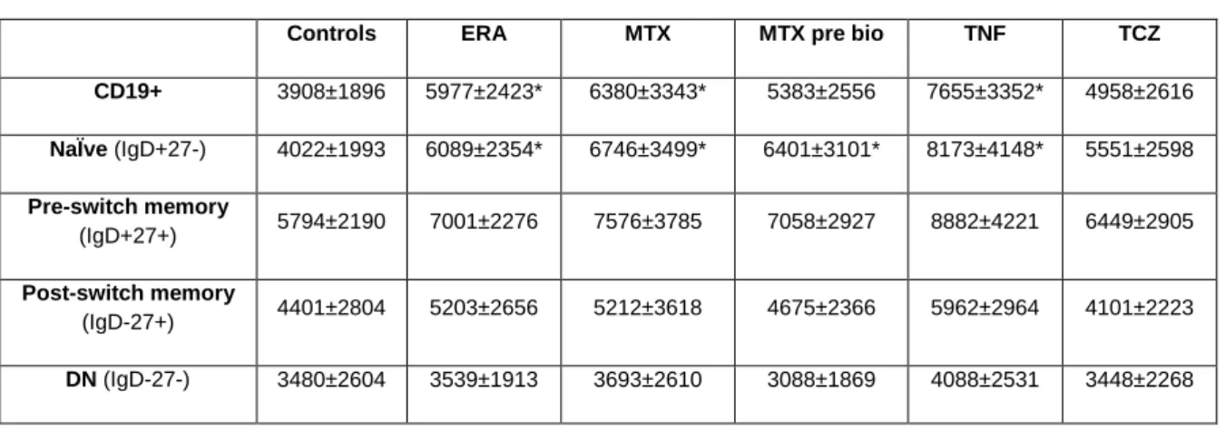
3.2. TNF inhibitors and TCZ treatment increase the expression of the activation marker HLA-DR on B cells, but have no effect on CD69 and CD86
In all groups studied, no significant differences were found in CD69 and CD86 MFI levels in all B cell subpopulations (Tables 5 and 6, respectively). Moreover, although no significant differences were observed in the frequencies of HLA-DR+ B cells in all groups (data not shown), alterations were found in HLA-DR MFI values in established RA patients treated with TNF inhibitors and TCZ in comparison with healthy controls and/ or in comparison with established RA treated with MTX pre-bio (Table 7). Indeed, it was observed that HLA-DR MFI values were increased in total CD19+ B cells, naïve, post-switch memory and DN B cells in established RA patients after treatment with TNF inhibitors in comparison with controls(p <
0.05). After TCZ treatment, HLA-DR MFI values were increased in total CD19+ B cells, naïve, pre-switch memory and DN B cells (p < 0.05). Additionally, both TNF inhibitors and TCZ treatments induced a significant increase in HLA-DR MFI values in total CD19+ B cells when compared with MTX pre-bio group (p < 0.05).
17
Table 5. MFI values of CD69 on B cell subpopulations. Represented values are mean ± SD.
Controls ERA MTX MTX pre bio TNF TCZ CD19+ 78±127 8±43 42±112 86±131 27±42 55±66
NaÏve (IgD+27-) 1±86 1±35 6±53 29±103 4±36 neg
Pre-switch memory (IgD+27+) 246±262 69±58 215±241 184±200 171±216 207±270
Post-switch memory (IgD-27+) 130±144 85±50 156±147 207±243 148±162 138±74
DN (IgD-27-) 108±142 52±63 136±154 119±115 78±65 221±103
Table 6. MFI values of CD86 on B cell subpopulations. Represented values are mean ± SD.
Controls ERA MTX MTX pre bio TNF TCZ CD19+ 69±136 65±66 111±143 144±102 67±136 95±69
NaÏve (IgD+27-) 48±124 12±110 38±152 13±227 13±131 16±95
Pre-switch memory (IgD+27+) 84±184 116±102 161±144 148±175 132±172 98±183
Post-switch memory (IgD-27+) 181±141 226±69 225±159 277±174 261±150 284±187
DN (IgD-27-) 125±90 163±54 169±92 169±89 158±102 110±64
Table 7. MFI values of HLA-DR on B cell subpopulations. Represented values are mean ± SD.
Controls ERA MTX MTX pre bio TNF TCZ
CD19+ 25596±8709 32803±10483 26643±14333 23869±11973 38693±11768*# 43917±1848*# NaÏve (IgD+27-) 31834±13161 38098±12499 31259±17522 28832±14606 44972±14006* 54894±23771*# Pre-switch memory (IgD+27+) 27497±13657 32426±9149 23627±10679 25547±13292 32238±12340 44302±21876* Post-switch memory (IgD-27+) 18012±6990 20544±6344 19199±10276 17262±8251 28359±7472*# 27368±15824 DN (IgD-27-) 22010±9004 27647±9225 22382±12687 21495±10344 33234±11366* 35312±11773*
* p < 0.05 in comparison with Controls # p < 0.05 in comparison with MTX pre-bio
18
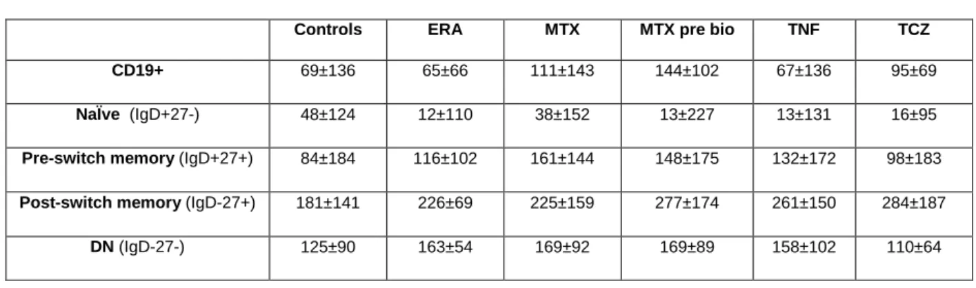
3.3. CXCR5 expression is increased since early RA
In all groups studied, no significant differences were found in the frequencies of CXCR5+ B cells in all B cell subpopulations (data not shown). Nevertheless, CXCR5 MFI values were significantly increased in total CD19+ B cells in ERA (p = 0.0276) and established RA patients after treatment with MTX and TNF inhibitors when compared to controls (p < 0.05) (Table 8). Moreover, it was observed that CXCR5 MFI values were significantly increased in naïve B cells in ERA and established RA patients treated with MTX, MTX pre-bio and TNF inhibitors in comparison with controls.
Table 8. MFI values of CXCR5 on B cell subpopulations. Represented values are mean ± SD.
Controls ERA MTX MTX pre bio TNF TCZ CD19+ 3908±1896 5977±2423* 6380±3343* 5383±2556 7655±3352* 4958±2616 NaÏve (IgD+27-) 4022±1993 6089±2354* 6746±3499* 6401±3101* 8173±4148* 5551±2598 Pre-switch memory (IgD+27+) 5794±2190 7001±2276 7576±3785 7058±2927 8882±4221 6449±2905 Post-switch memory (IgD-27+) 4401±2804 5203±2656 5212±3618 4675±2366 5962±2964 4101±2223 DN (IgD-27-) 3480±2604 3539±1913 3693±2610 3088±1869 4088±2531 3448±2268
* p < 0.05 in comparison with Controls
3.4. CD95 expression increases on memory B cell subpopulations after treatment with TNF inhibitors and TCZ
The analysis of the expression of CD95, a death receptor whose activation leads to apoptosis, revealed that no significant changes occur in CD95 MFI values on total CD19+ B cells in ERA and established RA patients irrespective of the treatment when compared to healthy controls. However, when analyzing B cell subpopulations, it was found that established RA patients treated with TNF inhibitors and TCZ have increased MFI values of CD95 in post-switch memory B cells in comparison to controls, but no significant differences where observed in the remaining B cell subsets (p < 0.05 )(Table 9).
19
Table 9. MFI values of CD95 on B cell subpopulations. Represented values are mean ± SD.
Controls ERA MTX MTX pre bio TNF TCZ CD19 453±322 231±176 434±319 371±377 585±244 821±495
NaÏve (IgD+27-) 313±438 123±263 179±318 99±219 372±224 549±489
Pre-switch memory (IgD+27+) 672±339 644±304 791±426 528±396 1020±458 1052±425
Post-switch memory (IgD-27+) 884±472 1005±470 919±796 981±740 2048±1693* 3243±2950*
DN (IgD-27-) 548±314 587±332 589±342 580±551 796±346 926±641
* p < 0.05 in comparison with Controls
3.5. IgM expression and the frequency of IgM+ B cells in circulation in ERA and established RA patients is similar to controls
In all groups of patients studied, no statistically significant differences were observed in the frequency of circulating IgM+ B cells (data not shown). Furthermore, no significant differences were found in IgM MFI values on total CD19+ B cells and all B cell subpopulations analyzed in all groups of patients when compared to healthy controls (Table 10).
Table 10. MFI values of IgM on B cell subpopulations. Represented values are mean ± SD.
Controls ERA MTX MTX pre bio TNF TCZ CD19 70±253 111±115 157±212 175±175 152±187 65±188
NaÏve (IgD+27-) 67±369 86±178 126±315 191±319 182±235 34±286
Pre-switch memory (IgD+27+) 523±515 532±418 679±676 569±390 744±613 483±588
Post-switch memory (IgD-27+) 74±71 56±51 55±47 91±49 66±37 39±36
20
3.6. ERA and established RA patients have alterations in the expression levels of CD5 and in the frequency of circulating CD5+ B cells
A significant decrease in the frequency of circulating CD5+ B cells was found in total CD19+ B cells, namely on naïve B cells, in all patients´ groups (ERA, established RA treated with MTX, MTX pre-bio, TNF inhibitors and TCZ) in comparison with controls (data not shown). However, the analysis of CD5 expression on B cell subpopulations has shown that both TNF inhibitors and TCZ treatment induce a significant increase in CD5 MFI values on pre-switch and post-switch memory B cells when compared to healthy controls (p < 0.05)(Table 10). Furthermore, significantly increased CD5 MFI values were also found on naïve B cells from TCZ treated RA patients in comparison with MTX and MTX pre-bio groups(p < 0.05).
Table 11. MFI values of CD5 on B cell subpopulations. Represented values are mean ± SD.
Controls ERA MTX MTX pre bio TNF TCZ
CD19+ 615±324 600±348 458±355 461±536 691±201 798±344
NaÏve (IgD+27-) 671±382 569±367 483±392 561±607 847±415 1112±224*#&
Pre-switch memory (IgD+27+) 695±360 1136±696 822±555 822±852 1530±1012* 1930±1290*#
Post-switch memory (IgD-27+) 284±291 532±393 406±297 391±475 643±344* 609±417*
DN (IgD-27-) 447±475 559±279 384±270 341±391 599±333 577±395
* p < 0.05 in comparison with Controls # p < 0.05 in comparison with MTX pre-bio
& p < 0.05 in comparison with MTX
3.7. TLR9 expression increases after TCZ treatment, but not after MTX or TNF inhibitors
A significant increase in TLR9 MFI values was found in total CD19+ B cells and all B cell subpopulations studied in established RA patients after treatment with TCZ in comparison not only with controls(p < 0.05), but also with MTX pre-bio group(p < 0.05)(Table 12). In addition, TLR9 MFI was significantly increased on post-switch memory and DN B cells in MTX treated patients when compared to controls. No significant differences were found in ERA or established RA patients treated with MTX pre-bio or TNF inhibitors in comparison with controls in all B cell subsets analyzed.
![Figure 2. Summary of biologic treatments used in rheumatoid arthritis. Adapted from [74]](https://thumb-eu.123doks.com/thumbv2/123dok_br/18490915.901070/23.892.211.686.451.690/figure-summary-biologic-treatments-used-rheumatoid-arthritis-adapted.webp)
![Figure 3: B cells development adapted from [83]](https://thumb-eu.123doks.com/thumbv2/123dok_br/18490915.901070/24.892.185.722.626.919/figure-b-cells-development-adapted.webp)