Agradeço à Professora Susana Pereira e ao Professor José Pissarra, pela oportunidade e confiança depositada. Pelo apoio e dedicação constantes, pelo entusiasmo e por todo o conhecimento partilhado. Obrigada.
Agradeço ao fantástico grupo do 2.61! Ao Alberto, ao Bruno e à Vanessa, pelas conversas sérias e pouco ou nada sérias, pelos sorrisos e risadas, pelo apoio constante, pela ajuda e companheirismo, que desde o primeiro dia me fizeram sentir em casa.
Um agradecimento muito especial à Cláudia. Pela dedicação e apoio incansáveis, mesmo que fosse pelo telefone, mensagem, email ou pombo correio, e mesmo quando tudo parecia impossível! Obrigada pela (muita) paciência, pela persistência e por acreditares sempre, até quando as coisas corriam menos bem. Obrigada!
A ti, Gabriel, obrigada por seres o meu porto seguro. Obrigada por tudo, sempre.
À minha família, o meu berço, obrigada pelo apoio incondicional.
Aos meus pais, o meu agradecimento mais doce. São a fonte de toda a minha força e sem eles com certeza não teria chegado até aqui.
In the past few years, the plant cell wall has been drawing more and more attention from the scientific community, particularly regarding the protein trafficking to and from this compartment. The secretion of soluble proteins has been for a long time thought to occur by bulk flow, following a default pathway to the extracellular matrix. However, data has been emerging on the possibility of the sorting to the cell wall is in fact regulated somehow, despite the absence of identified signals or receptors. Endocytic routes have also been suggested to be involved in the regulation of protein sorting to and from the cell wall. Moreover, the biogenesis of the cell wall itself depends on numerous intracellular trafficking and secretion events and it has been shown that those events are most probably controlled specifically and rigorously by the cell wall. From this perspective, as a vital compartment of the plant cell and holding a strategic position between the cell and the environment, the cell wall could in fact control and regulate certain cargo trafficking inside the cell, as well as important secretion events.
Cardosin A and cardosin B are aspartic proteinases first isolated from the flower tissues of cardoon (Cynara carduculus L.), which have been thoroughly characterised in our group. When transiently expressed in Nicotiana tabacum leaves, both cardosins have shown to accumulate in the lytic vacuole. Recently, however, experiments were conducted in N. tabacum mesophyll protoplasts and, curiously, cardosin B showed to be secreted to the culture medium, which could point towards a regulation by the cell wall. Moreover, the vacuole sorting determinants recently identified for cardosins – C-terminal peptides and the PSI domain – which are also worth investigating for a possible role in the regulation of sorting by the cell wall. The PSI from cardosin A, in particular, for the intriguing capability of directing proteins to the vacuole through a Golgi-independent route, seemed worth exploring in more detail.
Taking together the results of this study, the hypothesis of a role for the plant cell wall in the regulation of protein trafficking appears as plausible. The absence of cell wall in N. tabacum protoplasts expressing cardosins, which are typically vacuolar in this system, have originated partial protein secretion. The same pattern was observed for C-terminal peptides, which are known to follow the same intracellular route as whole cardosins; however, no secretion was detected for PSI A, supporting the Golgi-independent proposed route before reaching the lytic vacuole. As a whole, this study enhances the dynamic and active role of the cell wall in the protein sorting events and is the starting point of several studies exploring its participation on the regulation of intracellular sorting.
Nos últimos anos, a parede celular das plantas tem vindo a atrair a atenção da comunidade científica, particularmente no que diz respeito ao trânsito de proteínas de e para este compartimento. A secreção de proteínas solúveis foi considerada durante muito tempo como uma via default, transportando as proteínas para a matriz extracelular. Contudo, tem sido sugerida a possibilidade do transporte para a parede celular ser de facto regulado, apesar da inexistência de sinais ou recetores identificados. A via do endossoma é sugerida como estando envolvida na regulação do trânsito de proteínas de e para a parede celular. Por outro lado, a biogénese da própria parede celular depende de inúmeros processos de trânsito intracelular e secreção, e tem vindo a ser sugerido que estes são presumivelmente controlados pela parede celular de forma específica e rigorosa. Assim, sendo um compartimento vital à célula vegetal e detendo esta uma posição estratégica entre a célula e o meio exterior, a parede celular poderá de facto controlar e regular determinados transportes intracelulares de proteínas, assim como processos relevantes de secreção.
A cardosina A e a cardosina B são proteinases aspárticas que foram isoladas pela primeira vez dos tecidos da flor de cardo (Cynara cardunculus L.), e têm sido extensivamente caracterizadas no nosso grupo. Quando expressas de forma transiente em folhas de Nicotiana tabacum, ambas demonstraram acumular-se no vacúolo lítico. Recentemente, porém, foram efetuados estudos em protoplastos do mesófilo de N. tabacum e, curiosamente, verificou-se que a cardosina B era secretada para o meio de cultura, o que aponta para uma regulação por parte da parede celular. Recentemente, foram ainda identificados os determinantes vacuolares das cardosinas – a região C-terminal e o domínio PSI – cujo possível papel da parede celular na regulação do trânsito é também motivo de interesse. O PSI da cardosina A, em particular, pela sua intrigante capacidade de direcionar proteínas para o vacúolo por uma via independente do complexo de Golgi, suscitou especial interesse para um estudo mais detalhado.
De acordo com os resultados obtidos no presente estudo, a hipótese de a parede celular ter um papel na regulação do transporte de proteínas surge como plausível. A ausência de parede celular em protoplastos de N. tabacum a expressar cardosinas, tipicamente vacuolares neste sistema, originou a secreção parcial destas proteínas. O mesmo padrão é observado para os péptidos C-terminal, cuja rota intracelular é semelhante à das cardosinas; contudo, não foi observada secreção para o PSI A, suportando a rota para o vacúolo independente do complexo de Golgi. Em suma, este estudo realça o papel dinâmico e ativo da parede celular em processos de trânsito proteico e poderá ser um ponto de partida para diversos estudos, nomeadamente a exploração da sua participação na regulação do trânsito intracelular.
ACKNOWLEDGEMENTS ...1 SUMMARY ...3 RESUMO ...5 TABLE OF CONTENTS...7 LIST OF TABLES ... 11 LIST OF FIGURES ... 13 1. INTRODUCTION ... 19
1.1. The Endomembrane System ... 20
1.1.1. The Endoplasmic Reticulum ... 21
1.1.2. The ER – Golgi Interface ... 22
1.1.3. The Golgi apparatus ... 24
1.1.4. Post-GA transport ... 24
Protein Trafficking to the Vacuoles ... 25
Protein Trafficking to the Cell Wall ... 27
1.2. Cardosins – Models for Protein Sorting Studies ... 28
Cardosins – Aspartic Proteinases ... 28
Cardosins and Protein Sorting ... 29
1.3. Objectives of the Thesis ... 31
2. METHODOLOGY ... 32
2.1. Dissecting the role of the plant cell wall in the regulation of protein trafficking.
... 33
2.1.1. Germination and maintenance of Nicotiana tabacum SRI cv. Petit Havana
... 33
2.1.2. Isolation of protoplasts from Nicotiana tabacum leaves ... 33
2.1.3. Cell wall regeneration in Nicotiana tabacum protoplasts ... 33
2.1.4. Infiltration of Nicotiana tabacum leaves ... 34
2.1.5. Secretion assays ... 34
2.1.7. Preparation of proteins from protoplasts’ culture medium by
Trichloroacetic acid (TCA) precipitation ... 36
2.1.8. SDS-PAGE and Western blotting ... 36
2.1.9. Fluorescence Microscopy and Confocal Laser Scanning Microscopy ... 37
2.2. Unveiling the role of the PSI domain in the regulation of cardosin A trafficking
... 38
2.2.1. Primers design and site-directed mutagenesis ... 38
2.2.2. Addition of restriction enzyme adaptors ... 41
2.2.3. Cloning into pCR-Blunt vector ... 42
2.2.4. Plasmid miniprep and enzymatic restriction screening ... 42
2.2.5. Sub-cloning into pVKH18-En6 ... 43
2.2.6. Transient expression in Nicotiana tabacum ... 43
2.2.7. Expression analysis by CLSM ... 44
2.3. Maintenance and transformation of bacterial strains used along the work ... 44
2.3.1. Preparation of competent Escherichia coli ... 44
2.3.2. Transformation of Escherichia coli ... 45
2.3.3. Preparation of electrocompetent Agrobacterium tumefaciens ... 45
2.3.4. Transformation of Agrobacterium tumefaciens ... 45
3. RESULTS ... 46
3.1. Dissecting the role of the plant cell wall in the regulation of protein trafficking
...47
3.1.1. Protoplast culture and cell wall regeneration optimisation ... 47
3.1.2. Analysis of cardosins’ trafficking during protoplasting and cell wall
regeneration ... 50
3.1.3. The role of C-terminal region in cardosins trafficking during protoplasting
and cell wall reforming ... 54
3.1.4. The role of PSI domain in cardosins trafficking during protoplasting and
cell wall reforming ... 57
3.2. Unveiling the role of the PSI domain in the regulation of cardosin A trafficking
... 62
4. DISCUSSION ... 664.1.
Dissecting the role of the plant cell wall in the regulation of protein trafficking
………67
N. tabacum protoplasts regenerate a cell wall as early as 24h upon isolation
...67
Cardosins A and B became secreted upon protoplasting ... 68
The C-terminal VSD is responsible for cardosins secretion during protoplasting
... 71
The PSI:mCherry constructs are not secreted upon protoplasting ... 72
4.2.
Unveiling the role of the PSI domain in the regulation of cardosin A trafficking
………73
5. CONCLUSIONS AND FUTURE PERSPECTIVES ... 75 6. BIBLIOGRAPHIC REFERENCES ... 78Table 1 - Primers used in the Site-directed mutagenesis of cardosin A. ... 39 Table 2 - PCR reactions’ composition used for the Site-directed mutagenesis of cardosin A. .. 40 Table 3 - PCR conditions used for the Site-directed mutagenesis of cardosin A. ... 40 Table 4 - Primers used for the addition of restriction enzyme adaptors to AmutPSI construction.
... 41 Table 5 - PCR reactions’ composition used for the addition of restriction enzyme adaptors to AmutPSI construction. ... 41 Table 6 - PCR conditions used for the addition of restriction enzyme adaptors to AmutPSI construction. ... 42 Table 7 - Summary of the results obtained on the secretion assays performed, comprising the observations made through CLSM and Western blotting. ... 61
Figure 1 - Schematic representation of the cargo transport between the ER export sites and the Golgi apparatus. ... 22 Figure 2 - Schematic models of ER-Golgi protein transport. ... 23 Figure 3 - Schematic representation of soluble proteins transport to the vacuoles... 26 Figure 4 - Schematic representation of the proposed sequence of events during the processing of cardosin A. ... 29 Figure 5 - Schematic representation of the chimeric constructions used in this study. ... 35 Figure 6 - Monitorisation of cell wall regeneration in N. tabacum protoplasts maintained in
medium A. ... 48 Figure 7 - Monitorisation of cell wall regeneration in N. tabacum protoplasts maintained in
medium B. ... 49 Figure 8 - Confocal microscopy images of N. tabacum mesophyll protoplasts and the
respective leaf of origin, expressing cardosinA-mCherry during secretion assay and Western blot of the respective cell/medium protein fractions. ... 51 Figure 9 - Confocal microscopy images of N. tabacum mesophyll protoplasts and the
respective leaf of origin, expressing cardosinB-mCherry, during secretion assay and Western blot of the respective cell/medium protein fractions. ... 53 Figure 10 - Confocal microscopy images of N. tabacum mesophyll protoplasts and the
respective leaf of origin, expressing mCherry-CTermA, during secretion assay and
Western blot of the respective cell/medium protein fractions. ... 55 Figure 11 - Confocal microscopy images of N. tabacum mesophyll protoplasts and the
respective leaf of origin, expressing mCherry-CTermB, during secretion assay and
Western blot of the respective cell/medium protein fractions. ... 56 Figure 12 - Confocal microscopy images of N. tabacum mesophyll protoplasts and the
respective leaf of origin, expressing PSIA-mCherry, during secretion assay and Western blot of the respective cell/medium protein fractions. ... 58 Figure 13 - Confocal microscopy images of N. tabacum mesophyll protoplasts and the
respective leaf of origin, expressing PSIB-mCherry, during secretion assay and Western blot of the respective cell/medium protein fractions. ... 60 Figure 14 - Agarose gel electrophoresis of plasmid miniprep obtained from site-directed
mutagenesis of PSI cleavage sites. ... 62 Figure 15 - PCR products from restriction enzyme adaptors addition on AmutPSI_STP and
AmutPSI_noSTP. ... 63 Figure 16 - Restriction analysis of AmutPSI_STP and AmutPSI_noSTP after insertion into
Figure 17 - Restriction analysis of the insertion of AmutPSI_STP and AmutPSI_noSTP into pVKH18-En6 vector. ... 63 Figure 18 - Schematic representation of AmutPSI and AmutPSICh constructions. ... 64 Figure 19 - Confocal microscopy images of N. tabacum leaves expressing AmutPSICh three
and six days after agroinfiltration, and cardosinA-mCherry three days after
agroinfiltration. ... 65 Figure 20 - Schematic representation of the cell wall influence in the sorting of cardosins. ... 69 Figure 21 - Schematic representation of the close proximity between the secretory and
µg - microgram µL - microliter
APs – Aspartic Proteinases B5 – Gamborg’s B5 medium BiP - Binding protein
BSA – Bovine serum albumine BY2 – Bright Yellow 2 cells CCV – Clathrin Coated Vesicle
CLSM – Confocal Laser Scanning Microscopy C-ter – C-terminal peptide
ctVSD – C-terminal vacuolar sorting determinant d - day
DNA – Deoxyribonucleic acid
dNTPs – Deoxyribonucleotides tri-phosphate DV – Dense vesicle
EE – Early Endosome ER – Endoplasmic reticulum ERES – ER export sites GA – Golgi apparatus
GFP – Green Fluorescent Protein GTP - Guanosine-5'-triphosphate h - Hour
kDa - Kilodalton Kb - Kilobase
LB – Luria Bertani medium LE – late endosome LV – Lytic vacuole Min - minute mL – milliliter mM - milimolar MVB – Multivesicular Body
PAC – Precursor accumulating vesicles PCR – Polymerase Chain Reaction PDI – Protein disulfide isomerase PM – Plasma membrane
Pre – Signal peptide present in aspartic proteinases precursor Pro – Prosegment present in aspartic proteinases precursor
PSI – Plant Specific Insert PSV – Protein Storage Vacuole
psVSD – physical structure vacuolar sorting determinant PVC – Prevacuolar Compartment
Rpm – rotation per minute RT – Room Temperature
SDS – Sodium dodecyl sulphate
SDS-PAGE – SDS-Polyacrylamide gel electrophoresis
SNARE - soluble N-ethylmaleimide sensitive factor adaptor protein receptor SP – Signal Peptide
ssVSD – sequence specific vacuolar sorting domain TAE – Tris-acetate-EDTA buffer
TBS-T - Tris buffer saline with tween 20 TCA – Trichloroacetic acid
TEMED - N, N, N', N'-Tetramethylethylenediamine TGN – Trans-Golgi network
TIP – Tonoplast Intrinsic Protein UV - UltraViolet
V - volt
VSD – vacuolar sorting determinant VSR – vacuolar sorting receptor
The plant cell wall is a compartment with unique characteristics and relevant role in several cellular mechanisms. The synthesis and remodeling of the cell wall unquestionably implies numerous secretion and biosynthesis processes, though the protein trafficking pathways to and from this compartment, as well as its regulation, are yet poorly understood. Recently, new data has emerged regarding the existence of signals and intermediates to control those pathways, questioning the definition of the default pathway as a secretion route with no need for control mechanisms. Using the aspartic proteinases cardosin A and cardosin B as experimental models, our aim was to unveil the effects of the presence/absence of cell wall in the regulation of protein sorting, namely in vacuolar trafficking. Hence, the design of an experimental strategy required an overview of the endomembrane system, as well as the protein transport routes among organelles of the secretory and endocytic pathway and the cell wall. Furthermore, a quick survey on the main features of plant aspartic proteinases was needed, particularly on cardosin A and cardosin B, as a way of better understanding the protein models used.
1.1. The Endomembrane System
In contrast to prokaryotes, the eukaryotic cell is divided into compartments enclosed by membranes, allowing cells to specialise given that each compartment performs a different function. The functionality of this compartmentalisation relies on an efficient and well-organised exchange of cargo molecules between compartments, from synthesis to final destination. Hence, the endomembrane system is a key feature of eukaryotic cells, maintaining and controlling the trafficking of macromolecules as well as preserving the inner properties of intracellular organelles (Foresti & Denecke 2008; Carter et al. 2004). The endomembrane system consists of a set of specialised organelles, morphologically and functionally unique, which have important roles in the production and modification of macromolecules within the cell, as well as in their transport. The system comprises organelles from the secretory and endocytic pathways, including the endoplasmic reticulum (ER), the Golgi apparatus (GA), the trans-Golgi network (TGN), pre-vacuolar compartments (PVC), lytic and protein storage vacuoles (LV/PSV), the endosomes and the plasma membrane (Nebenführ 2002). As the secretory pathway manages the transport of newly formed molecules from the ER to their final destinations such as vacuoles or plasma membrane, the endocytic route leads molecules which have entered the cell for degradation/recycling. The system works as a whole, as a way of maintaining a dynamic equilibrium and functional integrity, upon which the life of the cell depends on (Satiat-Jeunemaitre et al. 1999; Hanton et al. 2005).
Apparently, the same machinery is used by plants and other systems, such as mammals or yeast, for the handling of macromolecules through the secretory pathway. Nevertheless, despite the identification of plant homologs for several transport-related proteins in other systems, plants seem to have evolved unique characteristics in order to optimise protein transport. For instance, the presence of protein storage vacuoles and a close connection between the endomembrane system and the cytoskeleton in plant cells represents a contrast with their mammalian and yeast counterparts. Furthermore, the ER of plant cells is pushed to the periphery by the large central vacuole contrastingly to the mammalian ER; however, its export sites (ERES) are highly motile despite the space constraint, whereas in mammals the ER is hardly motile. The absence of an intermediate organelle between the ER and the Golgi apparatus, the high motility and dynamics of the Golgi and the complexity and variety of cargo transport pathways, are also key features that distinguish the plant endomembrane system from similar ones (Hanton et al. 2006; Hanton et al. 2005; Nebenführ 2002).
1.1.1. The Endoplasmic Reticulum
The endoplasmic reticulum stands as the entry point into the secretory pathway. Mainly from the cytosol, newly synthesised soluble proteins enter the ER cotranslationally, led by an N-terminal signal peptide, which is subsequently cleaved by a luminal signal peptidase while the new polypeptide emerges into the ER lumen. Membrane proteins may also have a signal peptide or, in other cases, inner protein domains which regulate ER entrance. The translocation into the ER involves multiprotein aqueous pores, the translocon pores, through which the nascent polypeptide moves before emerging into the ER lumen. The polypeptide translocation across the ER membrane, the only membrane that needs to be crossed along the pathway, requires no energy. The folding of the newly synthesised polypeptide starts in the ER lumen, assisted by protein folding helpers, such as the BiP chaperone, calreticulin and protein disulfide isomerase (PDI). The N-glycosylation of many secretory proteins also occurs in the ER lumen, at the luminal side of the translocon po res (Vitale & Denecke 1999). If the conformational maturation cannot be successfully accomplished, the proteins are targeted for degradation in a process commonly known as “quality control” and dislocated in most cases to the cytosol for further disposal (Vitale & Ceriotti 2004).
1.1.2. The ER – Golgi Interface
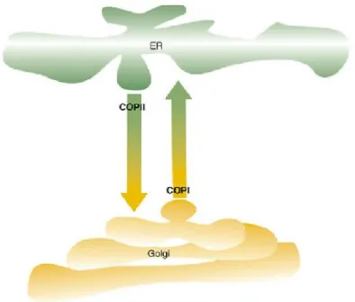
Protein transport between the ER and the Golgi apparatus is thought to be mediated mainly by coated protein complex (COP) vesicles, controlled by small GTPases (Fig 1). Export from the ERES to the Golgi requires the recruitment of COPII vesicles, which have the role of transporting newly synthesised proteins. The anterograde transport of molecules from ER to Golgi must be balanced with a backwards recycling transport. Hence, the retrograde transport, mediated by COPI vesicles, intents to retrieve from the Golgi specific molecules such as proteins that escape to the Golgi but are supposed to be resident within the ER, membrane lipids , or proteins that are actually involved in the anterograde transport but can be reclaimed back to the ER for further reuse (Hanton et al. 2005; Vitale & Ceriotti 2004).
Figure 1 - Schematic representation of the cargo transport between the ER export sites and the Golgi apparatus. The
transport from ERES to Golgi – anterograde transport – is mediated by COPII vesicles, while the backwards recycling transport – retrograde transport – is mediated by COPI vesicles. Adapted from Matheson et al. 2006.
The mechanisms behind the regulation of the COP vesicles recruitment are yet poorly understood. Is has been accepted that soluble proteins exit the ER via a bulk -flow mechanism, according to which the vesicle budding is spontaneous and constant, thus the retrograde transport becomes vital when it comes to retrieving ER resident prote ins (Vitale & Denecke 1999). However, the existence of receptors on the vesicles’ membrane cannot be excluded and an active transport model for anterograde transport is still a valid possibility (Matheson et al. 2006). The retrograde transport of soluble proteins, on the other hand, has been shown to be controlled by specif ic signals, such
as the carboxy-terminal H/KDEL motif. The latter interacts with the cis-Golgi, triggering the formation of COPI vesicles and the retrieval of ER resident proteins (Hanton et al. 2006).
The early plant secretory pathway has shown to be highly dynamic and so, the motility of the ER and Golgi stacks has originated a series of hypothesis tryin g to explain how exactly the protein transport occurs between the two organelles. The first hypothesis to be postulated was the “vacuum cleaner” model, according to which ERES are randomly distributed along the surface of the ER and the Golgi stacks sweep over the ER membrane, constantly capturing export vesicles (Fig 2, A). The stop-and-go model, on the other hand, proposes the existence of fixed ERES on the ER membrane and thus, by a yet unknown signal by ERES, the Golgi bodies would become temporarily detached from the actin filaments and halt their movement while the vesicles are transferred (Fig 2, B). More recently, a third model was hypothesised, the “mobile ERES”, where the cargo transport occurs in a continuous manner. The ERES and t he Golgi stacks would be closely linked and therefore move together, although the nature of this connection is not yet widely understood (Fig 2, C) (Hanton et al. 2005; Hanton et al. 2006). Furthermore, the ER-Golgi cargo transport is known not to require energy nor the intervention of the cytoskeleton (Brandizzi & Snapp 2002).
Figure 2 - Schematic models of ER-Golgi protein transport. A – “Vacuum cleaner” model. B – “Stop-and-go” model. C –
1.1.3. The Golgi apparatus
The Golgi apparatus holds a pivotal position among the organelles of the endomembrane system, for its importance in the assembly, processing and sorting of proteins going through the secretory pathway. It is involved in the synthesis of glycoproteins, glycolipids as well as cell wall matrix polysaccharides. The Golgi is commonly described as a tubulo-saccular structure with cisternal morphology, divided into individual and functionally independent stacks, which are able to travel through the cytoplasm along actin filaments. Due to the functional division into cis-face, medium-face and trans-face, each stack is considered a polarised structure, where the cis- generally corresponds to the entry face and the trans- to the maturing/exit face. Moreover, the Golgi enzymes follow a gradient distribution along the stack, changing activity gradually from cis- to trans-face to cope with metabolic events in a progressive manner (Hawes & Satiat-Jeunemaitre 2005; Nebenführ & Staehelin 2001).
The Trans-Golgi Network (TGN) is associated with the trans-face of Golgi stacks. The exact nature of this structure in plants is still a blur and controversy is yet a constant regarding its actual existence in plant cells. Despite the debate, the TGN seems to play a role in the packaging and targeting of cargo molecules leaving the Golgi apparatus, possibly working as an early-endosome (Kang et al. 2011; Park & Jürgens 2011).
1.1.4. Post-GA transport
The Golgi apparatus operates as a sorting station for cargo molecules travelling through the endomembrane system, from which trafficking routes diverge into different directions, including the vacuoles or the plasma membrane. The sorting of cargo molecules throughout the trafficking routes is dependent on carrier vesicles and the prevention of vesicle mislocalisation requires the recruitment of several proteins and factors, such as small GTPases and SNARE (soluble N-ethylmaleimide sensitive factor adaptor protein receptor) complexes (Jurgens 2004; Cai et al. 2007). A number of post-Golgi organelles have been identified as intermediate stations, comprising the trans-golgi network/early endosome, pre-vacuolar compartment/multivesicular body/late endosome, two types of vacuoles and the cell plate, even though the latter is a temporary organelle (Tse et al. 2004; Richter et al. 2009).
Protein Trafficking to the Vacuoles
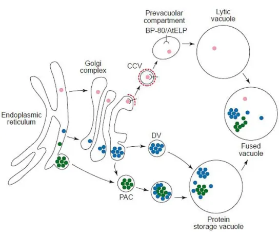
Vacuoles are true hallmarks of the plant cell, valuable in terms of versatility and functional diversity. The vacuoles are involved in several processes within the cell, such as protein degradation, maintenance of turgor pressure, accumulation of ions and secondary metabolites, programmed cell death and intracellular digestion of disposable components. Currently, two types of vacuoles are known to exist: the lytic vacuole (LV) and the protein storage vacuole (PSV). The latter is usually found in seeds or protein storage specific tissues for its ability to accumulate reserve material, while the LV is common in vegetative tissues. Due to its acidic and hydrolytic nature, the LV has a role in protein degradation and is frequently compared to the animal lysosome (Marty 1999). The outer membrane of each type of vacuole, named the tonoplast, is characterised by the presence of specific aquaporins, the Tonoplast Intrinsic Proteins (TIP). Thus, the presence of α-TIP indicates a protein storage vacuole, whereas the presence of γ-TIP indicates a lytic vacuole (Martinoia et al. 2007). It was recently shown that the two types of vacuoles may co-exist in the same cell. In mature cells, the LV and the PSV presumably merge when an isolated PSV is no longer needed, originating a large unique vacuole (Vitale & Raikhel 1999). The presence of the two types of vacuole in the same cell implies the existence of distinct sorting mechanisms to allow protein trafficking to each vacuole (Paris et al. 1996). Protein sorting to the vacuoles relies on specific vacuolar sorting determinants (VSD), which usually consist of N-terminal sequence specific VSD (ssVSD) or C-terminal VSD (ctVSD) cleavable signals. The ssVSD are known to be involved in the sorting to the lytic vacuole, whereas the ctVSD are generally found to lead proteins to the PSV. A third group of VSD is the physical structure VSD (psVSD), which comprises an internal region of the protein that is often cleaved during protein maturation. The psVSD are commonly found in storage proteins (Neuhaus & Rogers 1998).
The ssVSD is typically an NPIR motif or similar, which is recognised in the Golgi by BP80, a Golgi resident vacuolar sorting receptor (VSR). The BP80 receptor is involved in the recruitment of clathrin coated vesicles (CCV), in which the cargo molecules are transported from the Golgi to the PVC/MVB. The receptor is released from the cargo as soon as the vesicles reach the outer membrane of the PVC/MVB and is promptly recycled back to the Golgi apparatus. The delivery of the cargo proteins to the lytic vacuole is presumably accomplished by fusion of the PVC/MVB with the tonoplast (Richter et al. 2009).
When in route to the PSV, proteins are accumulated into dense vesicles (DV) in the Golgi apparatus. From the Golgi, DVs deliver their cargo directly into the PSV, fusing their membrane with the tonoplast. However, recent evidence suggests that CCV might also be involved in the sorting of storage proteins to the PSV, after interaction with BP80 receptor in
the Golgi apparatus (Hawes & Satiat-Jeunemaitre 2005). Another theory is that storage proteins might be transported to the PSV by means of precursor-accumulating vesicles (PAC), which can accumulate cargo directly from the ER or glycosylated cargo from the Golgi apparatus. Similarly to CCVs, PAC vesicles deliver their cargo by direct fusion with the PSV tonoplast. Moreover, it is currently uncertain if any post-Golgi compartment is involved in the sorting to the PSV, as the PVC/MVB or a similar compartment are suggested as hypothesis (Fig 3) (Jolliffe et al. 2005).
Figure 3 - Schematic representation of soluble proteins transport to the vacuoles. The route to the lytic vacuole is
represented by the pink dots. The soluble proteins in route to the lytic vacuole hold an ssVSD, which is recognised by the BP80 vacuolar sorting receptor in the Golgi apparatus. The BP80 receptor is involved in the recruitment of clathrin coated vesicles (CCV; clathrin coat is represented by the broken red line), which transport the cargo proteins to the prevacuolar compartment. The cargo is delivered to the lytic vacuole by fusion of the vesicles with the tonoplast, while the receptor is recycled back to the Golgi apparatus. Proteins in route to the protein storage vacuole hold a ctVSD and might be transported through different pathways, comprising the recruitment of dense vesicles (DV) at the Golgi apparatus, which deliver the protein cargo directly into the PSV, or the recruitment of precursor-accumulating vesicles (PAC), which can accumulate cargo directly from the ER to the PSV or glycosylated cargo from the Golgi to the PSV. In certain conditions, the lytic and protein storage vacuoles might merge, originating a large and unique vacuole (fused vacuole) which performs both lytic and protein storage functions. Adapted from Vitale & Raikhel 1999.
Protein Trafficking to the Cell Wall
The plant cell wall is a unique and dynamic structure with a vital role in cell division and growth, shape maintenance, cell defence and signalling (Szymanski & Cosgrove 2009). The architecture and biogenesis of this structure involves complex processes of biosynthesis and secretion; hence, the structural integrity of the cell wall and consequently the integrity of the plant cell count on the perfect regulation and synchronisation of these processes. Vesicular flow is mainly directed towards the cell wall, due to the need of transporting structural proteins and polysaccharides from the Golgi apparatus, such as wall-modifying enzymes, cellulose synthesis enzymatic complexes, pectins and hemicelluloses. Although the endomembrane system and the cell wall are intrinsically connected, the trafficking pathways of proteins to and from the plant cell wall are yet poorly understood (Rose & Lee 2010; De Caroli et al. 2011; Wightman & Turner 2010). The sorting of soluble proteins to the cell wall has been thought to follow a default route. Apart from the signal peptide needed for ER entrance, no signals or receptors were found to be involved in the regulation of this pathway and currently little is known regarding the intracellular route followed by the secretory vesicles before reaching the PM-CW complex (Denecke et al. 1990; Foresti & Denecke 2008; Rojo & Denecke 2008). In the last few years, the mechanisms that regulate the sorting to the cell wall as well as the routes taken by the cargo vesicles have become an important matter of discussion, as new data and plausible intermediates have been identified. Nonconventional routes to the cell surface have also been identified in eukaryotes, where proteins holding a signal peptide for entering the secretory pathway reach the plasma membrane by a COPII independent manner, presumably bypassing the Golgi (Nickel & Seedorf 2008; Nickel & Rabouille 2009). Routes for protein trafficking to the extracellular matrix involving the endosome have also been proposed. Plant endocytosis is a recently accepted process and so, endocytic compartments are yet to be thoroughly characterised (Müller et al. 2007). Nonetheless, a straight connection between the secretory and endocytic pathway has been suggested. Additionally, the two pathways would have organelles in common, where the routes would converge. The endocytosis process consists on the vesicle mediated uptake of cargo molecules from the extracellular matrix through the plasma membrane. The internalisation of the vesicles is followed by the transport of the cargo to endosomal compartments. The molecules are first delivered to the late endosome, presumably the PVC/MVB, from where they are either directed to a recycling pathway or to the lytic vacuole for degradation. The endocytic pathway, as well as its association with the secretory pathway, is still a matter of debate among the scientific community and many question marks are yet to be solved (Robinson et al. 2008; Samaj et al. 2005).
1.2. Cardosins – Models for Protein Sorting Studies
Cardosins – Aspartic Proteinases
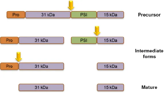
Cardosins are plant aspartic proteinases (AP) that were first isolated from the flowers of cardoon (Cynara cardunculus L.). Cardoon is quite an important plant in Portugal and Spain, where it is mainly distributed, due to the use of the milk clotting activity of APs to manufacture several types of cheese (Cordeiro et al. 1994). Although six more have been posteriorly identified, the first cardosins to be isolated – A and B – are beyond a doubt the better characterised, from molecular to biosynthesis and compartmentalisation levels (Veríssimo et al. 1996; Ramalho-Santos et al. 1998; Vieira et al. 2001; Pissarra et al. 2007). Despite all the research that has been developed during the past decade, the biological roles of these proteins are still unknown. Nonetheless, putative roles have been suggested, such as in the germination and development of seeds, storage protein conversion, flower senescence, defensive mechanisms and reproduction (Figueiredo et al. 2006; Pissarra et al. 2007; Pereira et al. 2008). Similarly to most APs, cardosins A and B are synthesised as preproenzymes, holding an N-terminal signal peptide, on which they rely for entering the secretory pathway through the ER, followed by a conserved pro-segment which is supposed to be involved in protein interaction with the active site (Kervinen et al. 1999). Apart from the signal peptide and the pro-segment, cardosins are divided into N-terminal and C-terminal domains, separated by a region of about 100 aminoacids – the Plant Specific Insert (PSI) domain (Ramalho-Santos et al. 1997). The PSI is a saposin-like domain, common to the most part of plant APs, which is thought to be involved in the proper folding and stability of the proteins, to interfere with the permeability of cell membranes and to be part of defence and signalling mechanisms (Mazorra-Manzano et al. 2010). During protein maturation along the secretory pathway, cardosins are processed through a series of events that lead to successive cleavages of the regions comprising the precursor (Fig 4). For cardosin A, the first step is the separation of the N-terminal domain from the PSI domain, followed by a total separation of the PSI. The pro-segment is the last region to be cleaved. For cardosin B, the process has not been thoroughly clarified, though its processing is thought to occur in the same way as for cardosin A (Ramalho-Santos et al. 1998; Vieira et al. 2001).
Figure 4 - Schematic representation of the proposed sequence of events during the processing of cardosin A. The first
cleavage separates the N-terminal region from the PSI domain, followed by the complete excision of the PSI and finally the Pro-segment is removed, originating the mature form.
Cardosins and Protein Sorting
Despite their high similarity, cardosins A and B are directed to different locations in the floral tissues of C. cardunculus. While cardosin A is an intracellular protein, mainly found in the vacuoles of the stigmatic papillae, cardosin B is found in the extracellular matrix of both stigma and style transmitting tissue (Duarte et al. 2008; Ramalho-Santos et al. 1997; Vieira et al. 2001). As cardoon is difficult to transform, heterologous systems such as Nicotiana tabacum or Arabidopsis thaliana are commonly used for trafficking studies in cardosins. The localisation pattern of these proteins is slightly modified when transiently expressed in leaf epidermis cells of N. tabacum, as both cardosins were observed accumulating in the lytic vacuole (Duarte et al. 2008; da Costa et al. 2010). Curiously, in N. tabacum mesophyll protoplasts expressing cardosin B, secretion assays performed in our group have shown secretion of the protein. So, the presence/absence of the cell wall might have an influence on the regulation of the sorting of cardosins (da Costa et al. 2010).
Recently in our group, two vacuolar sorting determinants were identified among the domains of cardosins A and B – the C-terminal peptide and the PSI domain. The C-terminal of cardosins was shown to efficiently target the protein to the lytic vacuole when placed at the terminus of the fusion protein. Moreover, similarly to the observed for other APs’ C-terminal peptides, it showed to be sufficient for the correct sorting of the proteins to the vacuole (Pereira et al. 2012, submitted). The C-terminal mediated transport has shown to follow a COPII dependent route from the ER to the Golgi apparatus, transporting the protein
through the classical pathway leading to the vacuole. Unlike the C-terminal, the PSI domain in not a common VSD among APs, although it has shown to successfully direct cardosins A and B to the vacuole. However, the PSIs from cardosin A and B direct the protein to the vacuole using different intracellular routes. While the PSI from cardosin B leads the protein to the vacuole through the Golgi apparatus, the PSI from cardosin A seems to bypass the Golgi, in a COPII independent manner (Pereira et al. 2012, submitted). Moreover, the association of the PSI with biological membranes has been demonstrated in vitro (Egas et al. 2000) and also in the plasma membrane/cell wall complex in cardoon seeds (Pereira et al. 2008). In the early stages of C. cardunculus reproduction, particularly during pollination, cardosin A needs to be transported to the cell wall in order to connect with a pollen receptor (Faro et al. 1999), an association presumably mediated by the PSI domain. Therefore, the involvement of a VSD, particularly the PSI domain, in the regulation of cardosins’ sorting in the presence or in absence of the cell wall is definitely a hypothesis worth exploring.
1.3. Objectives of the Thesis
The protein transport routes to and from the plant cell wall have been drawing more and more attention from the scientific community, as they are essential for the integrity of this structure and consequently for the functional maintenance of the plant cell. Evidence has emerged indicating that the cell wall, as one of the main components of the cell and an interface between the cell and the outside environment, could in fact be involved in the regulation of protein transport events. Recent results in our lab, using N. tabacum mesophyll protoplasts and cardosins B as experimental model, have indicated that the presence/absence of the cell wall might influence the destination of this protein. Hence, the main objective of this thesis was to unveil if the cell wall actually regulates protein sorting, using cardosins A and B as experimental models and N. tabacum mesophyll protoplast during cell wall regeneration. Mesophyll protoplasts isolated from N. tabacum leaves transformed with chimeric constructions of cardosin A and cardosin B fused to mCherry were used in secretion assays for the study of the cell wall regulation on the sorting of cardosins. The analysis was performed through confocal microscopy and biochemical analysis during cell wall regeneration.
Cardosin A and cardosin B are both destined for the vacuole in N. tabacum leaf, directed by two identified VSDs – the C-terminal region and the PSI domain. To pursue a possible connection between the identified VSDs and the sorting of cardosins in absence of the cell wall and during its regeneration was also an objective of this thesis. Chimeric constructions of the C-terminals and PSIs from cardosins A and B fused to mCherry were used for the transformation of N. tabacum leaves, for further protoplast isolation and secretion assays were performed in a similar way as for cardosins A and B. As protoplasts from N. tabacum during cell wall regeneration were used throughout the assays, an optimisation of protoplast culture and cell wall regeneration conditions are essential for the planning of the secretion assays.
Evidences have shown that the PSI from cardosin A is prone to interact with biological membranes, as it was shown in vitro as well as in cardoon seeds. It was also demonstrated that PSI A follows an intracellular route to the vacuole which is different from the usually followed by C-terminal peptides, bypassing the Golgi in a COPII independent manner. Hence, it is also our aim to better understand the maturation process of cardosin A and the role of the PSI domain in the sorting of this protein. A site directed-mutagenesis approach was chosen for this task, so as to impair the cleavage of the PSI domain during protein maturation and further analyse the eventual changes in protein processing and sorting. The planning of this experimental strategy was done in collaboration with the Theoretical Chemistry Group of the Faculty of Chemistry in Porto University.
2.1. Dissecting the role of the plant cell wall in the regulation
of protein trafficking
2.1.1. Germination and maintenance of Nicotiana tabacum SRI cv. Petit
Havana
N. tabacum seeds were allowed to germinate in Petri dishes on top of moistened filter paper. After 10-15 days at 22 ºC with 16 hours photoperiod, the seedlings were transferred individually to plant substrate (SIROPlant) and kept under the same conditions.
2.1.2. Isolation of protoplasts from Nicotiana tabacum leaves
Half of an infiltrated N. tabacum leaf was used for the isolation of protoplasts. The leaf was cut into small pieces and the lower epidermis was carefully removed with a tweezer. The small pieces of leaf were placed in Petri dishes with the lower epidermis facing down, floating in 8 mL of TEX medium [3 mM NH4NO3, 5 mM CaCl2.2H2O, 2.4 mM MES and 0.4 M sucrose, in B5 Gamborg medium (Duchefa)] containing 1% (w/v) cellulase and 0.25% (w/v) macerozyme, and incubated overnight in the dark, at room temperature. Subsequently to the digestion, the protoplasts were gently released from the leaf portions with a plastic pipette and recovered into a new Petri dish through a 100 µm nylon mesh, prior to being transferred to 15 mL centrifuge tubes. The protoplasts suspension was then overlaid with 1 mL of 0.4 M mannitol/W5 medium [W5 solution: 154 mM NaCl, 125 mM CaCl2, 5 mM KCl, 5 mM glucose and 1.4 mM MES, pH 5.6, filter sterilised (0.4 M mannitol/W5 solution, in a proportion of 4:1)] and centrifuged at 100 x g, acc/des 1, for 10 minutes. The living protoplasts, floating beneath the mannitol/W5 layer, were promptly recovered into a new 15 mL centrifuge tube containing mannitol/W5 (2/3 of the final volume, allowing the protoplasts to sink) and the two phases were gently mixed together. After a 5 minute centrifugation at 100 x g, acc/des 1, the supernatant was eliminated and the protoplasts stored at low temperature for further use.
2.1.3. Cell wall regeneration in Nicotiana tabacum protoplasts
The regeneration of the cell wall in N. tabacum leaves is a subject already described in the literature. However, the process of regeneration depends on numerous factors, such as temperature, light, pH, agitation, culture medium and even the health of the actual plants, among others. Thus, optimisation of culture conditions was required, in order to assure cell
viability along the following assays, as well as to define the duration of the assays and the time points for sample collecting.
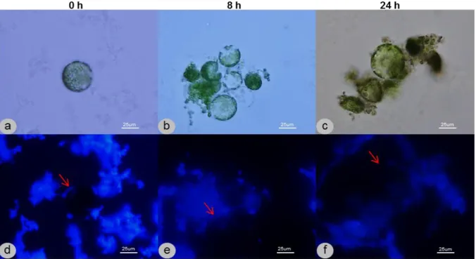
For optimization purposes, protoplasts of non-transformed N. tabacum leaves were isolated as previously described and placed on different culture media [medium A (3 mM NH4NO3, 5 mM CaCl2.2H2O, 2.4 mM MES and 0.4 M sucrose, in B5 Gamborg medium (Duchefa)) or medium B (0.7 M D-mannitol, 0.2 mM KH2PO4, 1 mM KNO3, 1 mM MgSO4, 10 mM CaCl2, 1 µM KI, 0.01 µM CuSO4, 1 µg/mL thiamine HCl, 100 µg/mL myo-inositol and 2 µg/mL glycine, pH 5.8, filter sterilized)]. Other factors were tested, such as the temperature, the presence/absence of light and the presence/absence of agitation. The viability of the cells was monitored by optical microscopy and the regeneration of the cell wall was observed over time by fluorescence microscopy, by means of a fluorescent stain – Calcofluor White ST (0.01%) – which strongly binds to cellulose, allowing the observation of de novo formation of cell wall, coloured white under UV light.
2.1.4. Infiltration of Nicotiana tabacum leaves
A. tumefaciens transformed cultures were inoculated into LB medium with the appropriate antibiotic and grown overnight at 28 ºC with 200 rpm orbital shaking. From the overnight culture, 1 mL was transferred to a 1.5 mL tube and centrifuged at 14000 x g for 1 minute. The cells were then resuspended in 1 mL of infiltration buffer (10 mM MgCl2, 10 mM MES, pH 5.6, filter sterilised) and centrifuged twice more, being however resuspended in infiltration buffer supplemented with 100 µM acetosyringone (4′-Hydroxy-3′,5′-dimethoxyacetophenone 97%, Sigma-Aldrich), in order to increase the virulence of the Agrobacterium. Absorbance measurements at 600 nm with 1/5 dilutions of cell suspensions preceded the infiltration. For each clone, an infiltration mixture was prepared with cell suspension and infiltration buffer with 100 mM acetosyringone, according to the following equation [(ODdesired/ODobserved).1000/5], where the desired OD was 0.1-0.3. The infiltration was performed in N. tabacum leaves, using a 1 mL plastic syringe (without needle) and placing the tip against the abaxial surface. The plunger was pressed gently, while supporting the adaxial surface with the finger, causing the suspension to diffuse through the leaf and into the mesophyllar air spaces. The plants were incubated at the same light/temperature conditions as previously described for germination and maintenance.
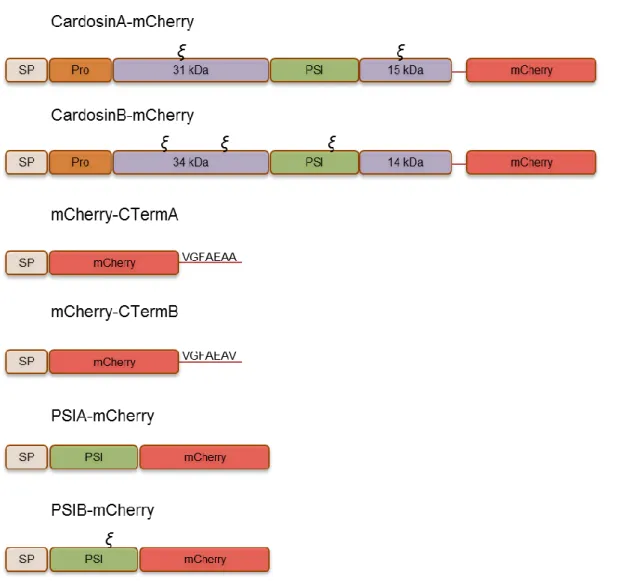
N. tabacum leaves were infiltrated with different cardosin A and cardosin B constructions fused to the fluorescent protein mCherry. The constructions used were already available in the laboratory and inserted into pVKH18En6 binary vector, ideal for transient expression in N. tabacum leaves.
Figure 5 - Schematic representation of the chimeric constructions used in this study. Constructions coding for cardosin A
and B, the PSI from cardosin A and B, and the C-terminal from cardosin A and B, fused to the fluorescent protein mCherry. The ξ symbol represents the glycosylation sites.
Approximately 24 hours after infiltration, to allow the transgene expression, protoplasts were isolated and incubated in 6 mL of medium B, in the dark at 22 ºC. At different time-points (0, 4, 8, 24, 30 hours), 1 mL samples were taken, separating the cells from the culture medium by letting the protoplasts afloat or sink, depending on the culture medium used. Different protein extraction treatments were applied to cells and culture medium fractions. Subsequently to the protein extractions, the samples were analysed by
Western Blotting. At the same sampling time-points, protoplasts and leaves were analysed under Confocal Laser Scanning Microscopy.
2.1.6. Protein extraction from protoplasts
To each sample of cells from the secretion assay, 75 µL of extraction buffer [50 mM sodium citrate pH 5.5, 5% (w/v) SDS, 0.01% (w/v) BSA, 150 mM NaCl, 2% (v/v) β-mercaptoethanol and 10 µL “Protease Inhibitor Cocktail” (Sigma) per 300 mg of fresh tissue] were added. Once mixed with the buffer, the samples were boiled for 10 minutes, prior to a 30 minute centrifugation at 14000 x g. The supernatant was then recovered to a new tube, and proteins quantified on QubitTM Fluorometer (Invitrogen) according to the manufacturer’s instructions and stored at -80 ºC.
2.1.7. Preparation of proteins from protoplasts’ culture medium by
Trichloroacetic acid (TCA) precipitation
To each culture medium sample from the secretion assay, one volume of 20% (w/v) TCA was added. After vortex homogenisation, the samples were incubated on ice for 15 minutes, preceding a 15 minute centrifugation at 14000 x g, in a refrigerated centrifuge (6 ºC). The resulting supernatant was discarded and 1 mL of ice-cold acetone was added, without resuspending the pellet, followed by another 15 minute centrifugation, similar to the previous one. The sediment was once more washed with acetone and air-dried at room temperature. Afterwards, the samples were resuspended in 75 µL of extraction buffer (same buffer as in 3.2.6), boiled for 5 minutes and centrifuged briefly at 14000 x g. The supernatant was then recovered to a new tube, and proteins quantified on QubitTM Fluorometer (Invitrogen) and stored at -80 ºC.
2.1.8. SDS-PAGE and Western blotting
The SDS-PAGE (sodium dodecyl sulphate polyacrylamide gel electrophoresis) is widely used for the separation of proteins according to their electrophoretic mobility, which diverges depending on the length of the polypeptide chain, protein folding and post-translational modifications. Samples containing approximately 15 µg of total protein were prepared with a 1:5 dilution of loading buffer [0,225 M Tris-HCl pH 8, 50% (v/v) Glycerol, 5% (w/v) SDS, 0.05% (w/v) Bromophenol blue and 0.25 M DTT] and by incubation at 65 ºC for 5 minutes. The proteins were then loaded in the gel electrophoresis system (Cleaver Scientific), with a 4% stacking gel [2,1 mL ddH2O, 495 µL acrylamide (30% Acrylamide/Bis Solution, BioRad),
375 µL 0.5 M Tris-HCl pH 6.8, 1.5 µL phenol red, 30 µL 10% (w/v) SDS, 30 µL 10% (w/v) APS (ammonium persulfate, Sigma) and 10 µL TEMED (N,N,N′,N′-Tetramethylethylenediamine, Sigma)] and a 12.5% resolving gel [3.14 mL ddH2O, 4.16 mL acrylamide (30% Acrylamide/Bis Solution, BioRad), 2.5 mL 1,5 M Tris-HCl pH 8.8, 100 µL 10% (w/v) SDS, 100 µL 10% (w/v) APS (ammonium persulfate, Sigma) and 10 µL TEMED (N,N,N′,N′-Tetramethylethylenediamine, Sigma)]. The molecular weight marker used was PageRuler Plus Prestained Protein Ladder (Thermo Scientific). Samples were electrophoresed by the application of an electric current of 100 V across the gel. After electrophoresis, the proteins were transferred onto a nitrocellulose membrane (Protran - Nitrocellulose Transfer Membrane, Whatman) for 1 hour at 100 V, using a Tris-glycine-methanol buffer (25 mM Tris, 192 mM Glycine, 20% (v/v) Methanol). Once the transfer was complete, the membrane was incubated in blocking solution [5% (w/v) milk powder (Molico, Nestlé), 1% BSA (Bovine Serum Albumin, Sigma-Aldrich) and 0.5% (v/v) Tween 20 in TBS-T buffer (50 mM Tris, 200 mM NaCl and 0.1% (v/v) Tween 20)] for 1 hour at room temperature, prior to an overnight incubation at 4 ºC with the primary antibody in blocking solution. For cardosin A, a specific antibody raised against the large subunit (CRAHSMYESSD) was used in a dilution of 1:1000. For cardosin B, an antibody raised against its large subunit was used (CVIHPRYDSGD) in a dilution of 1:1000. For C-terminal and PSI fusions with mCherry, an antibody raised against mCherry (Invitrogen) was used in a dilution of 1:1000. For the burst controls of secretion assays, an antibody raised against calreticulin was used in a dilution of 1:2000 (Denecke et al. 1995). The membrane was then washed three times (10min, 5min, 5min) with TBS-T and incubated with the secondary antibody [1:1000 dilution of anti-rabbit IgG conjugated to Alkaline Phosphatase (Vector)] for 30 minutes, at room temperature. The membranes were washed three times (10min, 5min, 5min) with TBT-T once more and the detection was performed using a chromogenic substrate for Alkaline Phosphatase (NBT/BCIP, Promega).
2.1.9. Fluorescence
Microscopy
and
Confocal
Laser
Scanning
Microscopy
For the monitoring of cell wall regeneration during time points optimization, protoplasts were observed through fluorescence microscopy. Prior to observation, a small portion of cell suspension was placed on a microscope slide and Calcofluor White ST at 0.01% was added. The images were acquired on a fluorescence microscope (Nikon OPTIPHOT-2) using an UV light emission filter.
During the secretion assays, the protoplasts were observed through Confocal Laser Scanning Microscopy. Thus, small portions of cell suspension were placed on microscope
slides for visualisation. On the same time points as the protoplasts’ visualisation, the transformed leaves of origin were also observed. Leaf pieces of approximately 1 cm2 were excised and placed on a glass slide with water, with the abaxial epidermis facing up. The images were acquired on a SP2 Leica Confocal Microscope (Leica Microsystems, Heidelberg) and the excitation wavelength used was 561 nm (for fluorescent protein mCherry).
2.2. Unveiling the role of the PSI domain in the regulation of
cardosin A trafficking
2.2.1. Primers design and site-directed mutagenesis
Polymerase Chain Reaction (PCR) was performed in order to originate point mutations in the cleavage sites of the PSI domain of cardosin A and thus, specific primers were designed for both cleavage sites, to block the PSI removal during protein processing (Table 1). During the design of the primers, the possibility of unwanted modifications in the conformation of the protein had to be minimised but, on the other hand, the aminoacidic modifications need to be drastic enough to meet our purpose. So, every mutation was carefully analysed in silico by molecular modulation methods. Although the crystallographic structure of the precursor form of cardosin A is not yet available, the modulation can be performed by homology. This work was made in collaboration with the Theoretical Chemistry Group from the Faculty of Sciences.
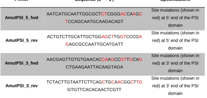
Table 1 - Primers used for the Site-directed mutagenesis of cardosin A.
Primer Sequence (5' → 3') Specifications
AmutPSI_5_fwd AATCATGCAATTGGCGCTCTCGGGACCAAGC
TCCAGCAATGCAAGACAGT
Site mutations (shown in red) at 5' end of the PSI
domain
AmutPSI_5_rev ACTGTCTTGCATTGCTGGAGCTTGGTCCCGA
GAGCGCCAATTGCATGATT
Site mutations (shown in red) at 5' end of the PSI
domain
AmutPSI_3_fwd AACGAGTTGTGTGAACACCAAGCCGTTGCAG
CTGAAGAATTACAAGTAGA
Site mutations (shown in red) at 3' end of the PSI
domain
AmutPSI_3_rev TCTACTTGTAATTCTTCAGCTGCAACGGCTTG
GTGTTCACACAACTCGTT
Site mutations (shown in red) at 3' end of the PSI
domain
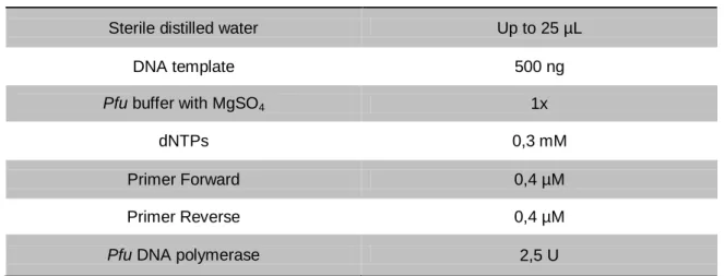
Two different reactions were performed for the mutation of the cleavage sites, as the 5' site was modified, sequenced and confirmed, prior to the mutation of the 3' one. In the first PCR reaction unmodified cardosin A (inserted in pBluescript II SK plasmid) was used as template while in the second reaction, the confirmed product of the first one was used as template DNA. In both reactions, Pfu DNA polymerase (Fermentas) was used, as a way to minimise the incorporation of errors, due to its proofreading activity. The PCR reactions’ composition and conditions were as described in tables 2 and 3, respectively.
Table 2 - PCR reactions’ composition used for the Site-directed mutagenesis of cardosin A.
Reagent Final concentration
Sterile distilled water Up to 25 µL
DNA template 500 ng
Pfu buffer with MgSO4 1x
dNTPs 0,3 mM
Primer Forward 0,4 µM
Primer Reverse 0,4 µM
Pfu DNA polymerase 2,5 U
Table 3 - PCR conditions used for the Site-directed mutagenesis of cardosin A.
Step Conditions Initialization 95 ºC – 5 min 16 cycles Denaturation 95 ºC – 30 sec Annealing 54 ºC – 30 sec Extension 72 ºC – 9 min
Final extension 72 ºC – 15 min
The resulting products of the Site-directed mutagenesis PCR reactions were digested with DpnI for 1 hour at 37 ºC followed by inactivation at 65 ºC for 15 minutes, as a way to eliminate the template DNA. Since DpnI, a frequent cutter, recognises Dam methylated DNA only, all the molecules originated in E. coli were degraded. Afterwards, the mutated DNA was used to transform competent E. coli and the screening was performed through sequencing (T7 and T3 universal primers; Eurofins MWG Operon), after DNA extraction using GenEluteTM Plasmid Miniprep Kit (Sigma-Aldrich), according to the manufacturer’s instructions.
2.2.2. Addition of restriction enzyme adaptors
To facilitate the subsequent cloning steps, restriction enzyme recognition sites had to be added to the AmutPSI construction obtained previously, which was accomplished through PCR reactions. Different combinations of restriction enzyme adaptors were used, in order to create AmutPSI_STP (with the stop codon at the end of the sequence) and AmutPSI_noSTP (without the stop codon at the end of the sequence) (Table 4). The construction AmutPSI_noSTP, when cloned into pVKH18-En6 vector, becomes in frame with mCherry, a fluorescent protein encoded by part of this vector, allowing further analysis through CLSM.
Table 4 - Primers used for the addition of restriction enzyme adaptors to AmutPSI construction.
Primer Sequence (5' → 3') Specifications
cardA 5' xba TCTAGAGCCGCCACCATGGGTACCTCAATCAAA
XbaI recognition site (in
red) added to 5' end of AmutPSI_STP and
AmutPSI_noSTP
ARnSTP_sal GTCGACGCTGCTTCTGCAAATCCAAC
SalI recognition site (in
red) added to 3' end of AmutPSI_noSTP
R1515_sac CTGAGCTCTCAAGCTGCTTCTGCAAAT
SacI recognition site (in red) added to 3' end of
AmutPSI_STP
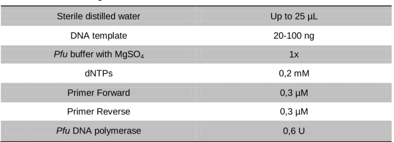
In these PCR reactions, Pfu DNA polymerase was used as previously described. AmutPSI was used as template DNA and the components and conditions were as follows in tables 5 and 6, respectively.
Table 5 - PCR reactions’ composition used for the addition of restriction enzyme adaptors to AmutPSI construction.
Reagent Final concentration
Sterile distilled water Up to 25 µL
DNA template 20-100 ng
Pfu buffer with MgSO4 1x
dNTPs 0,2 mM
Primer Forward 0,3 µM
Primer Reverse 0,3 µM
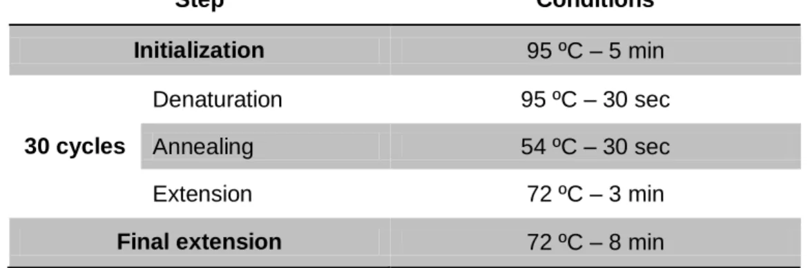
Table 6 - PCR conditions used for the addition of restriction enzyme adaptors to AmutPSI construction. Step Conditions Initialization 95 ºC – 5 min 30 cycles Denaturation 95 ºC – 30 sec Annealing 54 ºC – 30 sec Extension 72 ºC – 3 min
Final extension 72 ºC – 8 min
The products obtained were subjected to horizontal gel electrophoresis, in a 0.8-1% agarose gel prepared in 1x TAE buffer (40 mM Tris, 10% (v/v) acetic acid and 10 mM EDTA), containing 0.5 µg/mL Ethidium Bromide. The separation was held at 200 V in 0.25x TAE buffer and GeneRuler DNA Ladder Mix (Fermentas) was used as molecular marker. The DNA fragments were analysed under UV light. The DNA of interest was extracted and purified from the agarose gel using GenEluteTM Gel Extraction Kit (Sigma-Aldrich), according to the manufacturer’s instructions.
2.2.3. Cloning into pCR-Blunt vector
The blunt DNA fragments (AmutPSI_STP and AmutPSI_noSTP), obtained from the insertion of restriction enzyme adaptors through PCR, were subsequently cloned into pCR-Blunt vector (Zero pCR-Blunt Cloning Kit, Invitrogen), according to the manufacturer’s instructions. The molar ratio between insert and vector was 5:1 and the amount of insert was calculated using the equation bellow. Competent E. coli were then transformed with the product of the ligations.
2.2.4. Plasmid miniprep and enzymatic restriction screening
For screening purposes, plasmid DNA extraction from E. coli was performed by the boiling method. A single colony of transformed E. coli was inoculated in LB medium supplemented with kanamycin (50 µg/mL) and grown overnight at 37 ºC with 180 rpm orbital shaking. From the overnight culture, 1.5 mL were centrifuged 30 seconds at 14000 x g and cells were resuspended in 200 µL STET buffer (8% (w/v) sucrose, 0.1% (w/v) triton-100, 50
mM EDTA and 50 mM tris-HCl, pH 8.0), with 1.5 mg/mL lysozyme. After vortex homogenisation and a 5 minute incubation at room temperature, the cells were boiled for 45 seconds and centrifuged for 5 minutes at 14000 x g. The resulting pellet was removed using a toothpick and 200 µL of isopropanol was added for DNA precipitation. A gentle homogenisation preceded a 10 minute centrifugation at 14000 x g. The sediment was then washed with 200 µL 70% ethanol, air-dried and resuspended in 20 µL of ddH2O supplemented with RNAseA.
Once the DNA was isolated from the E. coli cultures, AmutPSI_STP and AmutPSI_noSTP were screened for putative positives clones by restriction enzyme digestion, using the enzymes correspondent to the previously added adaptors. Thus, for AmutPSI_STP XbaI and SacI were used, and for AmutPSI_noSTP XbaI and SalI were used. The restriction reactions occurred according to the enzymes manufacturers’ instructions and the results were analysed by agarose gel electrophoresis. The selected clones were confirmed by sequencing [M13 uni (-21) and M13 rev (-29) universal primers; Eurofins MWG Operon] prior to further cloning procedures.
2.2.5. Sub-cloning into pVKH18-En6
Once the sequences of AmutPSI_STP and AmutPSI_noSTP in pCR Blunt vector were confirmed, the DNA fragments were excised, using the restriction enzyme adaptors, and cloned into pVKH18-En6 (expression binary vector). The constructions were used to transform competent E. coli and, after DNA extraction, screening for positive clones by restriction analysis was performed, as previously described. One clone of each construction was used for the transformation of competent A. tumefaciens. Prior to N. tabacum leaf infiltration, the clones were screened once more by restriction analysis and only selected ones were further used.
2.2.6. Transient expression in Nicotiana tabacum
In an attempt to evaluate the expression and processing of AmutPSI_STP and AmutPSI_noSTP, N. tabacum leaves were infiltrated with A. tumefaciens cultures, as previously described. For AmutPSI_noSTP, N. tabacum leaves were similarly infiltrated and, at regular time points (3 and 6 days), leaf samples were analysed through CLSM. Concerning AmutPSI_noSTP, the analysis could not be performed in due time.