(Annals of the Brazilian Academy of Sciences)
Printed version ISSN 0001-3765 / Online version ISSN 1678-2690 www.scielo.br/aabc
Caspase-3 activation and increased procollagen type I in irradiated hearts
SAMARA C. FERREIRA-MACHADO1,2,3, CAMILA SALATA3, NAZARETH N. ROCHA4, ALEXANDRE F.S. CORRÊA5, SUZANA CÔRTE-REAL5, ANTÔNIO A.F. PEREGRINO3
,
VERA M.A. DE CAMPOS3, CHERLEY B.V. ANDRADE3, MARIO BERNARDO-FILHO6, JANUÁRIO B. CABRAL-NETO2 and CARLOS E. DEALMEIDA3
1Universidade Federal Fluminense, Instituto de Biologia, Departamento de Biologia Geral,
Rua Outeiro de São João Batista, s/n, Campus do Valonguinho, 24020-140 Niterói, RJ, Brasil 2
Universidade Federal do Rio de Janeiro, Instituto de Biofísica Carlos Chagas Filho,
Cidade Universitária, CCS, Laboratório de Radiobiologia Molecular, Ilha do Fundão, 21949-900 Rio de Janeiro, RJ, Brasil 3Universidade do Estado do Rio de Janeiro, Instituto de Biologia, Departamento de Biofísica e Biometria,
Laboratório de Ciências Radiológicas (LCR), Rua São Francisco Xavier, 524, Pavilhão Haroldo Lisboa da Cunha, Sala 136, Maracanã, 20550-900 Rio de Janeiro, RJ, Brasil
4
Universidade Federal Fluminense, Centro de Ciências Médicas, Rua Hernanni de Melo, 101, São Domingos, 24210-130 Niterói, RJ, Brasil
5Instituto Oswaldo Cruz/FIOCRUZ, Av. Brasil, 4365, Manguinhos, 21040-360 Rio de Janeiro, RJ, Brasil
6Universidade do Estado do Rio de Janeiro, Instituto de Biologia Roberto Alcantara Gomes, Laboratório de Radiofarmácia
Experimental, Av. 28 de setembro, 87 fundos, Vila Isabel, 20551-030 Rio de Janeiro, RJ, Brasil
Manuscript received on October 5, 2011; accepted for publication on December 19, 2011
ABSTRACT
The caspase-3-cleaved presence was evaluated in this study in the heart of irradiated rats, during the decline of ventricular function. Female Wistar rats were irradiated with a single dose of radiation (15 Gy) delivered directly to the heart and the molecular, histological and physiological evaluations were performed at thirteen months post-irradiation. The expressions of procollagen type I, TGF-ß1 and caspase-3-cleaved were analyzed using Western blotting. Cardiac structural and functional alterations were investigated by echocardiography and electron microscopy. In the irradiated group, the levels of procollagen type I, TGF-ß1 and caspase-3-cleaved are increased. Significant histological changes (degeneration of heart tissue and collagen deposition) and functional (reduced ejection fraction) were observed. Data suggest that the cardiac function decline after exposure to ionizing radiation is related, in part, to increased collagen and increased caspase-3-cleaved.
Key words: apoptosis, caspase-3, fibrosis, heart, ionizing radiation, TGF-β1.
Correspondence to: Samara Cristina Ferreira-Machado E-mail: [email protected]
INTRODUCTION
Technological advancement in treatment of cancer combined with early detection and diagnosis have improved the survival of patients suffering from
infarction. In clinical treatments part of the heart may be irradiated (Baker et al. 2009). Among other problems a diffuse myocardial fibrosis can be observed. It consists of diffuse proliferation of separating bands of collagen and/or replacing myocytes (Darby et al. 2010). Experimental studies show that this condition results in damage to the myocardial capillaries endothelium, causing a decrease in capillary density and can lead to fibrosis (Fajardo and Stewart 1973). If extensive, myocardial fibrosis may lead to congestive heart failure (Darby et al. 2010). An imbalance between vascular homeostasis and repair pathways, mediated in part by aberrant signaling by TGF-β1, has been implicated in this process (Stewart et al. 2010).
Loss of myocytes is a feature of the cardiomyopathic process that contributes to progressive decline in left ventricular function and congestive heart failure (Whelan et al. 2010). Studies have proposed that myocyte loss in cardiomyopathy can occur by apoptosis (Whelan et al. 2010). Apoptosis or programmed cell death is a highly regulated process, and as a final result of this gene regulation, it can be observed the chromatin condensation, cell shrinkage, nuclear and cellular fragmentation, ending with apoptotic bodies’ formations, which are subsequently phagocytosed (Goldspink et al. 2003).
Apoptosis may be the consequence of prolonged growth stimulation of adult myocytes, which presents a limited capacity of division. Growth stimulation initially occurs as a compensatory effort to meet chronically altered hemodynamic demands on the failing myocardium and is mediated by systemic and/or local up-regulation of mediators of adrenergic or rennin-angiotensin axes (Goldspink et al. 2003, Schlüter and Wenzel 2008, Whelan et al. 2010). Similarly, certain cytokines, like TGF-β1 can induce growth as well as apoptosis (Schröder et al. 2006). The induction of apoptosis as a form of cell death distinct from necrosis is associated with activation of aspartate-specifc cysteine proteases such as caspase-3 (Goldspink et al. 2003).
Cardiomyocyte apoptosis plays an important role in the progression of many cardiovascular disorders including heart failure. However, there is little information about apoptosis involvement in the evolution of radiation induced heart disease (RIHD). Thus, the main aim of this study is to investigate the involvement of caspase-3 activation in RIHD, and the cardiac tissue ultrastructural analysis.
MATERIALS AND METHODS
ANIMALS AND IRRADIATION
Female Wistar rats (n=14) were obtained at three months of age and weighing approximately 250 g. Rats were maintained on a 12-h light/dark cycle with food and water provided ad libitum. Animals were distributed into two groups (7 per group): control group and irradiated group (a single dose of irradiation of 15 Gy). Euthanasia was performed thirteen months post-irradiation. Before irradiation, animals were anesthetized with ketamine/xylazine (0.1 mg/kg) intraperitoneally injected (ip). Single dose of 15 Gy was calculated to be approximately equivalent to total fractionated doses of 30–50 Gy (2 Gy per fraction), according to the LQ model and an a/b ratio of 2:3 as described by Schultz-Hector (Schultz-Hector et al. 1992). Cardiac-specific irradiation was performed using a Varian Clinac 2100 C linear accelerator (Variant Medical Systems, CA, USA) with a 6 MV X-ray, and a dose rate of 240 centi-Gy/min. Individual rats were irradiated in the supine position using an anterior field size of 2x2 cm2 with bolus depth of 0.5 cm. The heart position was marked after CT simulation (HiSpeed CT/ Dual, GE Healthcare, USA). The study protocol was approved by the local ethical council (Universidade do Estado do Rio de Janeiro, UERJ, Brazil).
ECHOCARDIOGRAPHY
Echocardiographic analyses were performed using a two-dimensional echo Doppler cardiogram (Esaote, model CarisPlus, Firenze, Italy) equipped with a 10 MHz transducer. Measures included septum wall thickness, posterior wall thickness, left ventricle (LV) internal diameters and ejection fraction (EF). LV cardiac output was calculated by the following equation: Stroke volume (determined from aortic integral flow velocity and aortic valve area) x heart rate (HR). HR was obtained indirectly in M-Mode by measuring the interval between the systolic LV contraction waves at the level of the papillary muscles in the transverse axis. The parameters cited were obtained in accordance with the American Society of Echocardiography (Lang et al. 2005).
WESTERN BLOTTING
To extract total protein, LV samples were washed in phosphate buffer solution before homogenization in lysis buffer (1 mM NaHCO3 pH 8.0, 1 mM phenylmethanesulfonyl fluoride, 1 mM ethylene glycol tetraacetic acid, 4% nonyl phenoxyl polyethoxy ethanol-40, 1 µg/mL pepstatin, 10 µg/mL leupeptin, 2 µg/mL aprotinin and 3 mM Na3VO4). Lysates were resolved by sodium dodecyl sulfate polyacrylamide gel electrophoresis (8% or 15%) and subsequently transferred to nitrocellulose membranes. All chemicals were purchased from Sigma Aldrich (St Louis, MO, USA). The primary antibodies for proc type I (Santa Cruz Biotechnology, CA, USA), TGF-β1 (Santa Cruz Biotechnology), pro-caspase-3 (Santa Cruz Biotechnology, CA, USA) and α-tubulin (Clone b- 5-1-1 mouse) (Sigma Aldrich) were in dilutions of 1:50, 1:500, 1:500 and 1:5,000, respectively. The α-tubulin was used as a protein loading control. The proteins were detected using the Luminol system (Santa Cruz Biotechnology). Band densitometry was obtained by the ImageJ 1.45 software. Proc type I, TGF-β1 and pro-caspase-3 expression were calculated relative to α-tubulin expression in order to rule out the effects of divergent protein loading.
ELECTRON MICROSCOPY
The left ventricle heart tissue of the animals at 13 months (controls and irradiated with 15Gy) was processed and analyzed qualitatively with the use of transmission electron microscope Zeiss EM10C. The heart muscle fragments were washed in PBS and fixed for 1 hour in a solution containing 2.5% glutaraldehyde in sodium cacodylate buffer 0.1 M, pH 7.2 plus 3.5% sucrose. Then, the samples were washed for 10 min in the same buffer. This washing step was repeated 3 times. The material was post fixed for 1h with a 1% osmium tetroxide (OsO4) solution, in sodium cacodylate buffer 0.1 M, pH 7.2 plus 3.5% sucrose; dehydrated in acetone series (30, 50, 70, 90 and 100%) and embedded in Poly/Bed® 812 resin (Ted Pella, Inc). After polymerization, ultrathin sections were obtained and contrasted with uranyl acetate-lead citrate for ultrastructural observation.
STATISTICAL ANALYSIS
Echocardiogram and Western blotting parameters were compared using the Student’s unpaired t-test. In all analyses, P values < 0.05 were considered statistically significant. All results are expressed as mean ± standard deviation (SD).
RESULTS
ALTERATIONS IN THE HEART FUNCTION OF RATS SUBMITTED
TO IRRADIATION.
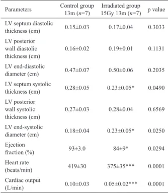
At thirteen months after irradiation, 15 Gy group showed reduced septum wall thickness and increased LV diameter (systole) in relation to control group (Table I). Functional analysis also revealed decrease in the EF, heart rate and cardiac output, featuring a picture of decompensated heart failure.
CARDIAC TGF-β1 AND PROC TYPE I PROTEINS WERE
INCREASED IN RATS SUBMITTED TO IRRADIATION.
irradiated heart tissue. As shown in Figure 1A and 1B, proc type I and TGF-β1 proteins expressions in the 15 Gy group were significantly increased compared to the control group.
PRO- CASPASE-3 LEVELS DECREASE IN RATS SUBMITTED TO
IRRADIATION.
The results demonstrated that the irradiated group showed reduced levels of protein pro-caspase-3 and concomitant increase in protein cleaved-caspase-3 (Figure 2). These results seem to indicate activation of this protein in the progression of heart failure caused by irradiation.
MORPHOLOGICAL ALTERATIONS IN THE CARDIAC TISSUE
AFTER IRRADIATION.
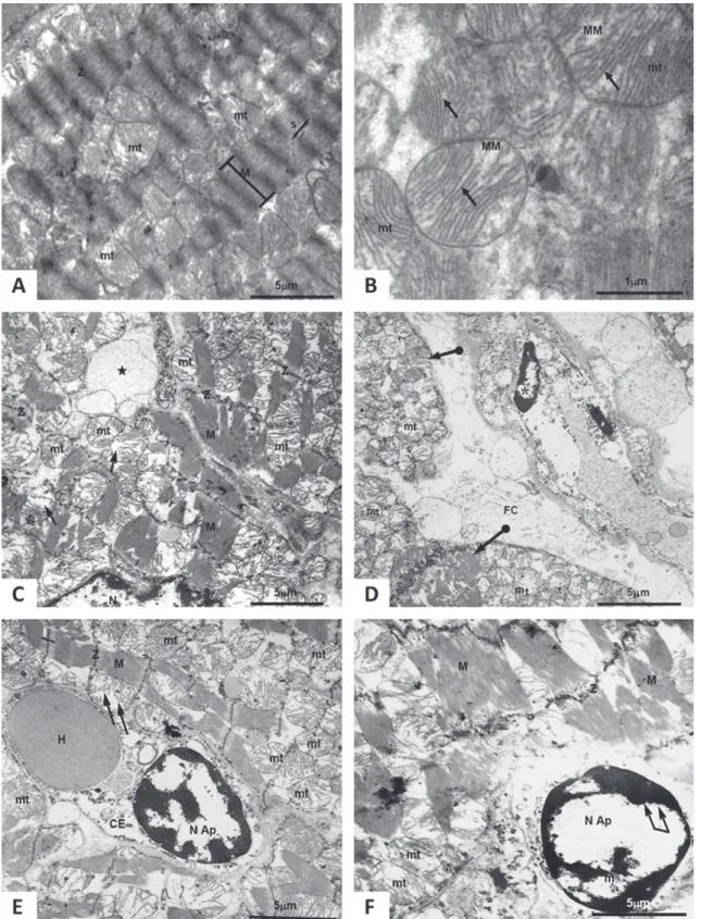
In control group (Figure 3A), it is possible to note the presence of myofibrils parallel to each other
Fig. 1 - Difference in expression of the proc type I and TGF-β1
proteins in the LV.
Panel A, representative Western blotting. of proc type I and TGF-β1. Panel B, densitometric analyses of the proc type I and TGF-β1 blots.
White bars represent control and black bars represent irradiated group. *p < 0.05.
Fig. 2 - Difference in expression of the pro-caspase-3 proteins in the LV.
Panel A, representative Western blotting. of pro-caspase-3. Panel B, densitometric analyses of the pro-caspase-3 blot. White bar represent control and black bar represent irradiated group. *p < 0.05.
Parameters Control group 13m (n=7)
Irradiated group 15Gy 13m (n=7) p value
LV septum diastolic
thickness (cm) 0.15±0.03 0.17±0.04 0.3033
LV posterior wall diastolic thickness (cm)
0.16±0.02 0.19±0.01 0.1131
LV end-diastolic
diameter (cm) 0.47±0.07 0.50±0.06 0.2035
LV septum systolic
thickness (cm) 0.28±0.05 0.23±0.05* 0.0490
LV posterior wall systolic thickness (cm)
0.27±0.03 0.28±0.04 0.6569
LV end-systolic
diameter (cm) 0.18±0.04 0.23±0.05* 0.0250
Ejection
fraction (%) 93±3.0 84±9* 0.0294
Heart rate
(beats/min) 419±30 375±35*** 0.0001
Cardiac output
(L/min) 0.10±0.03 0.05±0.02*** 0.0001
Values are mean ± SD. LV: left ventricle; 13m: 13th month. Each point
represents the mean of seven animals. Statistical significance was
accepted when p < 0.05 (*), using student’s t-test.
TABLE I
Effect of single dose irradiation (15 Gy) on cardiac examinations using echocardiography.
Fig. 3 - Transmission electron microscopy. (A) and (B) Control group; (C), (D), (E) and (F) Irradiated group.
mt: mitochondria, s: sarcomeres, one arrow: cristae, MM: mitochondrial matrix, M: myofibrils, asterisk: vacuoles containing cytoplasmic material, N: nucleus, arrow with end ball: cardiac muscle fibers, FC: collagen fibers, H: erythrocytes, Nap: apoptotic nuclei, CE: endothelial
(cristae mitochondrial) are intact. In the irradiated group, ultra structural changes of heart tissues are evident. Figure 3C demonstrates the existence of the extracellular matrix between the cardiomyocytes. Disorganized and degenerated myocytes are noted (Figure 3D). There was no default in alignment of cardiomyocytes myofibrils, leading to a structural impairment in contractile units. Another feature often observed in the irradiated group myocytes was the loss of mitochondrial integrity. These organelles are presented swollen, with disrupted membranes, highlighting the reduction of mitochondrial cristae and loss of electron-dense matrix. Besides the mentioned alterations, endothelial cells (Figure 3E) and myocytes (Figure 3F) were observed, displaying highly condensed chromatin associated with the nuclear envelope. This characteristic is an indication that they may be in the apoptosis process.
DISCUSSION
Increased interstitial fibrosis is an important event in the RIHD (Darby et al. 2010, Boerma and Hauer-Jensen 2010). This has been observed in patients exposed to radiation in the thoracic region and in heart irradiated animal studies. Tissue fibrosis is the excessive accumulation of collagen and other extracellular matrix (ECM) components following breakdown in the normal balance of ECM synthesis and degradation (Darby et al. 2010, Yarnold and Brotons 2010). Many experimental studies have demonstrated the involvement of TGF-β1 in this process. TGF-β1 is a cytokine involved in (myo) fibroblasts proliferation and activation. Its increased expression is followed by increased collagen deposition in irradiated tissue (Krüse et al. 2001, Boerma et al. 2002, Wynn 2008, Ferreira-Machado et al. 2010). TGF-β1 can also be the primary mediator for apoptosis (Schröder et al. 2006), but this relationship is unclear in the irradiated heart.
Animals or patients, studies have demonstrated increased apoptosis pathway, evidenced particularly by the presence of cleaved-caspase-3, especially in the
following heart diseases: ischemic cardiomyopathy, dilated cardiomyopathy, myocardial infarction, myocarditis, and arrhythmias (Condorelli et al. 1999, Feuerstein and Young 2000, Philipp et al. 2004, Kunapuli et al. 2006, Ferrari et al. 2009). Researches were driven by the intention of identifying the stimuli that induce cardiomyocyte apoptosis. Various stimuli can lead to myocytes apoptosis, and many can coexist in an advanced heart failure (Kang et al. 2000).
In this study, the expression of TGF-β1 and proc type I are concomitantly increased in irradiated group. These events coincide with increased cleavage of caspase-3 in cleaved-caspase-3 and the observation of cardiomyocytes with apoptotic characteristics. These data suggest that apoptosis in irradiated cardiac tissue is followed by collagen increases. These alterations, taken together, appear to contribute to the cardiac function decline, as it could be seen with the ejection fraction, cardiac output and heart rate reduction.
a muscle cell. When this organelle is altered, it may represent an energy deficit in the cell. This way, there may be a decline in myocyte function, consequently provoking a deficit in the heart functioning. In addition, this remodeling present in the mitochondrial cristae might lead to the release of cytochrome C, an important factor in the activation of procaspase in many models of apoptosis (Kang et al. 2000, Heath-Engel and Shore 2006).
The evidences of apoptosis, confirm the molecular assay findings, which the conversion of pro-caspase-3 to cleaved caspase-3 is noted. Although the electron microscopy did not reveal a constant chromatin condensation and nuclear fragmentation (cell death), the cardiomyocyte are showed very deteriorated (Communal et al. 2002). This could indicate that the myocytes are in a preapoptotic stage, while the morphologic nuclear changes do not happen (Narula et al. 1999). Dogan et al. (2010) also identified in rats, positive apoptotic cardiomyocytes in the heart irradiated with a dose of 20 Gy (Dogan et al. 2010), and in our study it was corroborated by western blotting , where it is possible to observe an increase cleaved caspase-3.
In the RIHD, the endothelial injury, appear to be the main event responsible for triggering late damage. Evidence shows that microvascular lesions (endothelial dysfunction) may be responsible for sustaining the chronic nature of the fibrosis induced by irradiation. Because of low blood perfusion, late myocardial degeneration has been observed by some authors and it is associated with progressive increase of fibrosis and cardiomyocytes apoptosis (Wang et al. 2007, Jelonek et al. 2011). That is why authors suggest that cleaved caspase-3 is more related to consequences of heart damages generated after long period of radiation therapy.
Besides the procollagen increase, this study showed an increased level of caspase-3 cleaved. Continued loss of myocytes may lead to myocardial dysfunction. Strategies that reduce the biological signals responsible for myocyte loss and chamber
remodeling should improve clinical outcomes, as decreased systolic function is a powerful predictor of adverse outcome in congestive heart failure.
ACKNOWLEDGMENTS
We thank Alfonso Meléndez, Anna Coimbra, Jorge Oliveira and Alessandra Peres for technical help. The research and authors were supported by grants from Conselho Nacional de Desenvolvimento Científico e Tecnológico (CNPq), Fundação Carlos Chagas Filho de Amparo à Pesquisa do Estado do Rio de Janeiro (FAPERJ), Universidade Federal do Rio de Janeiro (UFRJ), Universidade do Estado do Rio de Janeiro (UERJ) and Laboratório de Ciências Radiológicas (LCR).
RESUMO
Foi avaliada neste estudo a presença de caspase-3-clivada no coração de ratos irradiados durante o declínio da função ventricular. Ratas Wistar foram irradiadas com uma dose única de radiação (15 Gy) diretamente no coração e as análises molecular, histológica e fisiológica foram realizadas treze meses pós a irradiação dos animais. As expressões de procolágeno tipo I, TGF-ß1 e caspase-3-clivada foram analisadas usando Western blotting. Alterações cardíacas estruturais e funcionais foram investigadas por meio de ecocardiografia e microscopia eletrônica. No grupo irradiado, os níveis de procolágeno tipo I, TGF-ß1 e caspase-3-clivada estão aumentados Alterações histológicas importantes (degeneração do tecido cardíaco e deposição de colágeno) e funcionais (fração de ejeção reduzida) foram observadas. Dados sugerem que o declínio da função cardíaca após a exposição à radiação ionizante está relacionado, em parte, com aumento do colágeno e da caspase-3-clivada.
Palavras-chave: apoptose, caspase-3, fibrose, coração, radiação ionizante, TGF-β1.
REFERENCES
BOERMA M, BART CI AND WONDERGEM J. 2002. Effects of ionizing radiation on gene expression in cultured rat heart cells. Int J Radiat Biol 78(3): 219-225.
BOERMA M AND HAUER-JENSEN M. 2010. Potential targets for intervention in radiation-induced heart disease. Curr Drug Targets 11(11): 1405-1412.
COMMUNAL C, SUMANDEA M, DE TOMBE P, NARULA J, SOLARO RJ AND HAIJAR RJ. 2002. Functional consequences of caspase activation in cardiac myocytes. Proc Natl Acad Sci USA 99(9): 6252-6256.
CONDORELLI G, MORISCO C, STASSI G, NOTTE A, FARINA F, SGARAMELLA G, DE RIENZO A, RONCARATI R, TRIMARCO B AND LEMBO G. 1999. Increased cardiomyocyte apoptosis and changes in proapoptotic and antiapoptotic genes bax and bcl-2 during left ventricular adaptations to chronic pressure overload in the rat. Circulation 99(23): 3071-3078.
DARBY SC ET AL. 2010. Radiation-related heart disease: current knowledge and future prospects. Int J Radiat Oncol Biol Phys 76(3): 656-665.
DOGAN I, SEZEN O, SONMEZ B, ZENGIN AY, YENILMEZ E, YULUG E, ABIDIN I AND BAHAT Z. 2010. Myocardial perfu-sion alterations observed months after radiotherapy are rela-ted to the cellular damage. Nuklearmedizin 49(6): 209-215. FAJARDO LF AND STEWART JR. 1973. Pathogenesis of
radia-tion-induced myocardial fibrosis. Lab Invest 29: 244-257.
FERRARI R, CECONI C, CAMPO G, CANGIANO E, CAVAZZA C, SECCHIERO P AND TAVAZZI L. 2009. Mechanisms of remodelling: a question of life (stem cell production) and death (myocyte apoptosis). Circ J 73(11): 1973-1982. FERREIRA-MACHADO SC ET AL. 2010. Up-regulation of
angiotensin-converting enzyme and angiotensin II type 1 receptor in irradiated rats. Int J Radiat Biol 86(10): 880-887. FEUERSTEIN GZ AND YOUNG PR. 2000. Apoptosis in cardiac diseases: stress- and mitogen-activated signaling pathways. Cardiovasc Res 45(3): 560-569.
GIORDANO SH, KUO YF, FREEMAN JL, BUCHLOLZ TA, HORTOBAGYI GN AND GOODWIN JS. 2005. Risk of cardiac death after adjuvant radiotherapy for breast cancer. J Natl Cancer Inst 97(6): 419-424.
GOLDSPINK DF, BURNISTON JG AND TAN LB. 2003. Cardiomyocyte death and the ageing and failing heart. Exp Physiol 88(3): 447-458.
HEATH-ENGEL HM AND SHORE GC. 2006. Mitochondrial membrane dynamics, cristae remodelling and apoptosis. Biochim Biophys Acta 1763(5-6): 549-560.
JELONEK K, WALASZCZYK A, GABRYS D, PIETROWSKA M, KANTHOU C AND WIDLAK P. 2011. Cardiac endothelial cells isolated from mouse heart - a novel model for radiobiology Acta Biochim Pol 58(3): 397-404.
KANG PM, HAUNSTETTER A, AOKI H, USHEVA A AND IZUMO S. 2000. Morphological and molecular charac-terization of adult cardiomyocyte apoptosis during hypoxia and reoxygenation. Circ Res 87(2): 118-125. KRÜSE JJ, ZURCHER C, STROOTMAN EG, BART CI,
SCHLAGWEIN N, LEER JW AND WONDERGEM J. 2001. Structural changes in the auricles of the rat heart after local ionizing irradiation. Radiother Oncol 58(3): 303-311.
KUNAPULI S, ROSANIO S AND SCHWARZ ER. 2006. "How do cardiomyocytes die?" apoptosis and autophagic cell death in cardiac myocytes. J Card Fail 12(5): 381-391. LANG RM ET AL. 2005. Chamber Quantification Writing Group;
American Society of Echocardiography's Guidelines and Standards Committee; European Association of
Echocar-diography. Recommendations for chamber quantification:
a report from the American Society of Echocardiography's Guidelines and Standards Committee and the Chamber
Quantification Writing Group, developed in conjunction
with the European Association of Echocardiography, a branch of the European Society of Cardiology. J Am Soc Echocardiogr 18(12): 1440-1463.
NARULA J ET AL. 1999. Apoptosis in heart failure: release of cytochrome c from mitochondria and activation of caspase-3 in human cardiomyopathy. Proc Natl Acad Sci USA 96(14): 8144-8149.
OFFERSEN B, HøJRIS I AND OVERGAARD M. 2011. Radiation-induced heart morbidity after adjuvant radiotherapy of early breast cancer Is it still an issue? Radiother Oncol 100(2): 157-159.
PHILIPP S, PAGEL I, HOHNEL K, LUTZ J, BUTTGEREIT J, LANGENICKEL T, HAMET P, DIETZ R AND WILLENBROCK R. 2004. Regulation of caspase 3 and Fas in pressure overload-induced left ventricular dysfunction. Eur J Heart Fail 6(7): 845-851.
REAL SC AND DE MEIRELLES MN. 1991. Immunoelectron localization of mitochondrial F1 factor in cryosections of heart muscle cells. J Electron Microsc Tech 18(2): 192-196.
SCHLÜTER KD AND WENZEL S. 2008. Angiotensin II: a hormone involved in and contributing to pro-hypertrophic cardiac networks and target of anti-hypertrophic cross-talks. Pharmacol Ther 119(3): 311-325.
SCHRÖDER D, HEGER J, PIPER HM AND EULER G. 2006.
Angiotensin II stimulates apoptosis via TGF-β1
signalling in ventricular cardiomyocytes of rat. J Mol Med 84: 975-983.
SCHULTZ-HECTOR S, SUND M AND THAMES HD. 1992. Fractionation response and repair kinetics of radiation-induced heart failure in the rat. Radiother Oncol 23(1): 33-40.
STEWART FA, HOVING S AND RUSSEL NS. 2010. Vascular Damage as an Underlying Mechanism of Cardiac and Cerebral Toxicity in Irradiated Cancer Patients. Radiat Res 174: 865-869.
WANG J, BOERMA M, FU Q AND HAUER-JENSEN M. 2007.
Significance of endothelial dysfunction in the pathogenesis
of early and delayed radiation enteropathy. World J Gastroenterol 13(22): 3047-3055.
WHELAN RS, KAPLINSKIY V AND KITSIS RN. 2010. Cell Death in the Pathogenesis of Heart Disease: Mechanisms and
Significance. Annu Rev Physiol 72: 19-44.
WYNN TA. 2008. Cellular and molecular mechanisms of
fibrosis. J Pathol 214(2): 199-210.
YARNOLD J AND BROTONS MC. 2010. Pathogenetic mechanisms