• Nenhum resultado encontrado

J. Braz. Chem. Soc. vol.28 número11

N/A
N/A
Protected

Academic year: 2018

Share "J. Braz. Chem. Soc. vol.28 número11"

Copied!
12
0
0

Texto

(1)

Article

Printed in Brazil - ©2017 Sociedade Brasileira de Química 0103 - 5053 $6.00+0.00

*e-mail: hbnapolitano@gmail.com

The Influence of Methoxy and Ethoxy Groups on Supramolecular Arrangement of

Two Methoxy-chalcones

Jean M. F. Custodio,a Eduardo C. M. Faria,a Lóide O. Sallum,a Vitor S. Duarte,a

Wesley F. Vaz,a,b Gilberto L. B. de Aquino,a Paulo S. Carvalho Jr.a,c and

Hamilton B. Napolitano*,a

aCampus de Ciências Exatas e Tecnológicas, Universidade Estadual de Goiás,

75132-400 Anápolis-GO, Brazil

bInstituto Federal de Educação, Ciência e Tecnologia de Mato Grosso,

78455-000 Lucas do Rio Verde-MT, Brazil

cInstituto de Física de São Carlos, Universidade de São Paulo,

CP 369, 13560-970 São Carlos-SP, Brazil

The structures of two methoxylated chalcones, namely (E

)-1-(4-methoxyphenyl)-3-(3,4,5-trimethoxyphenyl)prop-2-en-1-one and (E)-3-(4-ethoxyphenyl)-1-(4-methoxyphenyl) prop-2-en-1-one, reveal the effect of the inclusion of the methoxyl and ethoxyl substituents of the conformation on methoxy-chalcone. Structural comparative study between two chalcones was done in this work and some effects on geometric parameters, such as planarity and dihedral angles, were described. In addition, intermolecular interactions responsible for crystalline packaging were investigated by Hirshfeld surfaces and the values of those interactions were analysed by comparing experimental and theoretical models. The molecular stability was expressed in terms of softness and hardness, both obtained from frontier molecular orbitals. Finally, there is a good agreement between calculated and experimental infrared spectrum, which allowed the assignment of the normal vibrational modes.

Keywords: methoxy-chalcones, X-ray diffraction, Hirshfeld surfaces, DFT calculations

Introduction

Chalcone molecule is a class of flavonoid intermediates that presents pharmacological importance due to their

presence in many pharmaceutical compounds.1-9 Chemically,

the molecular skeleton is characterized by aromatic rings moieties connected through three-carbon bridge

having a keto carbonyl group and one α,β-unsaturation

(Figure 1).5,10-13

Chalcones and chalcone derivatives are often obtained

from natural or synthetic sources.8,14 By synthesis perspective,

the Claisen-Schmidt condensation of aromatic aldehyde and aromatic acetophenone under either base or acid catalysis is a widely employed method to the synthesis of

chalcones.5,10-13 The versatility of class is evident from its

wide-ranging biological activities; in particular, antiviral, anthelmintic, amoebicidal, antibacterial, antiprotozoal,

antiulcer, cytotoxic, insecticidal, and anticancer.13,15-18 Also,

due to its chemical structure, several chalcones have been reported to exhibit non-linear optical (NLO) properties that

turn these compounds into potential functional materials.19-21

A study with methoxychalcone19 investigated the optical

properties of 3,4-dimethoxy-4’-methoxychalcone and it has shown promise for nonlinear optical applications. On the basis of these features, the investigation on structural and synthetic perspective assumes noteworthy importance for the extending and understanding of the applicability of molecule.

Figure 1. Chemical structures of (E

(2)

In the course of our studies of chalcone derivatives, we

have reported a detailed single crystal analysis for chalcone 2

((E

)-3-(4-ethoxyphenyl)-1-(4-methoxyphenyl)prop-2-en-1-one), and a structural comparison with the chalcone 1

((E)-1-(4-methoxyphenyl)-3-(3,4,5-trimethoxyphenyl)

prop-2-en-1-one) analogue. The supramolecular and crystal packing features of both structures have been characterized by Hirshfeld surfaces. In addition, electronic structure calculation was performed in order to explain differences due to solid and gas phases, to confirm site of interactions and to evaluate their chemical stability.

Experimental

Synthesis and crystallization

It was used 0.3 g (2 mmol) of 4-methoxyacetophenone with 0.39 g (2 mmol) of 3,4,5-trimethoxybenzaldehyde

to obtain chalcone 1 and 0.3 g (2 mmol) of

4-methoxyacetophenone with 0.30 g (2 mmol) of

4-ethoxybenzaldehyde benzaldehydes to obtain chalcone 2.

The substituted acetophenones was dissolved in 3 mL of methanol under stirring on ice bath. Then, 9 mL of a NaOH solution (50% m/v) was added after the substituted benzaldehydes. The resulting solution was stirred at room temperature for 24 h and then poured into ice water and neutralized with HCl solution 50%. The resulting precipitate was filtered, washed with water and purified

by recrystallization. Chalcone 2 crystallized through

slow evaporation of solvent, in which ethyl acetate

(CH3COOCH2CH3) was used. The process occurred at

10th day at a temperature of 25 °C with bottle semi-open.

A yellow prismatic single crystal with dimension of

0.34 × 0.32 × 0.26 mm was selected. Chalcone 1 was also

obtained by slow evaporation of solvent, however using

methanol (CH3OH) as solvent. The crystallization occurred

at 5th day at a temperature of about 2 °C, with open bottle.

For compound 2, a pale yellow prismatic single crystal

measuring 0.59 × 0.515 × 0.445 mm was selected.

Single crystal X-ray analysis

The diffraction data from chalcone 2 were obtained by the diffractometer KappaCCD model with monochromatic

radiation Mo Kα at room temperature. Then, the

software Saint22 was used for cell refinement and data

reduction. The structure was solved by direct methods and anisotropically refined with full-matrix least-squares

on F2 by the refinement program Shelxl-2014.23 All the

hydrogen atoms were placed in calculated positions and refined with fixed individual displacement parameters

[Uiso(H) = 1.2Ueq or 1.5Ueq] according to the riding model

(C–H bond lengths of 0.97 and 0.96 Å, for aromatic and methyl groups, respectively). The Ortep maps from asymmetric unit and the molecular representations were

obtained through the programs Ortep,24 Mercury25 and

Crystal Explorer.26 The possible H-bond were checked by

the Parst27 and Platon28 softwares. The crystallographic

information files of compound 2 were deposited in the

Cambridge Structural Data Base (CCDC)29,30 under the

code CCDC 1529799. Copies of the data can be obtained,

free of charge, via www.ccdc.cam.ac.uk. The compound 1

was previously synthesized and published, under the code

CCDC 841293, by our own researcher group31 and, in

order to get a better comparison, those information are also present in Table 1 and Figure 2. Additionally, Table 2 shows the atomic coordinates and equivalent isotropic

displacement parameters of compounds 1 and 2.

Hirshfeld surface analysis

Hirshfeld surface was used to visualize and interpret the potential intermolecular interactions of the compounds in study. The Hirshfeld surface can be understood as an attempt to define the occupied space by a molecule in a crystal where the electronic density is partitioned into

molecular fragments and a weight function wa(r) is defined

for each atom in a molecule as:

(1)

Table 1. Crystal data and structure refinement for chalcone 1 and

chalcone 2

Parameter C19H20O5 (1) C18H18O3 (2)

Formula weight 327.34 282.32

Temperature / K 298 (2) 298 (2)

Wavelength / Å 0.71073 0.71073

Crystal system, space group, Z monoclinic, P21/c, 4

orthorhombic, Pna21, 4

Unit cell dimensions a = 7.57700 (10) Å b = 16.2530 (5) Å c = 14.0850 (5) Å

α = γ = 90° β = 107.5280 (10)°

a = 6.32500 (10) Å b = 14.7150 (3) Å c = 16.2780 (3) Å α = β = γ = 90°

Volume / Å3 1654.02 (5) 1515.03 (7)

Calculated density / (mg m-3) 1.315 1.238

Absorption coefficient / mm-1 0.095 0.083

F (000) 692 600

Refinement method directed method directed method

Goodness-of-fit on F2 1.018 1.039

(3)

where ρiat(r) are spherically averaged electron densities of

the various atoms. Thus, the electron density of an atomic fragment can be defined as

(2)

where ρmol(r) indicates the molecular electron density. This

graphical tool represents all molecular interactions of a given compound and is important for studying molecular crystal structures because it describes the standards of molecular interactivity and it is possible to estimate the intermolecular contacts that can provide important information of molecular functions. In a Hirshfeld surface

the distance from the nearest atoms outside (de) and

inside (di) the surface and the normalized contact distance

based on these,

(3)

is symmetric in de and di, with rivdw and revdw being the van

der Waals radii of the atoms. In a graphical representation

of dnorm, close intermolecular distances are characterized

by two identically colored regions and this function

Figure 2. Ortep representations with 50% of probability showing

numbering scheme for chalcone 1 and 2.

Table 2. Atomic coordinates (× 10-4) and equivalent isotropic displacement parameters (A2× 103). U

eq is defined as one third of the trace of the orthogonalized

Uij tensor

Chalcone 1 Chalcone 2

Atom x y z Ueq Atom x y z Ueq

O(3) 5650(2) 2234(1) 6370(1) 60(1) O(2) –7040(8) 8757(4) 4602(3) 132(2)

O(5) 1609(2) 2627(1) 8267(1) 59(1) C(19) –977(9) 9053(6) 834(5) 116(2)

O(1) 7126(2) 200(1) 15087(1) 62(1) O(4) 976(4) 8776(2) 8708(2) 79(1)

C(6) 7884(2) 197(1) 12291(1) 44(1) C(13) –29(6) 8793(2) 7977(3) 68(1)

O(2) 9127(2) –301(1) 11051(1) 63(1) C(5) –2527(7) 9010(3) 3336(3) 85(1)

C(1) 8391(2) –492(1) 12898(1) 48(1) C(14) –1962(6) 9262(3) 7972(3) 80(1)

C(10) 5957(2) 1442(1) 8877(1) 45(1) C(3) –3327(7) 8686(2) 1944(3) 75(1)

C(9) 7125(2) 901(1) 9648(1) 47(1) C(17) 2966(6) 8315(4) 8757(3) 86(1)

O(4) 2518(2) 2803(1) 6546(1) 70(1) C(12) 689(6) 8382(3) 7259(3) 77(1)

C(3) 7444(2) 158(1) 14187(1) 48(1) O(1) –2883(6) 8661(3) 1130(2) 96(1)

C(14) 5303(2) 2063(1) 7249(1) 47(1) C(1) –5897(7) 8368(2) 2982(3) 80(1)

C(15) 6421(2) 1569(1) 7999(1) 47(1) C(8) –3432(9) 8693(3) 5115(4) 97(1)

C(11) 4386(2) 1820(1) 8996(1) 48(1) C(11) –513(7) 8438(3) 6558(3) 83(1)

C(12) 3237(2) 2289(1) 8233(1) 48(1) C(6) –4533(7) 8693(3) 3590(3) 80(1)

C(2) 8152(2) –524(1) 13833(1) 50(1) C(2) –5287(7) 8360(2) 2180(3) 79(1)

C(13) 3695(2) 2414(1) 7352(1) 50(1) C(10) –2496(7) 8896(3) 6565(3) 83(1)

C(4) 7008(2) 867(1) 13612(1) 53(1) C(4) –1933(7) 9016(3) 2533(3) 80(1)

C(7) 8074(2) 183(1) 11269(1) 47(1) C(7) –5136(9) 8723(4) 4432(3) 95(1)

C(5) 7205(2) 880(1) 12669(1) 50(1) C(20) 3802(9) 8455(5) 9605(4) 107(2)

C(8) 6902(2) 757(1) 10531(1) 51(1) C(15) –3140(6) 9300(3) 7284(3) 82(1)

C(16) 1014(3) 2440(1) 9115(1) 60(1) C(9) –3889(8) 8921(3) 5853(3) 91(1)

C(19) 7389(3) –528(1) 15679(1) 67(1)

C(17) 2136(3) 3645(1) 6632(2) 70(1)

(4)

highlights the donor and acceptor equally and it is therefore a powerful tool for analyzing intermolecular

interactions.32-34

GC-MS analysis

Gas chromatography coupled with mass spectrometry (GC-MS) was carried out on a Shimadzu QP2010-Plus mass spectrometer, in a non-polar columns (RTX-5 Restek, 30 m × 0.25 mm × 0.25 µm film thickness). The column oven was programmed to start at 80 °C for 5 min and

subsequently increased to 250 °C at a rate of 20 °C min-1

with a final hold of 10 min. The chromatogram of 1

(Figure S1) and 2 (Figure S2) are available in the

Supplementary Information (SI).

Spectroscopic characterization

Infrared spectroscopy (IR), mass spectroscopy and

nuclear magnetic resonance of hydrogen (1H NMR)

(Figures S3 and S4, SI) and carbon (13C NMR) (Figures S5

and S6, SI) were carried out. Infrared spectra were recorded

on a PerkinElmer Frontier in the range 4000-400 cm-1 using

the KBr pellet technique. 1H and 13C NMR spectra were

obtained on a Bruker 500 MHz spectrometer using CDCl3

and MeOD (Aldrich). Chemical shifts assignments were expressed as ppm using tetramethylsilane (TMS) as internal standard. Spectra visualization was performed through the Program ACD LABS 12.0.

Chalcone 1

Yellow powder, m.p. 131.1-132.5 °C; IR (KBr) ν / cm-1

1612 (C=O); MS: m/z (%) 328 [M]+ (100) (C19H20O5);

1H NMR (500 MHz, CDCl

3) d 3.92 (s, 3H, OCH3Ph),

3.93 (s, 3H, OCH3Ph), 3.95 (s, 6H, OCH3Ph), 6.89 (s,

2H, Ph), 7.02 (d, 2H, J 8.85 Hz,PhOCH3), 7.44 (d, 1H,

J 15.56 Hz, CHCO), 7.74 (d, 1H, J 15.56 Hz, CHPh), 8.07

(d, 2H, J 8.85 Hz, PhOCH3); 13C NMR (126 MHz, CDCl3)

d OCH3Ph (66.46, 66.91, 66.91), Ring Ph (103.29, 104.24,

11.89, 151.54, 159.18, 156.32, 165.31, 167.19), olefin (120.62, 145.10) 170.15 (C=O).

Chalcone 2

Yellow powder, m.p. 101.5-103.2 °C; IR (KBr) ν / cm-1

1692 (C=O); MS: m/z (%) 282 [M]+ (100) (C18H18O3);

1H NMR (500 MHz, CDCl

3) d 1.46 (t, 3H, J 7.0 Hz,

CH3CH2OPh), (s, 3H, OCH3Ph), 4.11 (q, 2H, J 7.0 Hz,

CH3CH2OPh), 6.97-6.88 (m, 2H), 7.04-6.96 (m, 2H), 7.45

(d, 1H, J 15.6 Hz), 7.61 (m, 2H), 7.80 (d, 1H, J 15.6 Hz),

8.08-8.01 (m, 2H); 13C NMR (126 MHz, CDCl

3) d 14.73

(CH3CH2OPh), 55.47 (OCH3Ph), 63.64 (Ar), 113.78,

114.87, 119.47, 127.66, 130.09, 130.68, 131.42, 143.88, 160.93, 163.25, 188.79 (C=O).

Computational procedures

For the theoretical calculations it was carried out

the molecular geometries of chalcones 1 and 2 from the

crystallographic information file (CIF) resulting from the data collection from crystalline samples by means of X-ray diffraction. Then this geometry was fully optimized using the density functional theory (DFT) implemented

in the Gaussian 09 package,35 with the Handy and

co-workers’36 long range corrected version of B3LYP

using the Coulomb-attenuating method, CAM-B3LYP as functional and, as basis set, we use the 6-311+g(d)

of Pople and co-workers.37 The wavefunction generated

using CAM-B3LYP/6-311+g(d) was used for molecular electrostatic potential map (MEP) and frontier molecular

orbitals calculations.38,39

Results and Discussion

Solid state studies

Obtained solids showed yellow coloration. Compound 1

had a yield of 84%, melting point range: 131.1-132.5 °C.

Molecular formula: C19H20O5 (328.13 g mol-1) while

compound 2 had a yield of 99%, melting point

range: 101.5-103.2 °C. Molecular formula: C18H18O3

(282.13 g mol-1).

Since the aromatic substituents have a significant influence on the structure and packing of chalcones,

the crystal structure of compounds 1 and 2 have been

investigated. Chalcone 1 is a methoxyl-chalcone with

three methoxy groups attached to C3, C4 and C5 atoms

from ring B. Meanwhile, chalcone 2 presents a similar

structure with only one ethoxyl substituent on the ring B

(Figures 1 and 2). The compound 1 has crystallized in a

monoclinic and space group P21/c, with Z’ = 1, while 2,31

unlike 1, crystallized in a orthorhombic crystal system40

and space group Pna21, also with four molecules per unit

cell measuring a = 6.32500 (10) Å, b = 14.7150 (3) Å,

c = 16.2780 (3) Å and α = β = γ = 90°.

(5)

by atoms of ring A. Furthermore, a difference of 1.88° appears for the angle formed by the C10−C15−C14 atoms of ring B, which is the aromatic ring that presents different substituents. Structurally, the difference that attracts the most attention is the difference in the dihedral

C19−O1−C3−C4, which causes the methoxy group methyl

to be disposed in opposite directions. The experimental

and theoretical parameters of chalcones 1 and 2, obtained

by X-ray diffractometry and DFT analysis are present in Table 3.

An overlap (ring A was used as fragment) of chalcones 1

and 2 showed the angle δ (δ1 for 1 and δ2 for 2) formed

between the aromatic rings of the two molecules (Figure 3).

This angle is 36.39° for 1 and 51.18° for 2, occurring

in opposite directions in each compound, resulting in a difference of about 90° in relation to plans formed by aromatic A.

When the chalcone 2 is analyzed taking as reference

the red plane, it is observed that the molecular planarity deviation arises as expressive form at C8 atom resulting in

a value of 36.39° in the aromatic ring. The chalcone 1 on

the other hand, although having a deviation of molecular planarity in an opposite direction, also starts the rotation

at C8 carbon. Chalcones 1 and 2 also differ by dihedral

angles C19−O1−C3−C4 (ω1), C8−C9−C10−C15 (ω2) and

C14−C13−O4−C17 (ω3). The values of ω1 (174.65° for

1 and −5.26° for 2)and ω3 (−120.16° for 1 and −179.54°

for 2) show that the methyl from methoxy groups and

Table 3. The experimental (X-ray) and theoretical (DFT) geometric parameters of chalcones 1 and 2

Bond distance / Å Bond angle / degree

Chalcone 1 Chalcone 2 Chalcone 1 Chalcone 2

X-ray DFT X-ray DFT X-ray DFT X-ray DFT

C19−O1 1.425 1.413 1.421 1.413 C19−O1−C3 118.07 118.75 119.77 118.75

O1−C3 1.361 1.353 1.355 1.353 O1−C3−C4 115.09 115.93 123.54 124.41

C3−C2 1.388 1.393 1.384 1.397 C4−C3−C2 120.18 119.66 119.86 119.70

C2−C1 1.384 1.385 1.361 1.375 C3−C2−C1 119.18 119.45 121.14 120.04

C1−C6 1.391 1.392 1.397 1.400 C2−C1−C6 121.59 121.64 120.48 121.19

C6−C5 1.394 1.398 1.414 1.390 C1−C6−C5 118.16 118.05 117.38 118.09

C5−C4 1.380 1.397 1.360 1.390 C6−C5−C4 120.98 121.03 122.08 121.44

C4−C3 1.391 1.394 1.390 1.390 C5−C4−C3 119.81 120.13 119.04 119.50

C6−C7 1.489 1.492 1.423 1.492 C1−C6−C7 119.49 117.89 121.82 117.90

C7−02 1.224 1.218 1.237 1.218 C6−C7−O2 120.83 120.00 118.53 119.81

C7−C8 1.478 1.482 1.549 1.481 O2−C7−C8 121.86 120.94 121.20 121.06

C8−C9 1.325 1.336 1.280 1.337 C7−C8−C9 123.59 120.00 120.59 120.02

C9−C10 1.468 1.462 1.456 1.459 C8−C9−C10 125.57 128.03 127.12 128.12

C10−C15 1.399 1.393 1.374 1.401 C9−C10−C15 119.09 117.76 119.17 118.94

C15−C14 1.393 1.392 1.346 1.378 C10−C15−C14 119.88 120.50 121.76 121.63

C14−C13 1.392 1.395 1.404 1.394 C15−C14−C13 120.60 119.92 120.48 120.03

C13−C12 1.401 1.400 1.392 1.394 C14−C13−C12 119.40 119.47 119.52 119.42

C12−C11 1.389 1.387 1.374 1.385 C13−C12−C11 120.02 120.45 119.39 119.73

C11−C10 1.394 1.396 1.424 1.394 C12−C11−C10 120.52 120.01 120.62 121.75

C12−O5 1.364 1.355 − − C13−C12−O5 116.14 115.13 − −

O5−C16 1.432 1.411 − − C12−O5−C16 117.33 118.52 − −

C13−O4 1.369 1.359 1.349 1.353 C14−C13−O4 118.38 119.96 115.12 116.10

O4−C17 1.411 1.421 1.432 1.421 C13−O4−C17 118.42 115.12 118.16 119.20

C14−O3 1.370 1.353 − − C15−C14−O3 124.37 124.37 − −

O3−C18 1.418 1.412 − − C14−O3−C18 117.76 118.46 − −

C17−C20 − − 1.492 1.509 O4−C17−C20 − − 107.31 107.69

Figure 3. Overlapping of 1 and 2, showing the angles formed by aromatic

(6)

methyl/ethyl from aromatic B have a different orientation, due to reversal of the rotation direction of the dihedral,

while ω2 (176.70° for 1 and 169.72° for 2) shows the

difference due to aromatic B rotation.

In the packing of compound 1 there are interactions

of type CH···O and CH···C, while the packing of

compound 2 presents only CH···C interactions. The main

interactions among 1 and 2 and their geometric parameters

are represented in Figure 4 and Table 4.

For chalcone 1, two C9−H9···O2, C16−H16A···O2

and C1−H1···C12 interactions form dimers (dimer 1 and dimer 2), increasing the structural stability of the compound [Figures 4(1a) and 4(1b)].

The C18−H18C···O1 interaction contributes to the increase in direction of c axis, stacking dimers 1, while C16−H16C···O4, C5−H5···O3 and C5−H5···C14 interactions stack dimers 2 along b and c axis. The interaction C17−H17B···C13 induces the position of

chalcone 2 in b axis direction while the C17−H17A···C15

and C17−H17A···C14 increase the packaging in direction to a and c axis (Figure 4(2)).

By analyzing the Figure 5a, it is possible to see that for the packaging of the first chalcone the angles formed between the molecules and the c axis are bigger (39.19°) than the existing angles in the second molecule that has a most planar layer on packing (4.07°).

This difference on packing corroborates the

non-planarity of compound 1 and hence, the greatest number

of intermolecular interactions. It can be explained by the presence of more electronegative group, such as the three methoxy on aromatic ring B. Moreover, the increase on electronegative groups also explains the difference noted

Table 4. Main observed intermolecular interactions for chalcones 1 and 2

D−H···A d(D−H) / Å d(H···A) / Å d(D···A) / Å d(D−H···A) / degree

Chalcone 1

C9−H9···O2i 0.937 2.504 3.417 164.49

C16−H16A···O2ii 0.962 2.555 3.483 162.21

C1−H1···C12ii 0.930 2.800 3.377 121.19

C18−H18C···O1iii 0.943 2.534 3.333 142.61

C5−H5···C14i 0.967 2.805 3.619 142.27

C5−H5···O3i 0.967 2.625 3.582 170.86

C16−H16C···O4i 0.963 2.602 3.290 128.59

Chalcone 2

C17−H17A···C15iv 1.076 2.674 3.730 167.06

C17−H17A···C14iv 1.076 2.835 3.724 139.90

C17−H17B···C13iv 1.045 2.762 3.584 135.54

i = −1 + x, y, z; ii = 1 − x, −y, −z; iii = x, y, −1 + z; iiii = 1 + x, y, z; iv = −½ + x, ½ − y, z.

Figure 4. Total interactions of chalcone 1 [(1a), (1b) and (1c)] and

chalcone 2. There are two independent dimers in the packing of chalcone

1 [(1a) and 2 (1b)].

(7)

in dihedral angle ω2, present near to carbonyl group and

olefinic portion.

The intermolecular interactions of 1 and 2 were

visualized and interpreted using Hirshfeld surface (HS) analysis. Hirshfeld surfaces are a valuable tool for the recognition of intermolecular interactions and were used in this study to promote analysis of the intermolecular

interactions present in 1 and 2. First, it involves distances

between the nucleus of an internal atom to HS indicating

a donor regions of intermolecular contacts (di), and the

distance between an extern nucleus to HS indicating an

acceptor regions of intermolecular contacts (de). Then, this

surface is denominated dnorm due to its normalization as a

function of van der Waals radius.34

Intermolecular interactions of compound 1 are

represented in Figures 6(1a) and 6(1b), followed by

intermolecular interactions of compound 2, in Figures 6(2a)

and 6(2b), where blue color indicates low intensity and red color indicates high intensity of contacts.

In chalcone 1, places indicated by (1r), (2r), (3r),

(7r), (4r), (5r) and (6r) are regions of higher de contacts,

corresponding to acceptor regions of C9−H9···O2, C16− H16A···O2, C1−H1···C12, C16−H16C···O4, C5−H5···C14, C5−H5···O3 and C18−H18C···O1 interactions, while places indicated by (1d), (2d), (3d), (7d), (4d), (5d) and (6d)

are regions of higher di contacts, corresponding to donor

regions of these same interactions, respectively. In contrast,

for chalcone 2, acceptor regions of C17−H17A···C15 and

C17−H17B···H13 interactions are recognized as red points in (1r) and (2r) in Figure 6(2a), while (1d) and (2d) in Figure 6(2b) represent donor regions of these interactions,

with high intensity of di. Note that the places (1r) and (2r)

in Figure 6(1a) has a stronger donor character than other

places of 1, while the interaction C17−H17A···C15 is

apparently stronger than the interaction C17−H17B···C13 of 2.

Hirshfeld surfaces also are a great tool for the

recognition of hydrophobic interactions (π···π and

C−H···π interactions). The shape index HS expresses

important information to understand the three-dimensional arrangement of the compounds studied here. Figure 6 shows

the π interactions observed in the crystal packing of 1

and 2.

Chalcone 1 and chalcone 2 are both stabilized by

C−H···π interactions (Figure 7). The effect of C−H···π

interactions from compound 1 involving Cg1 (gravity

center of aromatic ring formed by C1, C2, C3, C4, C5 and

C6 atoms) are shown in the Figure 7(1), with interaction

C19−H19C···Cg1, symmetry code 2 − x, −y, 1 − z, (H19C···Cg1 = 2.83 Å, D−H···A = 144.38°) forming a centrosymmetric dimer.

In contrast, chalcone 2 is stabilized by two C−H···π,

both involving Cg1. The Figures 7(2a) and 7(2b) show

interaction C1−H1···Cg1 (symmetry code ½ + x, ½ − y, z,

H1···Cg1 = 2.821 Å, D−H···A = 139.71°) and C15−H15···Cg1

(symmetry code 1 − x, −y, ½ + z, H15···Cg1 = 2.976 Å, D−H···A = 125.54°). Such interactions are indicated by large depressions above aromatic ring and red regions of

concave curvature.41

The distances di and de were combined on a

two-dimensional graph, representing a fingerprint of these functions. This combination of distances functions provides a mapping of all contacts present in the molecule,

making the fingerprints unique for each compound.42 The

fingerprints with respective percentage of each contact were

established for chalcone 1 and 2 (Figure 8).

In the fingerprints, the CH···π interactions are

represented by C···H contacts, while the π···π interactions

are recognized by C···C contacts. Due to the substitution

pattern, 2 has more CH···π interactions than 1. This

Figure 6. Hirshfeld surfaces indicating intermolecular interactions of 1

[(1a) and (1b)] and 2 [(2a) and (2b)].

Figure 7. Representation of C−H···π interactions present in (1),

involving C19−H19C···Cg1 and 2a and 2b, involving C1−H1···Cg1 and

(8)

can be explained by the steric hindrance of three methyl

groups in 1. Both structures do not have hydrophobic

interactions of the type π···π responsible for the crystal

packing. The O···H contact represents non-classic hydrogen, representing the C9−H9···O2, C16−H16A···O2,

C5−H5···O3, C18−H18C···O1 and C16−H16C···O4

interactions in chalcone 1. Both compounds are organic

molecules, therefore, the interactions index H···H is large; such interactions represent in percentage terms almost half of the total interactions, as indicated in Figure 8a.

Theoretical calculations

The root of the mean squared deviation (RMSD) values for internuclear distances and bond angles were

calculated for both molecules. It is noted that 1 has a higher

difference between theoretical and experimental models

(RMSD1 = 0.3569) than 2 (RMSD2 = 0.3045), as can be

seen in Figure 9.

Like the deviation of planarity, this higher value for

RMSD1 is due to stronger intermolecular interactions in 1.

The most variance value for chalcone 1 was observed for the

C7−C8−C9 angle, while C1−C6−C7 is the most variance

angle in chalcone 2. In general, large deviations between

these measurements exist because the X-ray results are in the solid phase, while the geometry was optimized for free molecule in vacuum.

MEP is useful for characterizing properties of chemical and biological systems, emphasizing the charge distribution

of molecules three-dimensionally.43 The MEP surface of

2 and 1 are shown in Figure 10. The MEP presents the

negative red regions and they are concentrated at the oxygen atoms showing the electrophilic sites of both molecules.

Blue regions show positive charge concentration areas, which are concentrated over the hydrogen atoms and methyl groups explaining the nucleophile sites of both molecules. The green region represents the zero potential regions. Therefore, when examining the Figure 10 it can be confirmed the existence of intra and intermolecular interactions of these molecules in solid state. The dipole moment is another parameter that predicts the polarized

nature of the molecule.44 The theoretical calculations show

that compound 2 (6.2954 D) has more than double of the

value of dipole moment of compound 1 (3.0503 D), being

the most polarized between the title compounds. We can see that the electron density on the O4 atom is different in

the compounds, for chalcone 1 the charge in O4 atom is

more negative than the same atom in chalcone 2.

The difference in energy of the highest occupied molecular orbital (HOMO) and lowest unoccupied molecular orbital (LUMO) orbitals is an important index for the chemical stability of molecules, these energies are directly related to the ability to donate and accept electrons. The energy difference between HOMO and LUMO is an important chemical stability index. A small HOMO-LUMO gap automatically means small excitation energies to the manifold of excited states and a large HOMO-LUMO gap implies high stability with respect to chemical reaction. They are also used to describe chemical

softness and hardness.6,45 The distribution and levels of

energy for HOMO and LUMO orbitals for chalcones 1

and 2 were calculated at the theory level

CAM-B3LYP/6-311+G(d) (Figure 11). For chalcone 1 the HOMO orbital

Figure 8. Quantification of different types of contacts (a) and the

fingerprints established for chalcones 1 and 2 (b and c).

Figure 9. Overlapping of theoretical (black) and experimental (gray)

models from chalcone 1 (a) and chalcone 2 (b).

Figure 10. Electrostatic potential maps for 1 and 2. Red color indicates

(9)

is localized entirely on the ring with the trimethoxy group and in the vinyl group, while the LUMO orbital is spread out throughout the molecule, except for two methoxy groups.

For 2 the HOMO and LUMO orbitals are spread out

throughout the molecule, except for methyl groups. The

high gap energy for chalcone 2 (6.4007 against 6.2611 eV

for chalcone 1) indicates that this compound has a slightly

high kinetic stability and low chemical reactivity. The

following formulas46 were used to calculate softness (σ)

and hardness (η), respectively:

(4)

(5)

The softness and hardness were calculated for 2

(σ = 0.3124 eV and η = 3.2003 eV) and for 1 (σ = 0.3194 eV

and η = 3.1305 eV). Thus we conclude the 1 has a higher

capacity to receive electrons while 2 has a higher capacity

to resist charge transference (i.e, resistance to change its electronic configuration).

Assignments

The infrared absorption spectra of chalcones 1 and 2 were

obtained in KBr, ca. 1% solution, on FTIR/IR Affinity-1 Shimadzu spectrophotometer, and the principal absorptions

bands (4000 to 400 cm-1) are represented in Table 5.

Figure 12 shows the theoretical and the experimental

infrared spectra of 1 and 2. As can be seen in Table 5, these

values are in line with each other and between the expected

experimental values, according to the literature.47

Figure 11. Frontier molecular orbitals of chalcones 1 and 2, followed by calculated homo and lumo band-gaps.

Table 5. Vibrational assignments, experimental and calculated wavenumbers in cm-1 of 1 and 2 at CAM-B3LYP/6-311+g(d)

IR assignments Unscaled IR frequencya / cm-1 Scaled IR frequencya / cm-1 I / (K mmol-1) FTIR / (K mmol-1)

ν(C=O) (1) 1765.20 1691.06 179.32 1612

(2) 1766.86 1692.65 146.59 1656

ν(C=C) (1) 1700.65 1629.22 46.80 1589

(2) 1695.85 1624.62 586.38 1607

νsym(CH3) (1) 3044.19, 3070.54 2916.33, 2941.57 52.61, 24.06 2914, 2948

(2) 3041.38-3047.98 2913.64-2919.96 47.59-59.83 2840-2927

νasym(CH3) (1) 3108.73, 3140.11 2978.16, 3008.22 34.84, 23.42 2967

(2) 3105.43-3115.88 2975.00-2985.01 33.12-28.89 2944-3000

ν(O−CAryl) (1) 1338.89, 1304.81 1282.65, 1250.00 56.05, 350.19 1360, 1305

(2) 1298.90, 1314.90, 1408.16 1244.34, 1259.67, 1349.01 91.72, 391.49, 20.57 1254, 1269, 1332

ν(O−C) (1) 1095.97, 1097.86 1049.93, 1051.74 59.68, 65.78 974-1019

(2) 1071.92-1108.25 1026.89-1061.70 212.72-3.80 996-1029

(10)

In order to make a better comparison between theoretical and experimental results, we apply in the theoretical values

a scaling factor of 0.95848 for the results obtained at

CAM-B3LYP/6-311+g(d) level of theory. This procedure corrects the systematic overestimation of the vibrational frequencies that is characteristic of the DFT methods and, besides, makes easier the assignments of the vibrational modes. In

this study the IR spectra were obtained for 1 and 2 dissolved

in KBr and theoretical measurements were made supposing they were in gas phase, which may explain the difference between results.

Experimentally the carbonyl group strongly absorbs

in the range from 1850 to 1650 cm-1. In chalcones we

must consider the conjugations effects that increase the single bond character of the C=O and C=C bonds in the resonance hybrid and hence lower their force constants, resulting in a lowering of the frequencies absorptions.

Generally, this effect results in a 25 to 45 cm-1 lowering

of the carbonyl frequency.49 These calculated frequencies

are 1691.06 cm-1 for chalcone 2 and 1692.65 cm-1 for

chalcone 1, in experimental IR spectra these values are 1612

and 1656 cm-1, respectively. For chalcones with methoxy

group, just like in both molecules, the C=O stretching

occurs, in average, around 1663 cm-1. The stretching modes

of vinyl group occur at 1660-1600 cm-1 though C=C stretch

appears at lower frequencies, with increased intensity,

when conjugated with carbonyl group.49 The calculated

wavenumbers for this mode are 1629.22 and 1624.62 cm-1

for chalcone 2 and chalcone 1, respectively, and in the

IR spectra these modes appear in 1589 and 1607 cm-1,

respectively. The O−C stretching vibration of the O-CH3

group appears in the wide region of 975 ± 125 cm-1 with

an intensity varying from weak to strong.47,50 The O-CH

3

stretching was calculated at 1049-1051 cm-1 for chalcone 2,

while experimental bands appears in 974-1019 cm-1.

In the range of 1026.89-1061.70 cm-1 for chalcone 1, it

can be observed as weak bands in IR at 996-1029 cm-1.

A methoxy group attached to an aromatic ring gives the

asymmetric stretching in the range 1310-1210 cm-1.50

The ab initio calculations give 1338.89 and 1304.81 cm-1

as methoxy stretching vibrations for chalcone 2, while

chalcone 1 presents these vibrations in 1244.34, 1259.67

and 1349.01 cm-1. IR spectra values for this vibrational

mode appear in 1360 and 1305 cm-1 for chalcone 2 and

1254, 1269 and 1332 cm-1 for chalcone 1. Electronic

effects such as back-donation and induction, mainly caused by the presence of oxygen atom adjacent to methyl group, can shift the position of CH stretching mode. In aromatic methoxy compounds the asymmetric mode are

expected in the regions 2985 ± 20 and 2955 ± 20 cm-1 and

the symmetric mode in the region 2845 ± 15 cm-1.50 For

chalcone 2 the computed wavenumbers of asymmetric

stretching of CH3 group are 2978.16 and 3008.22 cm-1,

while the bands in 2916.33 and 2941.57 cm-1 are assigned

as symmetric stretching mode. In IR spectra these values

are 2967 cm-1 for asymmetric stretching and 2914 and

2948 cm-1 as symmetric stretching mode. For chalcone 1

the computed wavenumbers of asymmetric stretching

of CH3 group are in the range of 2975.00-2985.01 cm-1,

while the bands in the range of 2913.64-2919.96 cm-1 are

assigned as symmetric stretching mode. In the IR spectra,

these values are 2944-3000 cm-1 and 2840-2927 cm-1 for

asymmetric and symmetric stretching modes, respectively.

Conclusions

Different substitution patterns of aromatic B caused significant variations in the crystal structure of the

chalcones studied. Chalcone 1, due to its greater number

of methoxy groups, has an increase in its electronegativity and, hence, has more intermolecular interactions than

chalcone 2. These interactions can be the planarity deviation

(11)

cause, observed by overlapping of both structure. In

contrast, the crystalline state of chalcone 2 is stabilized by

only C−H···C and C−H···π interactions, as can be seen by

the shape index Hirshfeld surfaces.

T h e t h e o r e t i c a l c a l c u l a t i o n s u s i n g CAM-B3LYP/6-311+g(d) systematically over-estimated the IR vibrational spectra and to avoid this we used a scale

factor48 of 0.958 with the objective of better convergence

with the experimental results. The spectroscopic data are consistent with the crystal structure. Moreover, the CAM-B3LYP functional showed to be a good option to obtain the vibrational spectra. The correlation between the experimental and theoretical structural values is very

good, the higher variance value in 1 was observed for the

C7−C8−C9 angle while in 2 the C1−C6−C7 angle was

the higher variance. With respect to the dipole moment,

compound 2 has more than the double of the value of

compound 1 which makes it the most polarized molecule

between the title compounds. Of the analysis of the frontier

molecular orbitals we have that the compound 2 has slightly

higher kinetic stability and lower chemical reactivity

compared to compound 1, furthermore, the softness and

chemical hardness energy, the results shown compound 1

has a higher capacity to receive electrons while compound 2

has a higher capacity to resist a charge transference.

Supplementary Information

Supplementary information (GC-MS chromatogram,

1H NMR and 13C NMR) is available free of charge at

http://jbcs.sbq.org.br as PDF file.

Acknowledgments

The authors are grateful to the Conselho Nacional de Desenvolvimento Científico e Tecnológico (CNPq, grant number 313070/2014-8). Also, the authors are thankful to Fundação de Amparo à Pesquisa do Estado de Goiás for financial support.

References

1. Bitencourt, H. R.; Guilhon, G. M. S. P.; Arruda, M. S. P.; Marinho, A. M. R.; Santos, A. S.; Brasil, D. S. B.; Santos, L. S.; J. Braz. Chem. Soc.2011, 22, 1333.

2. Gaikwad, P.; Priyadarsini, K. I.; Naumov, S.; Rao, B. S. M.; J. Phys. Chem. A2010, 114, 7877.

3. Isa, N. M.; Abdelwahab, S. I.; Mohan, S.; Abdul, A. B.; Sukari, M. A.; Taha, M. M. E.; Syam, S.; Narrima, P.; Cheah, S. C.; Ahmad, S.; Mustafa, M. R.; Braz. J. Med. Biol. Res.2012, 45, 524.

4. Rafiee, E.; Rahimi, F.; J. Chil. Chem. Soc.2013, 3, 2. 5. Sallum, L. O.; Napolitano, H. B.; Carvalho, P. S.; Cidade, A. F.;

de Aquino, G. L. B.; Coutinho, N. D.; Camargo, A. J.; Ellena, J.; de Oliveira, H. C. B.; Silva, V. H. C.; J. Phys. Chem. A2014,

118, 10048.

6. Vaz, W. F.; Custodio, J. M. F.; Silveira, R. G.; Castro, A. N.; Campos, C. E. M.; Anjos, M. M.; Oliveira, G. R.; Valverde, C.; Baseia, B.; Napolitano, H. B.; RSC Adv.2016, 6, 79215. 7. Yerragunta, V.; Kumaraswamy, T.; Suman, D.; Anusha, V.; Patil,

P.; Samhitha, T.; PharmaTutor2013, 1, 54.

8. Ghosh, R.; Das, A.; World J. Pharm. Pharm. Sci.2014, 3, 578. 9. Díaz-Tielas, C.; Graña, E.; Reigosa, M. J.; Sánchez-Moreiras,

A. M.; Planta Daninha2016, 34, 607.

10. Climent, M. J.; Corma, A.; Iborra, S.; Primo, J.; J. Catal.1995,

151, 60.

11. Ritter, M.; Martins, R. M.; Rosa, S. A.; Malavolta, J. L.; Lund, R. G.; Flores, A. F. C.; Pereira, C. M. P.; J. Braz. Chem. Soc.

2015, 26, 1201.

12. Siddiqui, Z. N.; Khan, T.; J. Braz. Chem. Soc.2014, 25, 1002. 13. Silva, W. A.; Andrade, C. K. Z.; Napolitano, H. B.; Vencato, I.;

Lariucci, C.; de Castro, M. M. R. C.; Camargo, A. J.; J. Braz. Chem. Soc.2013, 24, 133.

14. Bukhari, S. N. A.; Jasamai, M.; Jantan, I.; Ahmad, W.; Mini-Rev. Org. Chem.2013, 10, 73.

15. Rando, D. G.; Giarolla, J.; Pasqualoto, K. F. M.; Ferreira, E. I.; Braz. J. Pharm. Sci.2010, 46, 311.

16. Matos, M. J.; Vazquez-Rodriguez, S.; Uriarte, E.; Santana, L.; Expert Opin. Ther. Pat.2015, 25, 351.

17. Zhai, L.; J. Antimicrob. Chemother.1999, 43, 793. 18. Nowakowska, Z.; Eur. J. Med. Chem.2007, 42, 125.

19. Shettigar, V.; Patil, P. S.; Naveen, S.; Dharmaprakash, S. M.; Sridhar, M. A.; Shashidhara Prasad, J.; J. Cryst. Growth2006,

295, 44.

20. D’silva, E. D.; Podagatlapalli, G. K.; Rao, S. V.; Rao, D. N.; Dharmaprakash, S. M.; Cryst. Growth Des.2011, 11, 5362. 21. Prabhu, A. N.; Jayarama, A.; Subrahmanya Bhat, K.;

Upadhyaya, V.; J. Mol. Struct.2013, 1031, 79.

22. SAINT: Area-Detector Integration Software; Siemens Industrial Automation Inc., Madison, 1995.

23. Sheldrick, G. M.; Acta Crystallogr. Sect. C2015, 71, 3. 24. Farrugia, L. J.; J. Appl. Crystallogr.2012, 45, 849.

25. Macrae, C. F.; Edgington, P. R.; McCabe, P.; Pidcock, E.; Shields, G. P.; Taylor, R.; Towler, M.; van de Streek, J.; J. Appl. Crystallogr.2006, 39, 453.

26. Wolff, S. K.; Grimwood, D. J.; McKinnon, J. J.; Turner, M. J.; Jayatilaka, D.; Spackman, M. A.; Crystal Explorer (version 3.1); University of Western Australia, Crawley, Australia, 2012. 27. Nardelli, M.; J. Appl. Crystallogr.1995, 28, 659. 28. Spek, A. L.; Acta Crystallogr. Sect. D2009, 65, 148. 29. Groom, C. R.; Allen, F. H.; Angew. Chem., Int. Ed.2014, 53,

(12)

30. Battle, G. M.; Ferrence, G. M.; Allen, F. H.; J. Appl. Crystallogr.

2010, 43, 1208.

31. Carvalho-Jr, P. S.; Sallum, L. O.; Cidade, A. F.; Aquino, G. L. B.; Napolitano, H. B.; Acta Crystallogr. Sect. E Struct. Reports Online2011, E67, 0.

32. Soudani, S.; Ferretti, V.; Jelsch, C.; Lefebvre, F.; Ben Nasr, C.; Inorg. Chem. Commun.2015, 61, 187.

33. Hirshfeld, F. L.; Isr. J. Chem.1977, 16, 198.

34. McKinnon, J. J.; Spackman, M. A.; Mitchell, A. S.; Acta Crystallogr. Sect. B2004, 60, 627.

35. Frisch, M. J.; Trucks, G. W.; Schlegel, H. B.; Scuseria, G. E.; Robb, M. A.; Cheeseman, J. R.; Scalmani, G.; Barone, V.; Mennucci, B.; Petersson, G. A.; Nakatsuji, H.; Caricato, M.; Li, X.; Hratchian, H. P.; Izmaylov, A. F.; Bloino, J.; Zheng, G.; Sonnenberg, J. L.; Hada, M.; Ehara, M.; Toyota, K.; Fukuda, R.; Hasegawa, J.; Ishida, M.; Nakajima, T.; Honda, Y.; Kitao, O.; Nakai, H.; Vreven, T.; Montgomery Jr., J. A.; Peralta, J. E.; Ogliaro, F.; Bearpark, M.; Heyd, J. J.; Brothers, E.; Kudin, K. N.; Staroverov, V. N.; Kobayashi, R.; Normand, J.; Raghavachari, K.; Rendell, A.; Burant, J. C.; Iyengar, S. S.; Tomasi, J.; Cossi, M.; Rega, N.; Millam, J. M.; Klene, M.; Knox, J. E.; Cross, J. B.; Bakken, V.; Adamo, C.; Jaramillo, J.; Gomperts, R.; Stratmann, R. E.; Yazyev, O.; Austin, A. J.; Cammi, R.; Pomelli, C.; Ochterski, J. W.; Martin, R. L.; Morokuma, K.; Zakrzewski, V. G.; Voth, G. A.; Salvador, P.; Dannenberg, J. J.; Dapprich, S.; Daniels, A. D.; Farkas, Ö.; Foresman, J. B.; Ortiz, J. V; Cioslowski, J.; Fox, D. J.; Gaussian 09, Revision A.02; Gaussian Inc.: Wallingford CT, 2009. 36. Yanai, T.; Tew, D. P.; Handy, N. C. ; Chem. Phys. Lett.2004,

393, 51.

37. Krishnan, R.; Binkley, J. S.; Seeger, R.; Pople, J. A.; 1980, 72, 650.

38. Fukui, K.; Science1982, 218, 747.

39. Foresman, J. B.; Frisch, A.; Exploring Chemistry with Electronic Structure Methods: A Guide to Using Gaussian, 2nd ed.; Gaussian Inc.: Pittsburgh, USA, 1996.

40. Paufler, P.; Acta Crystallogr. Sect. A2004, 60, 641.

41. Spackman, M. A.; Jayatilaka, D.; CrystEngComm2009, 11, 19. 42. Spackman, M. A.; McKinnon, J. J.; CrystEngComm2002, 4,

378.

43. Galabov, B.; Nikolova, V.; Ilieva, S.; Chem. - Eur. J.2013, 19, 5149.

44. Gece, G.; Corros. Rev.2015, 33, 195.

45. Ternavisk, R. R.; Camargo, A. J.; Machado, F. B. C.; Rocco, J. A. F. F.; Aquino, G. L. B.; Silva, V. H. C.; Napolitano, H. B.; J. Mol. Model.2014, 20, 2526.

46. Ramaganthan, B.; Gopiraman, M.; Olasunkanmi, L. O.; Kabanda, M. M.; Yesudass, S.; Bahadur, I.; Adekunle, A. S.; Obot, I. B.; Ebenso, E. E.; RSC Adv.2015, 5, 76675.

47. Raj, A.; Raju, K.; Varghese, H. T.; Granadeiro, C. M.; Nogueira, H. I. S.; Yohannan Panicker, C.; J. Braz. Chem. Soc.2009, 20, 549.

48. Barnes, L.; Schindler, B.; Allouche, A.-R.; Simon, D.; Chambert, S.; Oomens, J.; Compagnon, I.; Phys. Chem. Chem. Phys.2015, 17, 25705.

49. Pavia, D. L.; Lampman, G. M.; Kriz, G. S.; Vyvyan, J. A.; Introduction to Spectroscopy, 5th ed.; Cengage Learning: Ohio, USA, 2015.

50. Panicker, C. Y.; Varghese, H. T.; John, M. A.; Harikumar, B.; Orient. J. Chem.2008, 24, 973.

Submitted: February 1, 2017

Imagem

Figure 1. Chemical structures of (E)-1-(4-methoxy-phen-yl)- (E)-1-(4-methoxy-phen-yl)-3 - ( (E)-1-(4-methoxy-phen-yl)-3 , 4 , 5 - t r i m e t h o x y - p h e n - y l ) p r o p - 2 - e n - 1 - o n e   (1 )   a n d  (E)-3-(4-ethoxyphenyl)-1-(4-methoxyphenyl)
Table 1. Crystal data and structure refinement for chalcone 1 and  chalcone 2
Table 2. Atomic coordinates (× 10 -4 ) and equivalent isotropic displacement parameters (A 2  × 10 3 )
Figure 3. Overlapping of 1 and 2, showing the angles formed by aromatic  A (red) and B (blue) rings.
+6

Referências

Documentos relacionados

In the present study, the thirty screened out phytochemicals were evaluated based upon DFT analysis and the lower band energy gap of molecular orbital energies

method, the highest occupied molecular orbital (HOMO) and lowest unoccupied molecular orbital (LUMO) energies were calculated to characterize global chemical reactivity

After the construction of 2D and 3D QSAR models, we performed theoretical calculations of some molecular properties, such as the maps of molecular orbitals (highest

When a molecule absorbs light in the UV-Vis region, an electron is promoted from the highest-energy occupied molecular orbital (HOMO) to the lowest- energy unoccupied

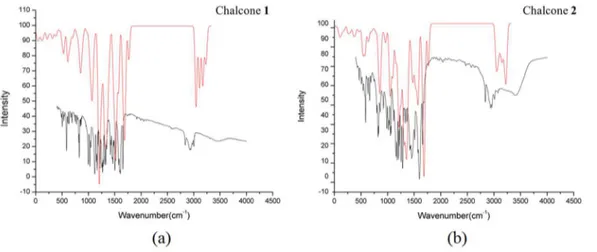
In the Raman spectrum, the intensity of the bands attributed to the hydroxyl units are far more intense than the bands assigned to water stretching vibrations. This is be- cause

The probability of attending school four our group of interest in this region increased by 6.5 percentage points after the expansion of the Bolsa Família program in 2007 and

Using a deformed exponential function and the molecular-orbital theory for the simplest molecular ion, two new analytical functions are proposed to represent the potential energy

Em geral, as propriedades são largamente determinadas pelas energias dos orbitais de fronteira : o orbital molecular ocupado de maior energia ( highest occupied