Gustavo Adolfo Watanabe-Kanno*, Jorge Abrão**
Study of the number of occlusal contacts in
maximum intercuspation before orthodontic
treatment in subjects with Angle Class I and
Class II Division 1 malocclusion
Objective: Define and compare numbers and types of occlusal contacts in maximum intercus-pation. Methods: The study consisted of clinical and photographic analysis of occlusal contacts in maximum intercuspation. Twenty-six Caucasian Brazilian subjects were selected before orth-odontic treatment, 20 males and 6 females, with ages ranging between 12 and 18 years. The subjects were diagnosed and grouped as follows: 13 with Angle Class I malocclusion and 13 with Angle Class II Division 1 malocclusion. After analysis, the occlusal contacts were classified according to the established criteria as: tripodism, bipodism, monopodism (respectively, three, two or one contact point with the slope of the fossa); cuspid to a marginal ridge; cuspid to two marginal ridges; cuspid tip to opposite inclined plane; surface to surface; and edge to edge. Results: The mean number of occlusal contacts per subject in Class I malocclusion was 43.38 and for Class II Division 1 malocclusion it was 44.38, this difference was not statistically signifi-cant (p>0.05). Conclusions: There is a variety of factors that influence the number of occlusal contacts between a Class I and a Class II, Division 1 malocclusion. There is no standardization of occlusal contact type according to the studied malocclusions. A proper selection of occlusal contact types such as cuspid to fossa or cuspid to marginal ridge and its location in the teeth should be individually defined according to the demands of each case. The existence of an ad-equate occlusal contact leads to a correct distribution of forces, promoting periodontal health.
Abstract
Keywords: Dental occlusion. Malocclusion. Orthodontics.
* MSc student in Orthodontics, School of Dentistry, University of São Paulo.
** Associate Professor of the Department of Orthodontics, School of Dentistry, University of São Paulo. How to cite this article: Watanabe-Kanno GA, Abrão J. Study of the number
of occlusal contacts in maximum intercuspation before orthodontic treatment in subjects with Angle Class I and Class II Division 1 malocclusion. Dental Press J Orthod. 2012 Jan-Feb;17(1):138-47.
intROduCtiOn
For a long time the basis for evaluation of success for an orthodontic treatment has been the establishment of a normal mesiodistal rela-tion of posterior teeth, ideal overbite and over-jet evaluated in a static manner. Along time, the orthodontics has assumed that only such fac-tors are not sufficient to achieve a satisfactory functional balance. Therefore, there is a trend among orthodontists to emphasize the im-portance in occlusal functional analysis before orthodontic treatment. The success of orth-odontic treatment is based on an optimal bal-ance between dental and skeletal components, and requires careful evaluation of occlusal con-tacts to have an efficient masticatory function.3 Maximum intercuspation (MI) is the most reproducible reference position. The teeth oc-clude in a position where there is maximum activity of the muscles.21 The MI position is morphologically determined by the shape and position of the teeth, periodontal propriore-ceptors, muscle memory and occlusal contacts. The nerve impulses enable the mandible to open and close, quickly and repeatedly in the same position. Most of the mandibular move-ments are functional (chewing) or parafunc-tional (bruxism), and occur in MI19, hence the importance of studying the occlusal contacts in that mandibular position. Also, it is impor-tant to locate the occlusal contacts in MI for maintaining the alignment of the teeth and the occlusal stability.10
On completion of orthodontic treatment, Andrews six keys of occlusion is one of the main purposes or a Class I malocclusion. Many times orthodontic treatments finish in Class II molar relationship, especially in Class II cases where molar distalizations are not part of the treatment plan. The clinician should be able to recognize and interpret the behavior of occlusal contacts in different malocclusions to obtain stability and function of the stomatognathic system, at the
beginning and most especially during the end of orthodontic treatment in cases that present ther-apeutic limitations. There is a great variety of studies on occlusal contacts in individuals with normal occlusion. Knowing that to understand what is a malocclusion one should understand a normal occlusion, these studies are used as refer-ence to establish means of comparison.
With the purpose of contributing for a bet-ter understanding of this subject, the aim of this study was to define and compare the number and types of occlusal contacts in maximum in-tercuspation in subjects with Angle Class I and Class II Division 1 malocclusions before orth-odontic treatment.
MAteRiAl And MethOds
The study sample consisted of 26 untreated subjects, 20 males and 6 females, Caucasians, with ages between 12 and 18 years, at begin-ning of orthodontic treatment. The patients were diagnosed and grouped into 13 with Angle Class I malocclusion and 13 with Angle Class II Division 1 malocclusion, from the Orthodontic Clinic of the Dental School of São Paulo Uni-versity, following these criteria: Complete per-manent dentition with erupted second molars, no caries lesions, no interproximal wear, no ex-tractions nor previous orthodontic treatment, healthy periodontal status and absence of tem-poromandibular joint dysfunction symptoms. This research project was approved by the Eth-ics Committee on Research of the University of São Paulo, report number 74/07.
A B
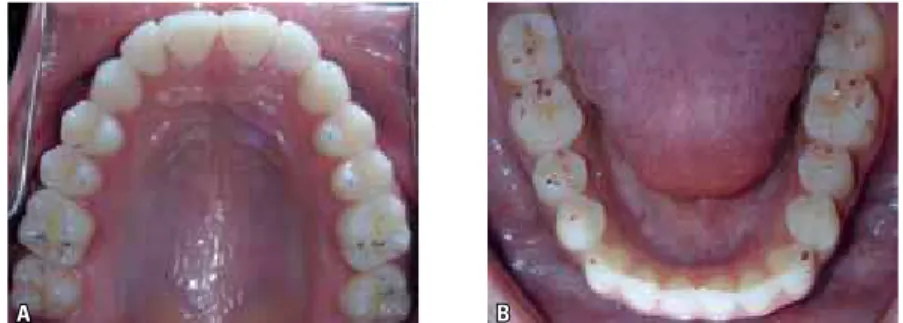
in MI and the occlusal contacts were assessed using 12 µm articulating film (Accu film II, Parkell™, Farmingdale, New York, USA). In this way, when in occlusion, the contacts were marked in black on the upper teeth and the lower teeth were marked in red. Using mouth retractors and an intraoral mirror, both arches were photographed (Fig 1).
Then polyvinyl siloxane-based occlusal reg-istrations (Re´Cord®, Bosworth, Illinois, USA) of the posterior occlusion were obtained
bi-laterally with the subjects in maximum inter-cuspation. Previously the subject was asked to swallow and then to close into maximum intercuspation. The bite registration material was applied to the occlusal surfaces of all lower canines, premolars, and molars both sides with a silicone gun. The subject was required to apply moderate pressure (Fig 2), comparable with the one employed for natural swallowing and chewing, to ensure that teeth were in con-tact, for 30 seconds. The reproducibility of this
FIGURE 1 - Intraoral photograph of MI contacts in the upper arch (A) and in the lower arch (B). FIGURE 2 - Occlusal registration in MI.
TABLE 1 - Types of occlusal contacts definitions.
Types of Occlusal Contacts Deinition
Tripodism Centric retention cusp contacts the perimeter of the slopes of the opponent fossa in three points.3,22
Bipodism Centric retention cusp contacts the perimeter of the slopes of the opponent fossa in two points.4
Monopodism Centric retention cusp contacts the fossa in one individual point.23
Cusp to marginal ridge Contact between the cusp tip and the opposite marginal ridge.23
Cusp to two marginal ridges Two contacts between the cusp tip and two opposite marginal ridges.22
Cusp to opposite inclined plane Individual contact of the cusp tip and the internal slope, external slope, mesial or distal slope on the opposite side.8
Surface to surface Individual contact between two opposing slopes.8
A B
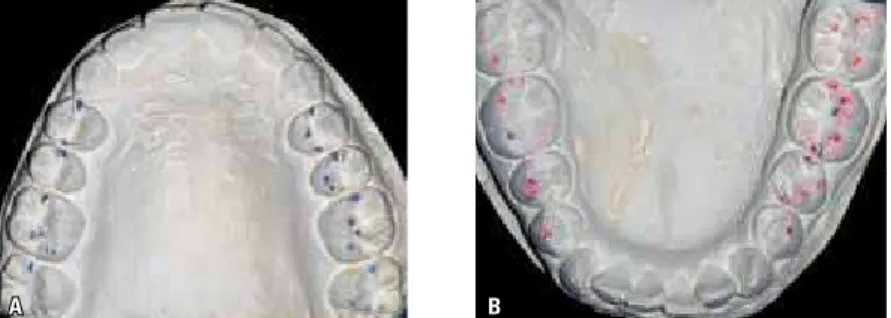
FIGURE 3 - Occlusal contacts in articulated study casts: A) upper and B) lower.
procedure was checked with the polyvinyl si-loxane bite record in maximum intercuspation position; the perforations of the record had to coincide with the occlusal contacts clinically marked with the articulating film. In addition, dental casts were mounted on semi-adjustable articulator (Bio-Art® 4000) in MI. This proce-dure was performed to facilitate the determi-nation of the occlusal contacts types (Table 1), using Arti-Fol 8 µm articulating film (Bausch, Köln, Germany) (Fig 3).
stAtistiCAl AnAlysis
Data was analyzed using the statistical soft-ware SPSS version 16.0 for Windows (SPSS Inc., Chicago, USA) and graphs were con-structed using Microsoft Excel 2007. Sample normality and homogeneity of variances were determined using Kolmogorov-Smirnov Z test. Student t-test was used to determine any sta-tistically significant differences in the observed number and location of occlusal contacts be-tween the different types of malocclusions (level of significance at 5%).
Results
The Kolmogorov-Smirnov test demon-strated that values had a normal distribution (p>0.05), therefore comparisons between mal-occlusion values were performed with para-metric tests based on the sample distribution.
number of occlusal contacts in maximum intercuspation (Mi)
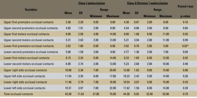
The comparison between the mean occlusal contacts in MI on the upper and lower arches, according to the malocclusion is described in Table 2, and demonstrated that:
» The mean number of total occlusal contacts in Angle Class I malocclusion was 43.38 contacts and in Class II Di-vision 1 it was 44.38. According to the paired t-test there was no statistical difference (p=0.79).
» The mean of occlusal contacts on the right side was 10.20 (range 7 to 22) and on the left side it was 11.5 (range 6 to 17) for Class I malocclusions. For the Angle Class II Division 1 malocclusion the mean of occlusal contacts was 11.8 (range 8 to 15) on the right side and 10.39 (range 5 to 15) on the left side. No significant difference was observed between the two types of malocclusion. » The means of occlusal contacts on lower
premolars in Class I and Class II Divi-sion 1 maloccluDivi-sions were 2.62 and 3.92, respectively. This difference was statistically significant (p=0.03).
distribution of occlusal contacts
Variables
Class I malocclusion Class II Division 1 malocclusion
Paired t test
Mean SD Range Mean SD Range
Minimum Maximum Minimum Maximum p-value
Upper first premolars occlusal contacts 3.38 2.29 0.00 9.00 4.38 0.87 3.00 6.00 0.16
Upper second premolars occlusal contacts 4.85 1.91 2.00 8.00 4.54 0.88 3.00 6.00 0.60
Upper first molars occlusal contacts 8.08 2.56 4.00 14.00 8.00 1.68 6.00 11.00 0.93
Upper second molars occlusal contacts 5.31 2.63 2.00 13.00 5.31 3.04 2.00 11.00 0.99
Lower first premolars occlusal contacts 2.62 1.80 0.00 6.00 3.92 0.76 3.00 5.00 0.03*
Lower second premolars occlusal contacts 5.00 1.58 3.00 9.00 4.77 1.36 2.00 7.00 0.69
Lower first molars occlusal contacts 8.15 2.34 5.00 14.00 8.23 1.69 6.00 12.00 0.92
Lower second molars occlusal contacts 6.00 2.74 2.00 13.00 5.23 2.68 2.00 10.00 0.48
Upper right side occlusal contacts 10.08 3.38 7.00 20.00 12.00 1.83 9.00 15.00 0.08
Upper left side occlusal contacts 11.54 3.26 6.00 17.00 10.23 2.42 5.00 14.00 0.26
Lower right side occlusal contacts 11.46 2.79 7.00 16.00 10.54 3.07 5.00 15.00 0.43
Lower left side occlusal contacts 10.31 3.97 7.00 22.00 11.62 1.56 8.00 14.00 0.28
Total occlusal contacts 43.38 11.63 27.00 75.00 44.38 6.02 32.00 53.00 0.79 TABLE 2 - Comparison between means of occlusal contacts in MI on the upper and lower arches.
TABLE 3 - General distribution of the types of occlusal contacts in absolute numbers. * Level of significance p<0.05.
A = Tripodism, B = Bipodism, C = Monopodism, D = Cusp to one marginal ridge, E = cusp to two marginal ridges, F = cusp tip to opposite slope, G = surface to surface, H = edge to edge.
Class I malocclusion
Types of occlusal contacts A B C D E F G H Total number
Number of occlusal contacts according to type 6 32 36 49 33 27 51 3 237
Total number of occlusal contacts 18 64 36 49 66 27 51 3 314
Class II Division 1 malocclusion
Types of occlusal contacts A B C D E F G H Total number
Number of occlusal contacts according to type 9 27 46 34 31 24 74 1 246
Total number of occlusal contacts 27 54 46 34 62 24 74 1 322
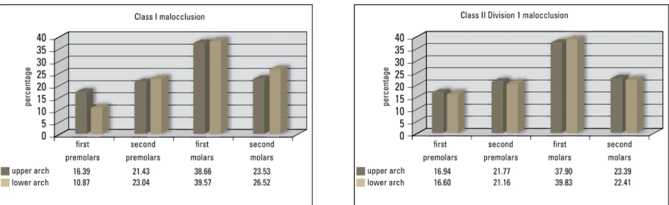
Division 1 a total of 246 contacts (Table 3). The highest concentration of the contacts was for the upper and lower first molars in both the Class I malocclusion and Class II Divi-sion 1, which had, respectively, an average of 39.12% and 38.87% of all contacts recorded (Fig 4). The second molars had an average of 25.03% and 22.9% of total contacts account-ed, respectively. The first lower premolar pre-sented the smallest number of contacts both in Angle Class I and Class II Division 1, reaching
FIGURE 4 - Distribution of occlusal contacts on the upper and lower arches, in percentage, according to the malocclusion. 40 40 first premolars first premolars
upper arch upper arch
Class I malocclusion Class II Division 1 malocclusion
16.39 21.43 38.66 23.53 16.94 21.77 37.90 23.39
10.87 23.04 39.57 26.52 16.60 21.16 39.83 22.41
lower arch lower arch
percentage percentage second premolars second premolars first molars first molars second molars second molars 35 35 30 30 25 25 20 20 15 15 10 10 5 5 0 0
FIGURE 5 - Distribution of occlusal contacts on the cusps, fossas and marginal ridges, in percentage, according to the malocclusion. 80 70 70 60 60 50 50 40 40 30 30 20 20 10 10 0 0 upper arch
Class I malocclusion Class II Division 1 malocclusion
cusps Fossas marginal ridges cusps Fossas marginal ridges
upper arch 45.07 48.34 54.93 51.66 47.83 45.68 52.17 54.32 71.58 66.23 28.42 33.77
lower arch lower arch
percentage percentage
FIGURE 6 - Distribution of types of occlusal contacts, in percentage, according to the malocclusion. 25 25 30 35 20 20 15 15 10 10
A B C D E F G H A B C D E F G H
5 5
0 0
A - Tripodism B - Bipodism C - Monopodism D - Cusp to one marginal ridge
E - cusp to two marginal ridges
Class II Division 1 malocclusion Class I malocclusion
F - cusp tip to opposite slope G - surface to surface H - edge to edge
percentage percentage
2.53 1.27 3.66
13,50 10.98 18.70 13.82 12.60 9.76 30.08 0.41 15.90 20.68 13.92 11.39 21.52
On the other hand, the marginal ridges of the upper teeth had the most contacts in both Class I and Class II Division 1 malocclusions, totaling 71.58% and 66.23%, respectively.
types of occlusal contacts
In Class I malocclusion (Fig 6), from a to-tal of 237 registered contacts, the higher fre-quency of occlusal contact types corresponded
A - Tripodism B - Bipodism C - Monopodism D - Cusp to one marginal ridge
to the surface to surface and a cuspid to mar-ginal ridge in 21.52% and 20.68%, respectively. Thus, the tripodism and edge to edge occlusal contacts registered the smallest percentage of total, with 2.53% and 1.27% respectively. On the other hand, in Angle Class II Division 1, from a total of 246 occlusal contacts, the most frequent contact types corresponded to the surface to surface (30.08%) and monopodism (18.70%). The lower frequency was shown by tripodism (3.66%) and edge to edge (0.41%).
disCussiOn
Comparing the occlusal contacts in maxi-mum intercuspation, between both maloc-clusions, there were no significant differences. There were no significant differences in most of the variables with the exception of the oc-clusal contacts on first mandibular premolars. Especially the Class II Division 1 malocclusion showed a higher average of contacts in that tooth, because of the skeletal and dental distal relationship typical characteristic of this maloc-clusion when compared to Class I malocmaloc-clusion.
These results are also related to the shape and function of the lower first premolar dur-ing mastication. The lower first premolar is the only posterior tooth with a lingual inclination in relation to the occlusal plane, it also has a larger buccal cusp in comparison to the lingual cusp. This shape variation is due to the primary function of the buccal cusp during mastication. This cusp is responsible for perforating food, establishing a primary contact, then the lingual cusp performs the second function which is to grind the food without contacting its antagonist tooth in MI.5,18
In our study, the shape of the lower premo-lar lingual cusp in Class I malocclusions was mostly smaller than in the Class II Division 1 malocclusion. This finding explains why the Class I malocclusion presented lower number of contacts in this tooth.
The total average of occlusal contacts in Class I malocclusion was 43.38 and in Class II Division 1 malocclusion was 44.38. Consider-ing the mean number of contacts per arch, it was observed that patients with Class I maloc-clusion had 21.69 contacts and in Class II Divi-sion 1, 22.19. This agrees with Gondim et al14 that registered an average of 23.20 contacts per arch and Oliveira22 with 20.5 contacts, 22 maxillary contacts and 19 mandible contacts in patients with natural normal occlusions. Atha-nasiou et al6 established an average of 23.8 con-tacts per arch in subjects with normal occlusion using the technique of photo-occlusion. Thus, Ricketts25 also recorded an average of 24 occlu-sal contacts for patients with normal occlusions. However, as for the number of dental occlusal contacts, Velmovitsky29 found in his study an average of 24.89 contacts in all patients—in dis-agreement with Hellman,16 which found 138 possible contacts in a normal occlusion, ranging from 90 to 103 for 28 teeth, and Anderson and Myers2 with 565 occlusal contacts evaluated on 32 subjects with an average of 17.7 contacts.
Considering that this research was per-formed in patients with Class I and Class II Di-vision 1 malocclusions, it can be inferred there-fore that small changes in individual and dental positions seem not to produce severe changes in the quantitative behavior of dental occlusal contacts in subjects with complete dentition, but does produce changes regarding their distri-bution and localization. This fact also explains that no significant differences were found be-tween the mean contact points bebe-tween the studied malocclusions.
an average of 18.15, and Taicher and Ehrlich10 with an average of 39.5, Garrido et al12 with an average of 19.43, and Gondim et al14 with an av-erage of 18.9. This avav-erage was relatively higher in comparison to other studies: Aoki et al4 with an average of 7.14, Gazit and Lieberman13 with an average of 9.30 (normal occlusion) and 7.60 (malocclusion), Korioth17 with an average of 14.0, McDevitt and Warreth19 with an average of 11.5, and Ferrario et al11 with an average of 13.0. Considering that these studies were developed in conditions of normal occlusion and using differ-ent methodologies as indirect determination of the occlusal contacts using interocclusal records, T-Scan or using different types of articulation papers, even so this research has reported results without significant differences when compared with other studies mentioned above.
In this study the occlusal contact types such as monopodism, bipodism and tripodism, in their majority concentrates on the first molars in descending order, both in Angle Class I and Class II Division 1 malocclusions. However, in Class I malocclusion, the monopodism occlusal contact type was located on distobuccal cus-pid and the central fossa of the first mandibu-lar momandibu-lar, and the central fossa and the palatal mesial cuspid of the first maxillary molar. In prosthodontics there is no established occlusal pattern and the occlusal types of contacts could be modified individually.7
Thus, the relationship cuspid-fossa is the most stable because it tends to direct the forces on the long axis of the teeth and with almost no lateral pressures and the relationship cuspid to one marginal ridge tends to separate the contact points and create an unstable occlusion.23 The first molars have greater physiological limit, to withstand an amount of load without damag-ing the periodontal ligament.15 When occlusal forces are applied to the long axis of the tooth it absorbs certain loads without raising the pro-prioceptive sensors. 4
The contact type cuspid to two marginal ridges was located mainly in the second premo-lar, with similar distribution between upper and lower arches. This type of occlusal relationship can also be considered according to the litera-ture as physiological, because it tends to distrib-ute the occlusal load close to the long axis of the teeth, with the disadvantage of promoting in some cases interproximal separation.1
According to the types of occlusal contacts in both malocclusions, Class I and Class II Di-vision 1, the type of occlusal contact surface to surface had the greater frequency. This type of occlusal contact does not promote stabili-zation of the mandible, creating tangential or horizontal forces on the supporting structures of the teeth, maintaining the muscles next to a very high level of activity. The anterior teeth that had occlusal contact occluded on an in-clined plane, considered clinically normal and stable. In anterior and posterior teeth with this type of occlusal contact there should be a balance in physiological forces of the tongue, lips, cheeks and occlusion, to maintain its rela-tive position.2 Posterior teeth are dependent on this same balance of forces for their physi-ological stability position. Through continu-ous eruption or modification in buccal forces, the posterior teeth can slide down on inclined planes to a new position, thus establishing its final occlusal stop. The vertical forces are more easily tolerated because they are directed to the apical region where there is a bone density, for example, cuspid-fossa contacts.27 Lateral forces are more destructive because they are directed against a buccal and lingual alveolar wall, which are fragile and very thin.
COnClusiOns
1. The average number of occlusal contacts per patient in Class I malocclusion was 43.38 and in Class II Division 1 malocclusion was 44.38, and this difference was not statistically significant. There is a variety of factors that in-fluence the number of occlusal contacts, such as small changes in individual tooth positions (ro-tations, infraocclusion, extrusion, linguoversions, buccal, mesial and distal displacements) and the anteroposterior and transverse relationship
between jaws and the occlusal morphology of teeth related to the mastication.
Contact address
Gustavo Adolfo Watanabe-Kanno
Av. Professor Lineu Prestes, 2227 – Cidade Universitária Zip code: 05508-000 – São Paulo/SP, Brazil
E-mail: [email protected] RefeRenCes
Submitted: February 5, 2009
Revised and accepted: February 10, 2010 1. Abrão J. Análise oclusal em pacientes ao término do
tratamento ortodôntico empregando-se a técnica de arco de canto. [tese]. São Paulo (SP): Universidade de São Paulo; 1991.
2. Anderson JR, Myers GE. Nature of contacts in centric occlusion in 32 adults. J Dent Res. 1971;50(1):7-13. 3. Brandão RCB, Brandão LBC. Ajuste oclusal na Ortodontia:
por que, quando e como? Rev Dental Press Ortod Ortop Facial. 2008;13(3):124-56.
4. Aoki H, Shimizu T, Shimizu Y, Yoshino R. Clinical evaluation of the occlusion of natural dentition by means of a semi-adjustable articulator. Bull Tokyo Dent Coll. 1970;11(4):211-21.
5. Arnold N, Frumker SC. Occlusal treatment: preventive and corrective occlusal adjustment. Philadelphia: Lea & Febiger;1976.
6. Athanasiou AE, Melsen B, Kimmel P. Occlusal tooth contacts in natural normal adult dentition in centric occlusion studied by photocclusion technique. Scand J Dent Res. 1989;97(5):439-45.
7. Beyron H. Occlusion: point of signiicance in planning
restorative procedures. J Prosthet Dent. 1973;30(4):641-52. 8. Ciancaglini R, Gherlone EF, Redaelli S, Radaelli G.
The distribution of occlusal contacts in the intercuspal position and temporomandibular disorder. J Oral Rehabil. 2002;29(11):1082-90.
9. Dawson PE. Avaliação, diagnóstico e tratamento dos problemas oclusais. São Paulo: Artes Médicas; 1993. 10. Ehrlich J, Taicher S. Intercuspal contacts of the
natural dentition in centric occlusion. J Prosthet Dent. 1981;45(4):419-21.
11. Ferrario VF, Serrao G, Dellavia C, Caruso E, Sforza C. Relationship between the number of occlusal contacts and masticatory muscle activity in healthy young adults. Cranio. 2002;20(2):91-8.
12. Garrido García VC, García Cartagena A, González Sequeros O. Evaluation of occlusal contacts in maximum intercuspation using the T-Scan system. J Oral Rehabil. 1997;24(12):899-903.
13. Gazit E, Lieberman MA. The intercuspal surface contact area registration: an additional tool for evaluation of normal occlusion. Angle Orthod.1973;43(1):96-106.
14. Gondim NFR, Paiva HJ, Paiva AMFV, Duarte ARC. Comportamento clínico dos contatos oclusais nas posições de máxima intercuspidação (PMI) e máxima intercuspidação habitual (MIH). Rev ABO Nac. 2003; 11(1):53-59.
15. Guichet NF. Biologic laws governing functions of muscles that move the mandible. Part II. Condylar position. J Prosthet Dent. 1977;38(1):35-41.
16. Hellman M. Variations in occlusion. Dent Cosmos. 1921; 63(6):608-19.
17. Korioth TW. Number and location of occlusal contacts in intercuspal position. J Prosthet Dent. 1990;64(2):206-10. 18. Kraus B, Jordan RE, Abrams L. Anatomia dental y oclusión.
México: Interamericana; 1972.
19. McDevitt WE, Warreth AA. Occlusal contacts in maximum intercuspation in normal dentitions. J Oral Rehabil. 1997;24(10):725-34.
20. McNamara DC, Henry PJ. Terminal hinge contact in dentitions. J Prosthet Dent. 1974;32(4):405-11. 21. Moller E. The chewing apparatus. An electromyographic
study of the action of the muscles of mastication and its correlation to facial morphology. Acta Physiol Scand Suppl. 1966;280:1-229.
22. Oliveira MLC. Estudo clínico e fotográico do número e
localização de contatos oclusais na posição de máxima intercuspidação (P.M.I) em oclusões normais [dissertação]. Recife (PE). Universidade de Pernambuco; 1996.
23. Pokorny DK. Current procedures in ixed prosthodontics.
Dent Clin North Am. 1971;15(3):685-710. 24. Ramfjord S, Ash MM. Oclusão. Rio de Janeiro:
Interamericana,1984. p. 329-337.
25. Ricketts RM. Occlusion: the medium of dentistry. J Prosthet Dent. 1969;21(1):39-60.
26. Riise C, Ericsson SG. A clinical study of the distribution of occlusal tooth contacts in the intercuspal position at light and hard pressure in adults. J Oral Rehabil. 1983;10(6):473-80.
27. Ross IF. Occlusal contacts of the natural teeth. J Prosthet Dent. 1974;32(6):660-7.
28. Simões WA. Ortopedia funcional dos maxilares através da reabilitação neuro-oclusal. São Paulo: Artes Médicas; 2003. 29. Velmovitsky L. Avaliação clínica comparativa de contatos
interoclusais entre o método computadorizado e o método
associado de itas plásticas e shimstock [dissertação]. São