Superficial morphology and mechanical properties of
in vivo aged orthodontic ligatures
Glaucio Serra Guimarães1, Liliane Siqueira de Morais2, Margareth Maria Gomes de Souza3, Carlos Nelson Elias4
Introduction:The degradation of elastic ligatures in the oral environment results in the need of periodic replacement
to maintain the optimal force during the orthodontic treatment. The purpose of this study was to perform a clinical pro-spective randomized study of the degradation of orthodontic elastomeric ligatures in the oral environment by scanning electron microscopy (SEM) and tensile strength test. Method: Two hundred elastic ligatures were randomly selected and
placed around the brackets of 5 volunteers and removed in groups of 10, at diferent times (1, 2, 3, and 4 weeks). The con-trol group was performed by another ity ligatures which were not submitted to the oral degradation. The analyses were done by scanning electron microscopy (SEM) and strength mechanical test. Results: The tensile strength test results
showed reduction in the ultimate strength values ater four weeks ageing in the oral environment and no statistical difer-ence in the yield strength values (p < 0.05). The orthodontic elastomeric ligatures surface was signiicantly degraded in the oral cavity ater four weeks. The elastomeric degradation began in the irst week when the increase in the roughness could be detected just in some areas. Aterwards, the surface became gradually rougher and, ater 4 weeks, it was totally rough with some crack areas. Conclusions: The elastic ligatures aged in the oral environment showed higher supericial
degradation and lower loss of mechanical properties ater the maximum experimental period.
Keywords:Elastomers. Elastic ligature. Degradation.
How to cite this article: Guimarães GS, Morais LS, Souza MMG, Elias CN. Supericial morphology and mechanical properties and of in vivo aged orthodontic ligatures. Dental Press J Orthod. 2013 May-June;18(3):107-12.
Submitted: May 29, 2010 - Revised and accepted: February 21, 2011
Contact address: Glaucio Serra Guimarães
Av. Nossa Senhora de Copacabana, 647 – Sl. 1108 – Copacabana CEP: 22.050-901 – Rio de Janeiro/RJ – Brazil
E-mail: [email protected]
1 Adjunct Professor, Fluminense Federal University. 2 PhD in Materials Science, IME.
3 Adjunct Professor of Orthodontics, Federal University of Rio de Janeiro. 4 Adjunct Professor, Militar Institute of Engineering.
» The authors report no commercial, proprietary or financial interest in the products or companies described in this article.
Introdução:a degradação das ligaduras elásticas no ambiente bucal resulta na necessidade de substituição periódica
para manter-se a força ótima durante o tratamento ortodôntico. Objetivo: a proposta desse trabalho foi realizar um
estudo clínico prospectivo randomizado da degradação de ligaduras elásticas ortodônticas envelhecidas no ambiente bucal por microscopia eletrônica de varredura e por ensaio de tração. Métodos: duzentas ligaduras foram
randomi-camente selecionadas e inseridas ao redor dos braquetes de 5 pacientes voluntários e, então, removidas, em grupos de 10, após diferentes tempos (1, 2, 3 e 4 semanas). O grupo controle foi formado por outras 50 ligaduras que não foram submetidas à degradação intrabucal. Resultados: o ensaio mecânico demonstrou diminuição no limite de resistência
à fratura após 4 semanas de degradação e não demonstrou diferença estatisticamente signiicativa no limite de elastici-dade. A análise por microscopia eletrônica de varredura revelou que a superfície dos elastômeros tornou-se signiicati-vamente degradada após 4 semanas. A degradação teve início na primeira semana, quando o acréscimo de rugosidade supericial pôde ser detectado em algumas áreas. Posteriormente, a superfície tornou-se gradativamente mais rugosa, sendo que após 4 semanas toda a região apresentou-se rugosa e com algumas fraturas supericiais. Conclusões: as
ligaduras elásticas envelhecidas no ambiente bucal apresentaram maior degradação supericial e menor perda das pro-priedades mecânicas após o período experimental máximo.
INTRODUCTION
Polymeric materials have been used in Orthodon-tics since 189314,15 and the elastomers are the main
polymers used in it. Orthodontic tooth movement is achieved by low force application for long periods, and the elastomers are the most used method to supply orthodontic force in the tooth movement.21 Its use is
related to the transformation of elastic potential energy in mechanical energy, resulting in tooth movement.3,12
Natural rubber (NR) was commercially produced in the beginning of the last century through cultiva-tion and puncture of the rubber tree (Haevea brasilien-sis), in Amazon, Brazil. The natural polymer synthe-sized from Haevea species has a high molecular weight average of nearly a million. It is composed of 3 trans-isoprene units at the end of the molecule and of several thousands of cis-isoprene units in the main chain.
Synthetic isoprene rubber (SR) is a polymer with cis-isoprene units combined by 1,4-linkages.4 The
SR has better properties than NR to orthodontic use.16 Synthetic rubbers are the most used
orthodon-tic polymer because they produce optimal force, are comfortable, are easy to clean and have low cost.19
Nevertheless, these materials have quick degradation in the oral cavity and, consequently, short lifetime, which is a notable disadvantage. The main causes of the quick degradation are the variation on pH and temperature, the humidity of the environment, the stress, and the bacterial action.7
The complex conditions present in the oral cav-ity cause degradation in elastomers. Some factors of this environment as humidity and temperature can be simulated in vitro, but others such as the presence of complex flora and its products can not be. These in vivo factors are able to induce substantial altera-tions in the structure and surface of elastomers.10
Thus, the purpose of the present work was to in-vestigate the degradation of orthodontic elastomeric ligatures in the oral environment by scanning elec-tron microscopy (SEM) and tensile strength test.
MATERIAL AND METHODS
Two hundred and fifty elastomeric ligatures ob-tained from manufacturer (TP Orthodontics, In-diana, USA) were randomly distributed in 5 equal groups composed by fifty test specimens each. Five volunteers who had not had any complicating
medi-cine and had not used antibiotics in the last 2 months were selected from patients about to start their orth-odontic treatment with fixed appliances in orthodon-tic department of Rio de Janeiro Federal University. The patients were invited to participate in the study during 6 weeks. After approval, written consent was given by patient in the moment of placement of ap-pliances. Forty elastomers were kept in the oral cav-ity of each patient for 1, 2, 3 or 4 weeks and the con-trol group were not placed in the oral environment.
At the first visit, 20 orthodontic elastomeric liga-tures were randomly selected and placed around the brackets in the oral cavity from the second premolar on one side to the second premolar on the opposite side, in both arches.
At the second appointment, ater one week, 10 lig-atures were pulled out (week-1 group). Five of them were evaluated by scanning electron microscopy to an-alyze the changes in the supericial morphology caused by oral degradation. The other ive were submitted to tensile strength test to analyze the changes in the me-chanical properties. At the same appointment, 10 new elastomeric ligatures were placed around the brackets which had the ligatures pulled out.
At the third visit, 10 ligatures which have been in the oral cavity for 2 weeks were pulled out (week-2 group) and analyzed in the same way of the first group. At this moment, 10 new ligatures were placed around the brackets.
After 2 weeks, in the fourth appointment, 10 liga-tures which have been in the oral cavity for 3 weeks were pulled out (week-3 group) and analyzed as the other groups.
In the last appointment, after 2 weeks more, the ligatures that have been in the oral cavity for 4 weeks were removed (week-4 group) and analyzed by scan-ning electron microscopy and tensile strength test.
The tensile test was performed in ambient tem-perature, in a universal test machine (Emic DL 10000, Brazil) with cross-head speed of 2 mm/min and load cell of 50 N. A pair of hooks was done with stainless steel (0.032-in diameter) and adapted in the test ma-chine11 (Fig 1). The yield strength and the ultimate
strength were used as parameters of the tensile test.21
For surface degradation analysis, the test speci-mens were coated with gold for 3 minutes at a cur-rent of 20 mÅ and vacuum of 200 mTorr. The im-ages were obtained by secondary electrons detector using a scanning electron microscope (JSM 5800, Jeol, Tokyo, Japan).23
Figure 1 - Tensile strength test adapted with two stainless steel hooks (0.032 inch).
Figure 2 - Box plot graph: diference in ultimate strength and yield strength among groups.
23.00 Mean Mean
Ultima
te Str
ength (N)
+ 1.00*SD + 1.00*SD
+ 1.96*SD + 1.96*SD
C 1w 2w C 1w 2w
13.2
13.0
12.8
12.6
12.4
12.2
12.0
11.8
11.6
11.4
3w 4w 3w 4w
24.00 25.00 26.00 27.00
Groups Groups
28.00 29.00 30.00 31.00
Yield Str
ength (N)
RESULTS
The analysis gave evidences of higher superficial degradation than mechanical properties loss. The tensile strength test results showed few mechanical properties changes in both parameters. The ultimate strength values demonstrated no statistical differ-ence among control, week-1, week-2, and week-3 groups. Although, the week-4 group had been sig-nificantly lower than the other groups. The values of yield strength did not show any statistical difference among all groups (Fig 2).
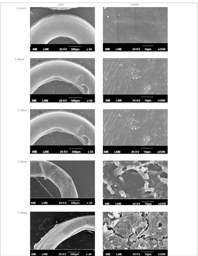
The surface analysis showed gradual degradation indicated by the increase of superficial roughness. Ef-fects of viscoelastic fluency were detectable after the first week and became more intense with increase of the oral permanency time. The superficial roughness raised in specific points in the week-1 group. The week-2 and week-3 groups demonstrated higher area of roughness. In the week-4 group, cracks were visi-ble in elastomeric surfaces and the entire elastomeric surface was rough (Fig 3).
DISCUSSION
Figure 3 - SEM surface morphology photomicrographs (electron secondary image). Control group: The supericial smoothness of the elastomer; Week-1 group: The efects of viscoelastic luency are apparent in some speciic points; Week-2 group: The increase of deformation points and the presence of bioilm above the elastomer are visible; Week-3 group: The supericial roughness is signiicantly higher and the thickness of the elastomer is decreased; Week-4 group: The degradation of the elastomer is clearly identiied for cracks and roughness in the total area.
50X 2500X
Control
1 Week
2 Week
3 Week
Some factors present in the oral environment as the intermittent stress of the elastomers, the pH, the oxygen content and the temperature variation are related to the relaxation. Although, the increase on the temperature is the most significant point of the degradation mechanism.16,18,22 De Genova et al,8
re-lated that the temperature in the oral cavity could vary from 0 to 64 °C. In this manner, one important aspect of oral degradation is the variation of the oral environment temperature and it depends on each in-dividual.11 In spite of the patients have different eating
habits, in the present work, it was observed similar viscoelastic fluency effects in the elastomeric ligatures of all patients. The increase in the internal diameter and reduction in the thickness of ligatures were clear-ly detected after the first week in the oral cavity. This degradation increased gradually, with the increase of permanency time in the oral environment (Fig 3).
Other important polymeric degradation mecha-nism occurs in the immersion in water solution as the saliva. This mechanism is pointed out by swell-ing and dissolution. The liquid is absorbed by the polymer, penetrating among the macromolecules. This produces an internal force that separates the chains and expands the polymer by breaking the sec-ondary links resulting on relaxation.4
The degradation of orthodontic elastomers can also occur by oxidation resulting in supericial cracks. The oxidation consists in the interaction among the elastomeric macromolecules with ozone and oxygen. The mechanism responsible for thechemical degrada-tion is the scission of macromolecules chains, in the couple bond between carbon atoms, preferentially at-tacked by ozone.4 In this study, it was observed cracks
on the surface of orthodontic elastomeric ligatures of week-4 group. These elastomeric ligatures were not to-tally immersed in oral luids during its use. Thus, they have partial contact with air and consequently with ozone and oxygen, that are factors of degradation.
The presence of bacterial flora is another impor-tant factor of degradation. Bode et al4 isolated a lot
of bacterium able to degraded natural and synthetic rubber. Most of them are present in the oral environ-ment, accelerating the biodegradation process.
The complex oral environment results in degra-dation with many variables like temperature, bac-This in-use biomaterial changes were caused by a
process known as aging. In the present study the elastic ligatures aging was characterized by the grad-ual increase in the surface roughness and by few changes in the mechanical properties of the mate-rial. Comparing the results of the tensile test and of the SEM analysis, one may suggest that elastomers have higher superficial degradation than loss of me-chanical properties.
The orthodontic elastomers are biodegraded in the oral environment mainly by hydrolyses. Their second-ary links are broken and the result is the relaxation,2,13
which is the main characteristic of degradation in or-thodontics elastomers. This phenomenon is respon-sible for the constant replacements of the elastomeric ligatures. The relaxation consequence is the decrease on mechanical energy transmitted to the tooth and i-nally,the reduction ofefectiveness of the tooth move-ment.2,11,13,14 Ater relaxation, the elastomers commonly
sufer the efects of viscoelastic luency.This phenom-enon consists in plastic deformation when the elasto-mer is submitted to forces below the plastic limit.6,17
Clinically, this efect could be observed in SEM ig-ures. Permanent deformities are visible in all ligatures submitted to the oral environment, although none of them was stressed above their plastic limit (Fig 3).
Hwang and Cha,14 in an in vitro study, observed
quick relaxation in elastic chains. The author described reduction of 50% on the initial force in the irst 24 hours. Andreasen and Bishara,1 in an in vivo study,
described reduction of 74% on initial force ater the same degradation time. Some important degradation mechanisms are present just in theoral ambient. In ex-ample, the accumulation of plaque on the elastomers and the prolonged contact with enzymes cause signii-cant degradation in these biomaterials.11 In the present
study, a gradual increase of supericial roughness oc-curred between 1 and 4 weeks. Eliades et al10 described
that elastomeric surfaces were modiied by irreversible adsorption of proteinaceous matter forming a bioilm ater 24 hours in oral cavity. Ater 3 weeks, this bioilm on elastomeric surface exhibited excessive mineraliza-tion increasing the surface roughness.10 In agreement,
terium, enzymes, pH, stress, chemical contents and others. More researches are necessary to produce new types of elastomers able to resist in this environment.
CONCLUSIONS
The elastic ligatures aged in the oral environment showed higher superficial degradation and lower loss
of mechanical properties after the maximum experi-mental period.
The clinically signiicant result is that ater the irst week in the oral cavity, the elastomeric ligatures al-ready presented signiicant supericial degradation and this process gradually increases resulting in signiicant supericial roughness, supporting plaque accumulation.
1. Andreasen GF, Bishara S. Comparison of alastik chains with elastics involved with intra-arch molar to molar forces. Angle Orthod. 1970;40(3):151-8.
2. Baty DL, Storie DJ, von Fraunhofer JA. Synthetic elastomeric chains: a literature review. Am J Orthod Dentofacial Orthop. 1994;105(6):536-42. 3. Beattie S, Monaghan P. An in vitro study simulating efects of daily
diet and patient elastic band change compliance on orthodontic latex elastics. Angle Orthod. 2004;74(2):234-9.
4. Bode HB, Kerkhof K, Jendrossek D. Bacterial degradation of natural and
synthetic rubber. Biomacromolecules. 2001;2(1):295-303.
5. Calado V, Montgomery DC. Projection of experiments using the Statistic.
1st ed. Rio de Janeiro: E-papers Services; 2003.
6. Callister WD. Materials science and engineering: an introduction. 50th ed. St. Louis: John Willey & Sons; 2000.
7. Cofelt MP. The efects of artiicial saliva and topical luoride treatments on the degradation of the elastic properties of orthodontic chains. Angle Orthod. 1992;62(4):265-74.
8. De Genova DC, Mclnnes-Ledoux P, Weinberg R, Shaye R. Force
degradation of orthodontic elastomeric chains: a product comparison study. Am J Orthod. 1985;87(5):377-84.
9. Eliades T, Bourauel C. Intraoral aging of orthodontic materials: the picture we miss and its clinical relevance. Am J Orthod Dentofacial Orthop. 2005;127(4):403-12.
10. Eliades T, Eliades G, Silikas N, Watts DC. Tensile properties of orthodontic elastomeric chains. Eur J Orthod. 2004;26(2):157-62.
11. Eliades T, Eliades G, Watts DC. Structural conformation of in vitro and in vivo aged orthodontic elastomeric modules. Eur J Orthod. 1999;21(6):649-58.
12. Graber TM, Vanarsdall RL. Orthodontics: current principles and techniques. 3rd ed. St Louis: Mosby; 2000.
13. Howard RS, Nicolai RJ. On the relaxation of orthodontic elastic threads. Angle Orthod. 1979;49(3):167-72.
REFERENCES
14. Hwang CJ, Cha JY. Mechanical and biological comparison of latex and silicone rubber bands. Am J Orthod Dentofacial Orthop. 2003;124(4):379-86.
15. Kanchana P, Godfrey K. Calibration of force extension and force degradation characteristics of orthodontic latex elastics. Am J Orthod Dentofacial Orthop. 2000;118(3):280-7.
16. Kersey ML, Glover K, Heo G, Raboud D, Major PW. An in vitro comparison of 4 brands of nonlatex orthodontic elastics. Am J Orthod Dentofacial Orthop. 2003;123(4):401-7.
17. Mano BS, Mendes LC. Introduction for polymers. 2nd ed. São Paulo: Edgard Blücher; 2001.
18. Nattrass C, Ireland AJ, Sherrif M. The efect of environmental factors on elastomeric chain and nickel titanium coil springs. Eur J Orthod. 1998;20(2):169-76.
19. Ferreira Neto JJ, Caetano MT. A degradação da força de segmentos de elásticos em cadeia de diferentes tamanhos: estudo comparativo in vitro. J Bras Ortodon Ortop Facial. 2004;9(51):225-33.
20. Proit WR, Fields HJ. Contemporary orthodontics. 2nd ed. St Louis: Mosby; 1986.
21. Russell KA, Milne AD, Khanna RA, Lee JM. In vitro assessment of the mechanical properties of latex and non-latex orthodontic elastics. Am J Orthod Dentofacial Orthop. 2001;120(1):36-44.
22. Stevenson JS, Kusy RP. Force application and decay characteristics of untreated and treated polyurethane elastomeric chains. Angle Orthod. 1994;64(6):455-64.