https://doi.org/10.1590/0004-282X20170052
ARTICLE
Malignant peripheral nerve sheath tumor with
and without neurofibromatosis type 1
Tumor maligno da bainha de nervo periférico com e sem Neurofibromatose tipo 1
Roberto André Torres de Vasconcelos1,2, Pedro Guimarães Coscarelli3, Regina Papais Alvarenga2, Marcus André Acioly2,4,5
1Instituto Nacional do Câncer, Departamento de Oncologia Cirúrgica, Divisão de Osso e Tecido Conjuntivo, Rio de Janeiro RJ, Brasil; 2Universidade Federal do Estado do Rio de Janeiro, Programa de Pós-Graduação em Neurologia, Rio de Janeiro RJ, Brasil; 3Universidade do Estado do Rio de Janeiro, Disciplina de Medicina Interna, Rio de Janeiro RJ, Brasil;
4Universidade Federal do Rio de Janeiro, Disciplina de Neurocirurgia, Rio de Janeiro RJ, Brasil; 5Universidade Federal Fluminense, Disciplina de Neurocirurgia, Niterói RJ, Brasil.
Correspondence: Roberto André Torres de Vasconcelos; Rua Equador, 831; 20220-410 Rio de Janeiro RJ; Brasil; E-mail: [email protected] Conflict of interest: There is no conlict of interest to declare.
Received 28 September 2016; Received in inal form 14 January 2017; Accepted 14 February 2017.
ABSTRACT
Objective: In this study, we review the institution’s experience in treating malignant peripheral nerve sheath tumors (MPNSTs). A secondary aim was to compare outcomes between MPNSTs with and without neuroibromatosis type 1 (NF1). Methods: Ninety-two patients with MPNSTs, over a period of 20 years, were reviewed. A retrospective chart review was performed. The median age was 43.5 years (range, 3–84 years) and 55.4% were female; 41 patients (44.6%) had NF1-associated tumors. Results: Mean tumor sizes were 15.8 ± 8.2 cm and 10.8 ± 6.3 cm for patients with and without NF1, respectively. Combined two- and ive-year overall survival was 48.5% and 29%. Multivariate analysis conirmed the association of tumor size greater than 10 cm (hazard ratio (HR) 2.99; 95% conidence interval (CI) 1.14–7.85; p = 0.0258) and presence of NF1 (HR 3.41; 95%CI 1.88–6.19; p < 0.001) with a decreased overall survival. Conclusion: Tumor size and NF1 status were the most important predictors of overall survival in our population.
Keywords: nerve sheath tumors; neuroibromatosis 1; survival.
RESUMO
Objetivo: Relatamos a experiência institucional no tratamento de tumores malignos da bainha de nervo periférico (TMBNP) e comparamos o prognóstico entre pacientes com e sem neuroibromatose tipo 1 (NF1). Métodos: Foram incluídos neste estudo 92 pacientes num período de 20 anos. Foi realizada uma análise retrospectiva dos prontuários, das características do tumor e do tratamento. A idade mediana era 43,5 anos (variação 3–84 anos) e 55,4% dos pacientes eram mulheres; 41 pacientes (44,6%) tinham tumores associados à NF1. Resultados: O diâmetro médio dos tumores era 15,8 ± 8,2cm e 10,8 ± 6,3cm para pacientes com e sem NF1, respectivamente. A sobrevida combinada em 2 e 5 anos foi de 48,5% e 29%. A análise multivariada conirmou que o tamanho do tumor acima de 10cm (hazard ratio (HR) 2.99; 95% intervalo de coniança (IC) 1.14–7.85; p = 0.0258) e a presença de NF1 (HR 3.41; 95%IC 1.88–6.19; p < 0.001) estão associados a uma pior sobrevida.
Conclusões: O tamanho do tumor e a associação com NF1 foram os preditores mais importantes de sobrevida na nossa população.
Palavras-chave: neoplasias de bainha neural; neuroibromatose tipo 1; sobrevivência.
Malignant peripheral nerve sheath tumors (MPNSTs) cor-respond to 5–10% of all soft tissue sarcomas1,2. he MPNST
originates from Schwann cells or Schwann cell precursors and was previously known as neuroibrosarcoma, neurogenic sarcoma, malignant schwannoma, and malignant neurilem-moma3. he MPNSTs occur mainly in adults with a median
age of 35 to 44 years2,4,5,6, and occasionally afect children2.
hese are aggressive tumors with a signiicant susceptibility to metastasize early in their course4,5,6.
Malignant peripheral nerve sheath tumor pathogenesis is yet to be fully understood, but human and experimental
studies have indicated the combination of cell transformation (skin-derived precursor cell) through loss of tumor suppres-sors (PTEN, p53 and p16INK4A) and hyper-activation of growth
factor receptor signaling, such as EGFR and PDGFR, together with extracellular matrix-mediated interactions, particularly mast cell pro-inlammatory signaling7.
Malignant peripheral nerve sheath tumors have a widely-recognized association with neuroibromatosis type 1 (NF1) in up to half of all cases2,4,5,6,7,8. his association is
MPNST5,7. A third group has recently been included as a con
-sequence of radiation therapy (RT-induced MPNST), which accounts for approximately 10% of the patients reported2,5.
It is still unclear, however, how outcomes are inluenced by diferent etiologies5. Radiation therapy-induced MPNST,
for instance, seems to trend towards a worse disease spe-ciic survival compared with NF1-associated and sporadic tumors5. However, the classical association with a worse
prognosis has been the NF1-associated group. Several stud -ies have addressed this issue2,4,5,6,8, which remains
controver-sial. A recent meta-analysis done by Kolberg et al.8 collected
more than 1,800 non-overlapping MPNST patients with a speciic question: whether NF1 status had any efect on sur -vival. Interestingly, their results demonstrated a signiicantly worse outcome in NF1 patients, but with a recent trend in the last decade towards similar results in the sporadic group. In addition, they found no diference in survival when analyz -ing their own MPNST patients8.
Due to their rarity and diiculty in diagnosis, MPNST series are rarely reported in the literature, mainly com-ing from US, Europe and Asian populations. In this study, we sought to report the irst Latin American series of MPNST patients, who were treated consecutively over 20 years, in order to investigate patient and treatment char-acteristics, as well as prognostic factors in our popula-tion. A secondary aim was to compare outcomes between MPNSTs with and without NF1.
METHODS
All patients with a MPNST, treated from January 2, 1990 to December 30, 2010, were included in the study popula -tion. he institutional review board approved the terms and conditions of the present study. he histological diag -nosis was conirmed by experienced institutional patholo -gists, but not re-reviewed for this study. Both NF1-associated and sporadic MPNSTs were included. Tumors were consid-ered NF1-associated if the patient had the clinical diagnosis of NF1, based on two or more of the National Institutes of Health (NIH) criteria9.
Based on this initial selection, 92 consecutive patients were treated at our institution and composed our study population. A retrospective chart review of patient, tumor and treatment characteristics was performed. Clinical data included age, gender, ethnicity, time of disease, NF1 status, tumor location (head and neck, trunk, and extremities), tumor size (main diameter), disease stage (American Joint Committee on Cancer [AJCC] staging system)10, surgical
resection, treatment goal (whether curative or palliative), and the use of chemotherapy and/or radiation therapy.
he median age at our study population was 43.5 years (range, 3–84 years) and 55.4% were female. Forty-one patients (44.6%) had NF1-associated MPNSTs.
Statistical analysis
Categorical variables were compared by using chi-square analyses, while continuous variables were compared using the Student t-test and Mann-Whitney non-parametric test, when necessary. he estimated overall and disease-free survivals were derived from the Kaplan-Meier method and Mantel-Haenszel log-rank test: survdif. he Cox proportional-hazard model was used to investigate signii -cant prognostic factors. We used Epi InfoTM (version 7;
avail-able at www.cdc.gov/epiinfo/index.html) and the R software (version 2.15.2; available at www.r-project.org). he level of signiicance was set at p < 0.05.
RESULTS
Patient characteristics and treatment modality In the NF-1 associated group, patients were commonly male (p = 0.046), younger (p = 0.001) and had larger tumors (p = 0.003). Mean time to presentation was 9.6 months (range, 2-48 months) in the NF1-associated group and 16.7 months (range, 1-96 months) in the sporadic group (p = 0.6). Mean tumor sizes were 15.8 ± 8.2 cm (range, 3–47 cm) and 10.8 ± 6.3 cm (range, 2–25 cm) for patients with and without NF1, respec-tively. Tumor distribution was similar between groups in the way that tumors were located in the extremities for most of the cases, followed by trunk, and head and neck lesions. Six patients (6.5%) presented with distant metastases, all of whom were afected by NF1. Patient demographics and treatment charac -teristics of the 92 patients are detailed in Table 1.
Curative treatment was commonly implemented in the sporadic group (78% vs 39% in the NF1-associated group, p = 0.01). Twenty-two patients (23.9%) were treated with neoadjuvant radiation therapy, 15 (16.3%) were treated with adjuvant radiation therapy and one patient (1.08%) was treated with brachytherapy. Palliative radiation therapy was given in the other 16 patients (17.4%). Overall, radiation therapy was given in 48.8% of NF1-associated and 66.7% of sporadic tumors. External beam therapy was given to these patients with doses in the range from 8-30 Gy for palliative means and 50-66 Gy for adjuvant purposes. Chemotherapy was used in 14 patients (15.2%; doxorubicin in seven, doxo -rubicin/ifosfamide in six; rescue regimens in one), mostly with a palliative intention (13 of 14 patients). Overall, chemo-therapy was a component of treatment in 35.7% and 64.3% of NF1-associated and sporadic MPNST patients, respectively.
Local recurrence and survival analysis
in the sporadic group, 20 patients (39.2%) had no evidence of disease, one (2%) was alive with disease, 21 (41.2%) were dead from the disease, one (2%) died of other causes, and eight (15.7%) were lost to the follow-up.
he analysis of local recurrence was conined to the 56 patients with localized disease. In this group, 15 patients (26.8%) developed a local recurrence 1-192 months after surgery. Eight patients were re-operated upon after recur-rence, four in each MPNST group. After a mean follow-up of 40 months, two patients (25%) were alive without evidence of disease, two (25%) were alive with disease, and four (50%) died from the disease.
he two- and ive-year overall survival was 21% and 18% for NF1-associated and 76% and 40% for sporadic MPNST, respectively. Overall survival was signiicantly lower in the NF1-associated group (p < 0.0001), while disease-free survival was not statistically diferent between the groups (p = 0.17) (Figures 1 and 2). Age, gender and tumor location had no impact on the MPNST outcome (Table 2). On the other hand, tumor size and the treatment goal exerted a signiicant impact on the course of disease (Figures 3 and 4).
Table 1. Patient characteristics.
Characteristics NF1-associated Sporadic p-value
Gender
Male 23 (56.1%) 18 (35.3%) 0.046
Female 18 (43.9%) 33 (64.7%) NS
Age at diagnosis, yr, (median) 6–68 (36.2) 3– 84 (48.9) 0.001 Presentation
Mass 29 (70.8%) 37 (72.5%)
NS
Pain 11 (26.8%) 8 (15.7%)
Ulceration 1 (2.4%) 3 (5.9%)
Hemorrhage - 2 (3.9%)
Skin macula - 1 (2%)
Tumor size
< 5 cm 3 (7.3%) 11 (21.6%)
0.003
5–10 cm 7 (17.1%) 16 (31.4%)
> 10 cm 30 (73.2%) 18 (35.3%)
Missing 1 (2.4%) 6 (11.8%)
Tumor location
Extremity 24 (58.5%) 33 (64.7%) NS
Trunk 13 (31.7%) 11 (21.6%)
Head and neck 4 (9.8%) 7 (13.7%) AJCC grade
IA 1 (2.4%) 3 (5.9%)
NS
IB 1 (2.4%) 4 (7.8%)
IIA 2 (5%) 6 (11.8%)
IIB 1 (2.4%) 3 (5.9%)
III 30 (73.2%) 35 (68.6%)
IV 6 (14.6%)
-Treatment goal
Curative 16 (39.1%) 40 (78.4%)
0.01 Palliative 25 (60.9%) 11 (21.6%)
Follow-up, mo, mean 24.8 46.5 0.0001
NF1: neuroibromatosis type 1; yr: year; AJCC: American Joint Committee on Cancer; Mo: months; NS: not signiicant.
Table 2. Prognostic factors for overall survival and disease-free
survival in the univariate analysis.
Prognostic factor OS (mean)
(mo) p-value
DFS (mean) (mo) p-value
NF1-status
With 47.4
<0.0001 52.5 0.17
Without 104.6 76.5
Age
< 45 yr 90.2
0.42 78.4 0.54
> 45 yr 82.8 60.7
Gender
Male 69.6
0.12 53.2 0.11
Female 91.6 81.6
Tumor location
Trunk 66.4
0.16
68.7
0.53
Extremity 107 51.5
Head and neck 37.1 36.2
Tumor size
< 5 cm 75.8
0.005
75.1
0.041
5–10 cm 94.5 75.5
> 10 cm 56.2 60.8
Treatment goal
Palliative 13.8
0.0001 - -
Curative 134.3
-NF1: neuroibromatosis type 1; OS: overall survival; DFS: disease-free survival; yr: year; mo: months; NS: not signiicant.
50
0 100 150 200 250
0.0 0.2 0.4 0.6 0.8 1.0
months
survival probability
Mantel−Haenszel test: p = 0,012 NF1 w/o NF1
Figure 1. Kaplan-Meier curves for cumulative overall survival from
MPNST patients with and without neuroibromatosis type 1 (NF1).
50
0 100 150
0.0 0.2 0.4 0.6 0.8 1.0
months
disease-free survival probability Mantel−Haenszel test: p not significant NF1 w/o NF1
Figure 2. Kaplan-Meier curves for cumulative disease-free
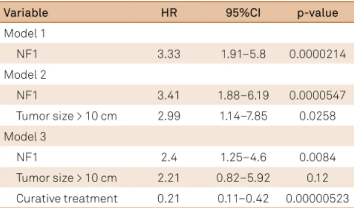
In the univariate analysis, increasing tumor size (greater than 10 cm) and the presence of NF1 were associated with a worse overall survival (Table 2). Multivariate analysis con -irmed that a tumor size greater than 10 cm (HR 2.99; 95%CI 1.14–7.85; p = 0.0258) and the presence of NF1 (HR 3.41; 95%CI 1.88–6.19; p < 0.001) were associated with a decreased overall survival (Table 3).
DISCUSSION
To the best of our knowledge the current study repre -sents the irst Latin American MPNST series reported in the literature. Here, we report on a population of 92 patients who underwent treatment at our institution during a 20-year period. From an epidemiological standpoint, our patients were mostly female, which is similar to that observed by Stucky et al.2, but most of the published series indicate at
least a slight male predominance4,5,6,8. his is especially true
for NF1-associated tumors, in which patients are generally male, younger, and have larger tumors at presentation4,5,6,8.
Our results also corroborated previous reports concerning
the occurrence of NF1 in approximately 45% of the patients and tumor distribution afecting mostly the extremities2,4,5,6,7,8.
In our series, the presence of NF1 and tumor size had a sig-niicant negative impact on overall survival. he higher risk of mortality among NF1 patients has been a very conlicting issue in the literature. As mentioned, Kolberg et al.8 found
inconsistent results between their own series and the results of their seminal meta-analyses, which pointed towards similar survival in the last decade. hey described at least four pos -sibilities for which NF1 patients may carry a worse prognosis, 50
0 100 150 200 250
0.0 0.2 0.4 0.6 0.8 1.0
months
survival probability
Mantel−Haenszel test: p = 0,012 NF1 w/o NF1
Figure 3. This graph illustrates the overall survival from
malignant peripheral nerve sheath tumor (MPNST) patients according to tumor size.
50
0 100 150 200 250
0.0 0.2 0.4 0.6 0.8 1.0
months
survival probability
Mantel−Haenszel test: p < 0,001
A
B
C
palliative
curative
50
0 100 150 200 250
0.0 0.2 0.4 0.6 0.8 1.0
months
survival probability
Mantel−Haenszel test: p = 0,231 curative NF1 curative w/o NF1
50
0 100 150 200 250
0.0 0.2 0.4 0.6 0.8 1.0
months
survival probability
Mantel−Haenszel test: p = 0,002 palliative NF1 palliative w/o NF1
Figure 4. Kaplan-Meier curves for cumulative overall
survival from malignant peripheral nerve sheath tumor (MPNST) patients according to treatment goal (A). In B, the graph
demonstrates the overall survival for curative treatment according to tumor etiology (patients with and without neuroibromatosis type 1 [NF1]). In C, the graph illustrates the overall survival for palliative treatment according to tumor etiology.
Table 3. Multivariate analysis of prognostic factors associated
with MPNST overall survival.
Variable HR 95%CI p-value
Model 1
NF1 3.33 1.91–5.8 0.0000214
Model 2
NF1 3.41 1.88–6.19 0.0000547
Tumor size > 10 cm 2.99 1.14–7.85 0.0258
Model 3
NF1 2.4 1.25–4.6 0.0084
Tumor size > 10 cm 2.21 0.82–5.92 0.12
namely: 1) NF1-associated MPNSTs are inherently aggressive, 2) tumoral defense systems are less it to control tumor growth, 3) delayed diagnosis resulting in advanced tumors, and (4) dif-ferent treatment strategies in sporadic MPNST patients. he irst two assumptions represent biological characteristics, which do not ind any support in the molecular data known today8. he latter two represent clinical characteristics.
Interestingly, NF1 patients presented within 9.6 months from the onset of symptoms, while non-NF1 patients sought medical attention with a mean time of 16.7 months (p = 0.6). According to our results, neither delayed diagnosis nor difer -ent treatm-ent strategies could burden the responsibility for a worse outcome in the NF1-associated group, converse to their assumptions. Our NF1 patients presented earlier with larger tumors and advanced disease suggesting other patho-genetic mechanisms for such an aggressive behavior.
In addition, we found a combined ive-year overall sur -vival of 29%. Considering only sporadic MPNST patients, the ive-year overall survival was 40%, which is very similar to other large series4,6,8. Of note, however, is the observation that
73.2% of the NF1-associated tumors and 35.3% of the sporadic tumors in our series had lesions greater than 10 cm, making it the largest MPNST cohort of giant tumors reported to date. As previous authors, and we, have demonstrated, tumor size is one of the most important prognosticators of survival in patients with MPNST4,5,6,8. his could have contributed to our
dismal results in the NF1 population. Moreover, it is worth mentioning that six patients (14.6%) of the NF1-associated tumor group presented with distant metastasis, which per se
bore a fourfold increased risk of mortality6.
Finally, as a soft tissue sarcoma, MPNSTs have been gener -ally stratiied by the AJCC staging system, which is considered the standard11. Tumor size, regional lymph node status,
occur-rence of distant metastasis and histological grade are collected in order to stage tumors and direct treatment10,11. Since it is used for
stratifying all soft tissue sarcomas, which comprise a wide variety of histological subtypes with diferent biological behavior, several limitations of the current staging system have been addressed11.
For categorizing tumor size (T-stage), for instance, the dichot-omous division into less than, and greater than, 5 cm is usually efective in capturing the impact on outcome only for truncal sar -coma11. Our current analysis demonstrated that tumors greater
than 10 cm, regardless of the NF1 status, carry a signiicantly worse prognosis in comparison with patients having intermedi-ate-sized tumors (between 5–10 cm). his is in line with previous indings4,8, suggesting that the AJCC staging system has signii
-cant limitations for the subset of MPNSTs, at least in the T-stage, and can relect inaccurate prognostic information.
Even though MPNST survival is still dismal, it has con-sistently improved over time8, which has raised an
increas-ing interest in knowincreas-ing and improvincreas-ing survivor experi -ence. In this regard, there is a recent recommendation by the Center for Medical Technology Policy to include patient reported outcomes and measures of health-related quality of life into prospective clinical studies in the onco-logic population12. This is extremely important in order
to incorporate the patients’ perspectives into treatment decision-making, thereby permitting a better understand -ing of the impact of disease and treatment on the patients’ quality of life (QOL)13.
here is a paucity of data in literature regarding QOL in patients sufering from MPNSTs. A recent national sur -vey done in England included only two patients afected by MPSNTs, from their responders14. heir results were ana
-lyzed together with bone sarcomas and other soft tissue tumors. Interestingly, patient QOL was not related to ampu -tation level or to histological diagnosis. Pain, on the other hand, afected approximately 90% of the patients in difer -ent levels, having a signiicant negative impact on QOL and physical function. Davidson et al.13 found similar results,
in things that concern the patient, to the impact of type of resection and radiation therapy among patients sufering from soft tissue sarcomas. Even though there was a high level of health-related quality of life one year after treatment, the anxiety/depression domain was associated with a sig -niicant change in the long-term, indicating that clinicians should be aware of the emotional impact of treatment13.
Finally, half of the MPNST patients have NF1-associated tumors, which per se determine a decreased QOL in com -parison to the general population15.
here are limitations to this study. his is a retrospective investigation, in which methods of data entry and character-ization change over time, thereby resulting in fragmentation of data in some cases.
In conclusion, MPNSTs are rare, aggressive tumors, which demonstrate a propensity to recur locally and disseminate early in their course, in spite of combined multimodality therapy. From the epidemiological standpoint, we observed a similar distribution of gender, tumor location and NF1 sta -tus, compared to previous reports in the literature using a more homogeneous population. Tumor size was consider-ably larger in the current study, suggesting that more efective screening programs should be developed in order to detect these tumors at an early stage, even for patients not afected by NF1. he presence of NF1 and tumor size had a signiicant negative impact on overall survival.
References
1. Pisters PW, Leung DH, Woodruff J, Shi W, Brennan MF. Analysis of prognostic factors in 1,041 patients with localized soft tissue sarcomas of the extremities. J Clin Oncol. 1996;14(5):1679-89. https://doi.org/10.1200/JCO.1996.14.5.1679
3. Scheithauer BW, Louis DN, Hunter S, Woodruff JM, Antonescu CR. Malignant peripheral nerve sheath tumor (MPNST). In: Louis DN, Ohgaki H, Wiestler OD, Cavenee WK, editors. WHO Classiication of tumours of the central nervous system. 3rd ed. Geneva: WHO Press; 2007. p. 160-2.
4. Anghileri M, Miceli R, Fiore M, Mariani L, Ferrari A, Mussi C et al. Malignant peripheral nerve sheath tumors: prognostic factors and survival in a series of patients treated at a single institution. Cancer. 2006;107(5):1065-74. https://doi.org/10.1002/cncr.22098
5. LaFemina J, Qin LX, Moraco NH, Antonescu CR,
Fields RC, Crago AM et al. Oncologic outcomes of sporadic, neuroibromatosis-associated, and radiation-induced malignant peripheral nerve sheath tumors. Ann Surg Oncol. 2013;20(1):66-72. https://doi.org/10.1245/s10434-012-2573-2
6. Zou C, Smith KD, Liu J, Lahat G, Myers S, Wang WL et al. Clinical, pathological, and molecular variables predictive of malignant peripheral nerve sheath tumor outcome. Ann Surg. 2009;249(6):1014-22. https://doi.org/10.1097/SLA.0b013e3181a77e9a
7. Durbin AD, Ki DH, He S, Look AT. Malignant peripheral nerve sheath tumors. Adv Exp Med Biol. 2016;916:495-530. https://doi.org/10.1007/978-3-319-30654-4_22
8. Kolberg M, Høland M, Agesen TH, Brekke HR, Liestøl K, Hall KS et al. Survival meta-analyses for >1800 malignant peripheral nerve sheath tumor patients with and without neuroibromatosis type 1. Neuro-oncol. 2013;15(2):135-47. https://doi.org/10.1093/neuonc/nos287
9. National Institutes of Health Consensus Development Conference Statement: neuroibromatosis; Bethesda, Md, USA, July 13-15, 1987. Neuroibromatosis. 1988;1(3):172-8.
10. Edge SB, Byrd DR, Compton CC, editors. AJCC cancer staging manual. 7th ed. New York: Springer; 2010.
11. Massarweh NN, Dickson PV, Anaya DA. Soft tissue sarcomas: staging principles and prognostic nomograms. J Surg Oncol. 2015;111|(5):532-9. https://doi.org/10.1002/jso.23851
12. Basch E, Abernethy AP, Mullins CD, Reeve BB, Smith ML, Coons SJ et al. Recommendations for incorporating patient-reported outcomes into clinical comparative effectiveness research in adult oncology. J Clin Oncol. 2012;30(34):4249-55. https://doi.org/10.1200/JCO.2012.42.5967
13. Davidson D, Barr RD, Riad S, Grifin AM, Chung PW, Catton CN et al. Health-related quality of life following treatment for extremity soft tissue sarcoma. J Surg Oncol. 2016;114(7):821-7. https://doi.org/10.1002/jso.24424
14. Furtado S, Grimer RJ, Cool P, Murray SA, Briggs T, Fulton J et al. Physical functioning, pain and quality of life after amputation for musculoskeletal tumours: a national survey. Bone Joint J. 2015;97-B(9):1284-90. https://doi.org/10.1302/0301-620X.97B9.35192