Primary Localization and Tumor Thickness as Prognostic
Factors of Survival in Patients with Mucosal Melanoma
Tarun Mehra1,4*, Gerd Gro¨zinger2, Steven Mann2, Emmanuella Guenova3, Rudolf Moos4, Martin Ro¨cken1, Claus Detlef Claussen2, Reinhard Dummer3, Stephan Clasen2, Aline Naumann5., Claus Garbe1.
1Department of Dermatology, Eberhard-Karls-University, Tu¨bingen, Germany,2Department of Diagnostic and Interventional Radiology, Eberhard-Karls-University, Tu¨bingen, Germany,3Department of Dermatology, University Hospital of Zu¨rich, Zu¨rich, Switzerland,4Medical Directorate, Universita¨tsSpital Zu¨rich, Zu¨rich, Switzerland, 5Institute for Clinical Epidemiology and Applied Biometry, Eberhard-Karls-University, Tu¨bingen, Germany
Abstract
Background:Data on survival with mucosal melanoma and on prognostic factors of are scarce. It is still unclear if the disease course allows for mucosal melanoma to be treated as primary cutaneous melanoma or if differences in overall survival patterns require adapted therapeutic approaches. Furthermore, this investigation is the first to present 10-year survival rates for mucosal melanomas of different anatomical localizations.
Methodology:116 cases from Sep 10 1984 until Feb 15 2011 retrieved from the Comprehensive Cancer Center and of the Central Register of the German Dermatologic Society databases in Tu¨bingen were included in our analysis. We recorded anatomical location and tumor thickness, and estimated overall survival at 2, 5 and 10 years and the mean overall survival time. Survival times were analyzed with the Kaplan-Meier method. The log-rank test was used to compare survival times by localizations and by T-stages.
Principal Findings:We found a median overall survival time of 80.9 months, with an overall 2-year survival of 71.7%, 5-year survival of 55.8% and 10-year survival of 38.3%. The 10-year survival rates for patients with T1, T2, T3or T4stage tumors were 100.0%, 77.9%, 66.3% and 10.6% respectively. 10-year survival of patients with melanomas of the vulva was 64.5% in comparison to 22.3% of patients with non-vulva mucosal melanomas.
Conclusion: Survival times differed significantly between patients with melanomas of the vulva compared to the rest (p = 0.0006). It also depends on T-stage at the time of diagnosis (p,0.0001).
Citation:Mehra T, Gro¨zinger G, Mann S, Guenova E, Moos R, et al. (2014) Primary Localization and Tumor Thickness as Prognostic Factors of Survival in Patients with Mucosal Melanoma. PLoS ONE 9(11): e112535. doi:10.1371/journal.pone.0112535
Editor:Soheil S. Dadras, University of Connecticut Health Center, United States of America ReceivedJune 12, 2014;AcceptedOctober 8, 2014;PublishedNovember 10, 2014
Copyright:ß2014 Mehra et al. This is an open-access article distributed under the terms of the Creative Commons Attribution License, which permits unrestricted use, distribution, and reproduction in any medium, provided the original author and source are credited.
Data Availability:The authors confirm that, for approved reasons, some access restrictions apply to the data underlying the findings. Due to an ethical restriction, raw data are unsuitable for public deposition as they contain patient medical records. Anonymized data are available upon request to Dr. Tarun Mehra (tarun.mehra@usz.ch).
Funding:This work was partially supported by the Deutsche Forschungsgemeinschaft (Grant number GU1271/2-1) for EG. The Deutsche Forschungsge-meinschaft did not participate in the design or analysis of the study, nor did participate in any form in the drafting of the manuscript. The additional Funding was provided by the Eberhard-Karls-University-Hospital of Tu¨bingen in Germany (salaries and publication costs) as well as the Universita¨tsSpital Zu¨rich, Switzerland (salaries). The funders had no role in study design, data collection and analysis, decision to publish, or preparation of the manuscript.
Competing Interests:The authors have declared that no competing interests exist. * Email: tarun.mehra@usz.ch
.These authors contributed equally to this work.
Introduction
Primary mucosal melanoma is a rare neoplasm, accounting for approximately 1% of all melanomas [1]. Mucosal melanoma has been associated with a poorer prognosis than cutaneous melanoma [2] with 5-year disease-specific survival rates roughly a third of those seen in cutaneous melanoma (25.0% vs 80.8%) [1]. Adequate data allowing for the establishment of a reliable prognostic staging system are still sparse, although data from larger patient cohorts especially of those affected with mucosal melanoma of the head and neck have started to be published [3]. We here present data from 116 patients with mucosal melanoma from multiple anatomic sites with the aim of establishing prognostic markers as to help establish a classification system for
primary mucosal melanomas. To our best knowledge, our sample of patients with primary mucosal melanoma is the largest published so far for mucosal melanomas of various anatomical locations and the only study publishing corresponding 10-year survival.
Materials and Methods
Patients
The Comprehensive Cancer Center database and the Central Register of the German dermatological Society both in Tu¨bingen, were searched for cases of primary mucosal melanoma (malignant melanoma with a primary site being a mucosal epithelium of any anatomical region). The database of the Comprehensive Cancer
Mucosal Melanoma
Total Vulva Vagina Penis Upper Airway Conjunctiva GI-Tract
Number of Patients (%) 116 (100%) 41 (35.3%) 6 (5.2%) 8 (6.9%) 36 (31.0%) 5 (4.3%) 20 (17.2%)
Female Sex (%) 85 (73.3%) 41 (100%) 6 (100%) 0 (0.0%) 23 (63.9%) 4 (80.0%) 11 (55.0%)
Age (years)
Median 66.5 67.0 61.0 72.5 64.0 65.0 67.0
Range 20–89 20–83 46–68 32–80 41–89 43–80 45–80
Thickness (n = 65, mm)
Median 2.9 2.0 6.5 2.0 5.0 0.7 7.0
Range 0.1–30.0 0.2–9.0 1.2–12.0 0.36–4.0 0.1–21.0 0.3–1.1 2.1–30.0
Thickness* (n = 116, %)
TX 35 (30.2%) 5 (12.2%) 0 0 20 (55.6%) 2 (40.0%) 8 (40.0%)
Tis 2 (1.7%) 2 (4.9%) 0 0 0 0 0
T1 10 (8.6%) 6 (14.6%) 0 2 (25.0%) 1 (2.8%) 1 (20.0%) 0
T2 18 (15.5%) 11 (26.8%) 2 (33.3%) 2 (25.0%) 2 (5.6%) 1 (20.0%) 0
T3 24 (20.7%) 9 (22.0%) 0 4 (50.0%) 4 (11.1%) 1 (20.0%) 6 (30.0%)
T4 27 (23.8%) 8 (19.5%) 4 (66.7%) 0 9 (25.0%) 0 6 (30.0%)
Lymph Node* (n = 116, %)
NX 17 (14.7%) 5 (12.2%) 0 0 8 (22.2%) 0 4 (20.0%)
N0 81 (69.8%) 30 (73.2%) 5 (83.3%) 6 (75.0%) 25 (69.4%) 5 (100%) 10 (50.0%)
N1–3 18 (15.5%) 6 (14.6%) 1 (16.7%) 2 (25.0%) 3 (8.3%) 0 6 (30.0%)
Metastasis* (n = 116, %)
MX 9 (7.8%) 3 (7.3%) 0 0 5 (13.9%) 0 1 (5.0%)
M0 102 (87.9%) 38 (92.7%) 6 (100%) 8 (100%) 27 (75.0%) 5 (100%) 18 (90.0%)
M1 5 (4.3%) 0 0 0 4 (11.1%) 0 1 (5.0%)
Resection (n = 102, %)
Total 102 40 6 8 26 3 19
RX 44 (43.1%) 11 (27.5%) 4 (66.7%) 2 (25.0%) 17 (65.4%) 1 (33.3%) 9 (47.4%)
R0 44 (43.1%) 25 (62.5%) 0 4 (50.0%) 7 (26.9%) 1 (33.3%) 7 (36.8%)
R1–2 14 (13.7%) 4 (10.0%) 2 (33.3%) 2 (25.0%) 2 (7.7%) 1 (33.3%) 3 (15.8%)
Relapse 42 (36.1%) 16 (39.0%) 4 (66.7%) 3 (37.5%) 12 (33.3%) 1 (20.0%) 6 (30.0%)
(n = 116, %)
Survival (n = 116)
2 years 71.7% 91.2% 53.3% 100% 59.0% 75.0% 47.4%
5 years 55.8% 78.6% 53.3% 83.3% 40.58% 75.0% 24.4%
10 years 38.3% 64.5% 53.3% 83.3% 21.31% 0.0% 0.0%
Prognostic
Factors
of
Mucosal
Melanoma
ONE
|
www.ploson
e.org
2
November
2014
|
Volume
9
|
Issue
11
|
Center yielded 48 cases and the database of the German dermatological Society 118 cases of mucosal melanoma, diagnosed or treated in Tu¨bingen in a period from Sep 10 1984 until Feb 15 2011. After merging the two databases and excluding duplicates as well as cases with insufficient data, 116 cases were analyzed (Table 1). The observation period started on Sep 10 1984 and finished on Dec 22 2011. All cases analyzed had a clinically confirmed diagnosis. We recorded location, age, sex, tumor thickness, lymph node involvement, resection status, relapse and estimated overall survival at 2, 5 and 10 years and its mean.
Clinical staging, follow-up and treatment were done at the Department of Dermatology, radiological staging and follow-up were done at the Department of Diagnostic and Interventional Radiology, both at the Eberhard-Karls-University of Tu¨bingen, Germany.
Follow-up was carried out according to guidelines for cutaneous melanoma [4]. Maximum follow-up time was 297 months.
Ethics
IRB approval was provided by the Ethics committee of the Medical Faculty of the University of Tu¨bingen. The aforemen-tioned IRB specifically approved this study. The study consisted in the retrospective analysis of already present clinical data of previously treated patients at our institution. No consent, written or oral, was obtained retrospectively. The aforementioned IRB specifically approved this consent procedure, as mentioned in the written IRB statement submitted as supplementary material together with the manuscript.
Statistical analysis
The statistical analysis was done using JMP 10.0 for Mac [5] and R 3.1.0 using the survival-package [6] respectively. Survival time was defined as being the duration between the date of diagnosis and the date of death from any cause (overall survival) and was assessed with the Kaplan-Meier method [7]. The pointwise 95% confidence intervals from the estimated survival probability at 2-, 5- or 10-years are based on a log-log-transformation, described as log-transfor-mation by Klein and Moeschberger [8]. The log-rank test was used to compare survival times by T-stage, using different subgroups of patients (all or N0M0) and by localization (vulva vs non-vulva).
Assuming an overall significance level ofa= 0.05, we adjusted for multiple testing (n = 3) by only considering results as ‘‘significant’’ if p,0.017 (Bonferroni correction).
Where applicable, findings were classified according to AICC 7th Edition, 2009.
Results
Patients and tumor localization
116 patients were included (Table 1). Of these, 85 (73.3% of total) were female. The median age at diagnosis was 66.5 years, ranging from 20 to 89 years. The anatomical sites of the primary were vulva (41 cases, 35.3% of total), vagina (6 cases, 5.2%), penis (8 cases, 6.9%), upper airway including nasal/paranasal sinuses as well as the oral cavity (36 cases, 31.0%) conjunctiva (5 cases, 4.3%) and the gastrointestinal tract (GI-tract) including anus (20 cases, 17.2%). The sojourn time of patients in the study ranged from 1 to 297 months; its median was 28.5 months with an interquartile range of 12.5 to 70.5 months.
Tumor thickness
Of 116 patients, 65 had a documented tumor thickness of the primary at the time of diagnosis, the median tumor thickness was 2.9 mm and ranging from 0.1–30.0 mm.
Table 1. Cont. Mucosal Melanoma Total Vulva Vagina Penis Upper Airway Conjunctiva GI-Tract Mean (Months) 93.3 133.3 11.5 30.8 46.5 58.7 38.5 Median (Months) 80.9 185.2 N/A N /A 48.7 71.5 19.5 Range (Months) 0.7–296.4 0.9–296.4 2.8–30.7 2.9–125.2 2.1–186.6 19.8–71.5 0.7–96.3 OD (n, %) 49 (42.2%) 11 (26.8%) 2 (33.3%) 1 (12.5%) 20 (55.6%) 2 (40.0%) 13 (65.0%) CO (n, %) 67 (57.8%) 30 (73.2%) 4 (66.7%) 7 (87.5%) 16 (44.4%) 3 (60.0%) 7 (35.0%) *Findings at d iagnosis, classified according to A ICC 7 th edition (2009). % indicates % of g roup, except Number o f P atients, where % indicates % o f total. Thickness: thickness of primary tumor. Resection: pathological d escr iption o f resection status. Survival: overall survival. OD: observed deaths. CO: censored outcomes, N /A: not applicable (median could not be calculated, as o ver half the cases in this group were censored). doi:10.1371/journal.pone. 0112535.t001
Prognostic Factors of Mucosal Melanoma
Mucosal melanomas situated in the GI-tract had the highest median tumor thickness (7.0 mm), followed by tumors of the vagina (6.5 mm) and of the upper airway (5.0 mm). Melanomas of the conjunctiva had the lowest median tumor thickness (0.7 mm), followed by the penis and the vulva (both 2.0 mm).
Staging at time of diagnosis
The tumor of two patients (1.7% of total) were classified as Tis, 10 as T1(8.6%), 18 as T2(15.5%), 24 as T3(20.7%) and 27 as T4
(23.8%). The remaining 35 patients (30.2%) had an unknown tumor thickness or T-Stage at the time of diagnosis and were classified as TX. Both Tis melanomas were melanomas of the vulva. The site with the highest percentage of T4- tumors was the
vagina (4 from 6, 66.7%, the 2 others being classified as T2)
followed by the GI-tract (6 of 20, 30.0%) and the upper airway (9 from 36, 25.0%). Primary mucosal melanomas of the upper airway had the highest percentage of undocumented primary tumor thickness at the time of diagnosis (55.6%, 20 of 36 cases), followed by primaries of the GI-tract (8 of 20 cases, 40.0%) and of the conjunctiva (2 of 5 cases, 40.0%).
At the time of diagnosis, 81 patients (69.8%) did not have an involvement of the regional lymph nodes whereas 18 patients (15.5%) did and 17 patients (14.7%) had an unknown nodal status. At the time of diagnosis, 5 cases of metastatic disease (M1, 4.3%)
were reported, 102 patients did not have detectable metastases (M0, 89.7%) and 9 cases were of unknown metastatic state (MX,
7.8%).
Of the 116 patients, 102 were operated. Of these, 44 had an unknown resectional status (RX, 43.1%), 44 were classified as R0
(43.1%) and 14 as R1(13.7%).
During follow- up 42 of 116 patients suffered a relapse (36.1%).
Site of primary tumor and survival
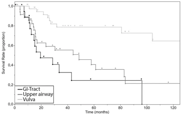
The overall survival times, combining patients in any anatom-ical site averaged 93.3 months, with a median overall survival of 80.9 months, ranging from 0.7 to 296.4 months (Figure 1, Figure S1). The group of patients with the lowest mean overall survival had a tumor at the vagina (11.5 months) followed by the penis (30.8 months) and GI-tract (38.5 months). The group of patients with the highest mean overall survival had a tumor at the vulva (133.3 months) followed by conjunctiva (58.7 months) and upper airway (46.5 months). The overall 2-year survival of patients with all sites of the primary was 71.7% (95% confidence interval (CI): 61.7% to 79.6%), 5-year survival was 55.8% (95% CI: 44.5% to 65.7%) and 10-year survival was 38.3% (95% CI: 24% to 51.3%). We also performed a survival analysis with the Kaplan-Meier method of the three cohorts with the highest number of cases (vulva, upper airway and GI-tract) (Figure 2). A clear distinction between the curve of the survival rate of patients with a primary of the vulva and the survival curve of the two other groups was observed. We did not test for significance due to overcrossing Kaplan-Meier curves of the survival rate of cases with primary tumors of the upper airway and of the GI-tract. As the data suggested a marked difference in survival times between vulva and non-vulva mucosal melanomas, we subsequently analyzed the survival of the cohort with a primary of the vulva (11 events in 41 Figure 1. Overall 10-year survival of all cases of primary mucosal melanoma included in this study (n = 116).
cases) compared to the rest (38 events in 75 cases). 5 and 10-year overall survival were 78.6% (95% CI: 55.2% to 89.2%), 64.5% (95% CI: 37.6% to 81.4%) and 42.9% (95% CI: 29.2% to 55.9%), 22.3% (95% CI: 7.9% to 39.1%) for patients with a melanoma of the vulva and non-vulva respectively with corresponding median survival times of 185.2 months and 44.3 months (Figure 3). The survival times of patients with a melanoma of the vulva were significantly different from that of the rest (p = 0.0006).
Impact of tumor thickness on survival
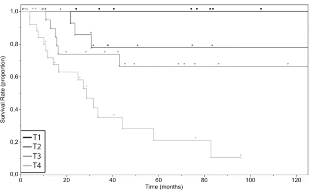
All the patients with a primary tumor classified as Tis or T1
(n = 2 and n = 10 respectively) had a 10-year overall survival of 100% (lower limit of 95% CI: 100%). For patients with tumors classified as T2(4 events in 18 cases), the 5- and 10-year survival
were 77.9% (95% CI: 35.4% to 92.3% and 25.9% to 92.3% respectively). The 5- and 10-year survival rate of patients with T3
-Figure 2. Overall 10-year survival of cases of primary mucosal melanoma according to the localization of the primary tumor.GI-Tract (n = 20), Upper airway (n = 36), Vulva (n = 41).
doi:10.1371/journal.pone.0112535.g002
Figure 3. Overall 10-year survival of the 116 cases of primary mucosal melanoma grouped according to the localization of the primary tumor.Vulva (n = 41), Non-vulva (n = 75).
doi:10.1371/journal.pone.0112535.g003
Prognostic Factors of Mucosal Melanoma
staged primaries (6 events in 24 cases) were both 66.3% (95% CI: 35.6% to 83.5% and 15.7% to 83.5% respectively). The 5-year survival rate of patients with T4-staged primaries (17 events in 27
cases) was 21.1% (95% CI: 5.8% to 42.7%). 10-year survival for patients with T4 tumors could not be estimated. The last living
patient was censored after 96 months when the survival curve was at the level of 10.6%. Mean overall survival times were 118.3, 35.4 and 36.5 months for patients with T2, T3or T4tumor respectively
(Table 2). Survival times of the groups were significantly different (p,0.0001) (Figure 4).
To exclude possible sources of bias, we restricted the following comparison of T1(0 events in 10 cases), T2(4 events in 16 cases),
T3(1 event in 15 cases) and T4(13 events in 21 cases) to patients of
subgroup N0M0(no involvement of the regional lymph nodes and
no distant metastasis). 5 and 10-year overall survival were 100.0% (lower limit of 95% CI: 100%) for T1N0M0, 76.2% (95% CI:
30.5% to 91.7% and 25.5% to 91.7% respectively) for T2N0M0,
88.9% (95% CI: 33.7% to 98.4% and 3% to 98.4% respectively) for T3N0M0. 10-year survival for patients with T4N0M0 tumors
could not be estimated. The last living patient was censored after 96 months when the survival curve was at the level of 12.4%, the 5-year overall survival was 24.9% (95% CI: 6.6% to 49.1%). Mean overall survival times were 116.2, 43.0 and 38.3 months for T2N0M0, T3N0M0and T4N0M0cohorts (Table 3, Figure S2). The
differences in survival time between groups was significant (p,
0.0001).
Impact of lymph node involvement, metastasis and resectional status on survival
Out of 99 of 116 cases with a documented status of regional lymph node metastasis, 81 did not have (N0) and 18 had an
involvement of the regional lymph nodes (N1–3). Patients staged as
N0had an overall 5-year survival of 56.2% and a 10-year survival
of 42.9%. Those with a tumor burden in the regional lymph nodes had an overall 5-year survival rate of 44.5% (Figure S3). 10-year survival for patients with a tumor burden in the regional lymph nodes could not be estimated. The last living patient was censored Table 2.Characteristics of the patient cohorts classified according to their T stage.
Total Tis T1 T2 T3 T4
Number of Patients (%) 81 (100.0%) 2 (2.5%) 10 (12.3%) 18 (22.2%) 24 (29.6%) 27 (33.3%)
Female Sex (%) 64 (79.0%) 2 (2.5%) 8 (9.9%) 15 (18.5%) 18 (22.2%) 21 (25.9%)
Age (years)
Median 64 66 50 64.5 70 70
Range 43–89 49–83 20–72 36–80 22–83 46–89
Lymph Node* (n = 81, %)
NX 0 0 0 0 0 0
N0 67 (82.7%) 2 (100.0%) 10 (100.0%) 16 (88.9%) 18 (75.0%) 21 (77.8%)
N1–3 14 (17.3%) 0 0 2 (11.1%) 6 (25.0%) 6 (22.2%)
Metastasis* (n = 81, %)
MX 0 0 0 0 0 0
M0 78 (96.3%) 2 (100.0%) 10 (100.0%) 18 (100.0%) 21 (87.5%) 27 (100.0%)
M1 3 (3.7%) 0 0 0 3 (12.5%) 0
Resection (n = 81, %)
Total 77 (100.0%) 2 (100.0%) 10 (100.0%) 18 (100.0%) 21 (87.5%) 26 (96.3%)
RX 29 (37.7%) 0 5 (50.0%) 6 (33.3%) 5 (23.8%) 13 (50.0%)
R0 36 (46.8%) 2 (100.0%) 4 (40.0%) 11 (61.1%) 13 (61.9%) 6 (23.1%)
R1–2 12 (15.6%) 0 1 (10.0%) 155.6%) 3 (14.3%) 7 (26.9%)
Relapse 32 (39.5%) 0 3 (30.0%) 8 (44.4%) 9 (37.5%) 12 (44.4%)
(n = 81, %) Survival (n = 81)
2 years 76.6% 100% 100% 85.7% 73.7% 62.9%
5 years 59.0% 100% 100% 77.9% 66.3% 21.1%
10 years 55.0% 100% 100% 77.9% 66.3% N/A
Mean (Months) 92.5 N/A N/A 118.3 35.4 36.5
Median (Months) 144.6 N/A N/A N/A N/A 28.7
Range (Months) 0.7–296.4 1.0–245.6 0.9–296.4 2.9–186.6 2.0–137.6 0.7–95.9 OD (n, %) 27 (33.3%) 0 (0.0%) 0 (0.0%) 4 (22.2%) 6 (25.0%) 17 (63.0%) CO (n, %) 54 (66.7%) 2 (100.0%) 10 (100.0%) 14 (77.8%) 18 (75.0%) 10 (37.0%) T stage according to tumor thickness at diagnosis, classified according to AICC 7th edition (2009).
% indicates % of group, except Number of Patients, where % indicates % of total. Thickness: thickness of primary tumor. Resection: pathological description of resection status. Survival: overall survival. OD: observed deaths. CO: censored outcomes, N/A: not applicable (median could not be calculated, as over half the cases in this group were censored).
after 75 months when the survival curve was at the level of 29.7%. A trend towards increased survival for N0patients could be found.
Of 107 cases with an established status for distant metastasis, 102 did not have (M0) and 5 had distant metastasis (M1) at
diagnosis. Patients staged as M0had an overall 2-year survival rate
of 73.9%, a 5-year survival of 58.1% and a 10-year survival of 41.4%. Those classified as M1had an overall 2-year survival rate
of 40%. At 5 years from diagnosis, no M1 patient remained in
observation; the last patient died after 49 months (Figure S4). A trend towards increased survival for M0patients could be found,
although the very few cases of M1patients (n = 5) has to be taken
into consideration.
Of 58 cases with a documented tumor resection, 44 had tumor free borders (R0) and 14 had resection borders which were not
(R1–2). R0-patients had an overall 5-year survival of 71.3% and a
10-year survival of 61.1%. R1–2-patients had an overall 2-year
survival rate of 62.9% and a 5-year survival of 52.4% (Figure S5). 10-year survival for R1–2-patients could not be estimated. The last
living patient was censored after 96 months when the survival curve was at the level of 38%.
Then we retrospectively staged cases according to the Mucosal Melanoma Staging System published by Iversen et al. in 1980 [9,10], which groups cases into regional disease (any T, N0, M0,
Stage I, n = 74), involvement of the regional lymph nodes (any T, N1–3, M0, Stage II, n = 18) and distant metastasis (any T, any N,
M1, Stage III, n = 5). Patients classified as Stage I had an overall
2-year survival of 75.2%, 5-2-year survival of 59.4% and 10-2-year survival of 44.4%. Corresponding survival for Stage II patients were 61.1% and 44.5%. 10-year survival for Stage II patients could not be estimated. The last living patient was censored after 75 months when the survival curve was at the level of 29.7%. 2-year survival for Stage III patients was 40%; at 5 2-years from diagnosis, no Stage III patient remained in observation; the last patient died after 49 months (Figure 5).
The trace of the Kaplan-Meier curve showed a trend towards a lower survival for the cohort of cases with extensive disease (Stage I) in comparison towards the cohort with regional disease (Stage II/III) (Figure S6).
Discussion
Published 5-year survival for mucosal melanoma varies between 17 and 40% [11–14]. We observed an overall 5-year survival of 55.8%. Our survival rates were higher for patients of all anatomic regions except for those with a primary in the GI-tract [11]. Especially striking is our cohort of melanomas of the vulva and the penis, which have survival rates which approach those seen in cutaneous melanoma [15]. We report considerably higher overall survival rates for patients with mucosal melanoma than those reported by Kim et al. who found a 2-year survival rate of 59.7% and a 5-year survival rate of 31.9% [11]. The distribution of the anatomical sites was similar to our cohort, reporting a generally lower 5-year survival rate per group. Nonetheless, the sites of occurrence of the primaries were differently grouped, as our research combined anorectum and GI to GI-tract and nasal, oral and maxillary sinus to upper airway, but split genitourinary into penis, vulva and vagina. In accordance with the literature, our findings confirm the lower survival rates of mucosal melanoma [14]. Indeed, 5-year and 10-year survival for patients with cutaneous melanoma are approximately 80% and 70–80% respectively [14,15].
It seems that independently of lymph node involvement and distant metastasis, survival times between T-stage groups are different. However, survival of mucosal melanoma patients in comparison to similar cohorts of cutaneous melanoma does not appear to be markedly different in the absence of lymph node involvement or metastasis, with the exception of mucosal melanomas with a high tumor thickness (.4.0 mm or T4). Figure 4. Overall 10-year survival of the 79 cases of primary mucosal melanoma classified as T1(n = 10), T2(n = 18), T3(n = 24), T4 (n = 27), according to the tumor thickness of the primary at diagnosis.Tumors classified according to the AICC, 7th edition (2009). doi:10.1371/journal.pone.0112535.g004
Prognostic Factors of Mucosal Melanoma
Table 3.Characteristics of the patient cohorts classified according to their T stage, all N0M0.
Mucosal Melanoma
Total N0M0 TisN0M0 T1N0M0 T2N0M0 T3N0M0 T4N0M0 Number of Patients (%) 64 (100.0%) 2 (3.1%) 10 (15.6%) 16 (25.0%) 15 (23.4%) 21 (32.8%)
Female Sex (%) 51 (79.7%) 2 (3.9%) 8 (15.7%) 13 (25.5%) 12 (23.5%) 16 (31.4%)
Age (years)
Median 66.5 66 50 64.5 73 68
Range 20–89 49–83 20–72 36–80 22–83 46–89
Resection (n = 64, %)
Total 62 (100.0%) 2 (100.0%) 10 (100.0%) 16 (100.0%) 14 (100.0%) 20 (100.0%)
RX 24 (38.7%) 0 5 (50.0%) 5 (31.3%) 4 (28.6%) 10 (50.0%)
R0 29 (46.8%) 2 (100.0%) 4 (40.0%) 11 (68.8%) 8 (57.1%) 4 (20.0%)
R1–2 9 (14.5%) 0 1 (10.0%) 0 2 (14.3%) 6 (30.0%)
Relapse 21 (32.8%) 0 3 (30.0%) 6 (37.5%) 3 (20.0%) 9 (42.9%)
(n = 64, %) Survival (n = 64)
2 years 83.4% 100% 100% 84.6% 100.0% 63.2%
5 years 66.0% 100% 100% 76.2% 88.9% 24.9%
10 years 61.6% 100% 100% 76.2% 88.9% N/A
Mean (Months) 101.5 N/A N/A 116.2 43.0 38.3
Median (Months) 144.6 N/A N/A N/A N/A 27.3
Range (Months) 0.7–296.4 1.0–245.6 0.9–296.4 2.9–186.6 2.0–137.6 0.7–95.9 OD (n, %) 27 (33.3%) 0 (0.0%) 0 (0.0%) 4 (25.0%) 1(6.7.0%) 13 (61.9%) CO (n, %) 54 (66.7%) 2 (100.0%) 10 (100.0%) 12 (75.0%) 14 (93.3%) 8 (38.1%) T stage according to tumor thickness at diagnosis, classified according to AICC 7th edition (2009).
% indicates % of group, except Number of Patients, where % indicates % of total. Thickness: thickness of primary tumor. Resection: pathological description of resection status. Survival: overall survival. OD: observed deaths. CO: censored outcomes, N/A: not applicable (median could not be calculated, as over half the cases in this group were censored).
doi:10.1371/journal.pone.0112535.t003
Figure 5. Overall 10-year survival of the 97 cases of primary mucosal melanoma which could be classified according to the Mucosal Melanoma Staging System[9].Stage I: local disease (any T, N0, M0, n = 74), Stage II: regional lymph node involvement (any T, N1–3, M0, n = 18),
Stage III: distant metastasis (any T, any N, M1, n = 5).
The 5-year survival rates for patients with regional metastatic disease of mucosal melanoma seem to be poorer than those of cutaneous melanoma (stage II in our classification, 44.5% vs. 78%, 59% and 40% for IIIA, IIIB, IIIC respectively) [16].
The one censored case considered apart, the four other cases with a stage M1at diagnosis died before reaching the 5-year
time-point; the last patient died after 49 months (vs. 5-year survival of 10–20% for M1band M1ccombined) [16]. Therefore it seems that
the poorer prognosis of mucosal melanoma can be attributed to a more aggressive metastatic behavior, which coincides with the author’s clinical observations.
We chose not to stage our cases with the TNM staging system, although it would have made results more comparable, as the cohorts were too small for the different TNM sub-splits.
One astonishing result was the significantly superior survival of patients with a mucosal melanoma of the vulva in comparison to the rest. Nonetheless, in this region the epithelium changes from mucosal to squamous, so it is arguable how many of these melanomas were actually mucosal in sensu stricto. However, the significantly superior survival rate could be influenced by differences in the tumor thickness: although the percentage of T3and T4stage primaries between the two groups was similar (T3:
22.0% vs. 20.0% and T4: 19.5% vs. 25.3% for vulva vs.
non-vulva), the relative amount of Tis, T1 and T2stage tumors was
higher in the primaries localized at the vulva (4.9%, 14.6% and 26.8% for vulva, 0.0%, 5.3% and 9.3% for non-vulva) with a markedly higher proportion of TX tumors for non-vulval localizations (vulva: 12.2% vs. non-vulva: 40.0%). Anyhow, survival rates were similar to those seen in cutaneous melanoma [15]. We therefore would welcome a discussion on whether vulval melanoma should be considered mucosal or if it should be classified as a cutaneous malignancy.
In summary, we strongly advocate including tumor thickness and a distinction between vulval and non-vulval mucosal melanoma when designing a staging system if it is to have a prognostic value. We also advocate considering the establishment of separate therapeutic regimens for mucosal melanoma due to a more aggressive systemic disease. We further suggest considering melanoma of the vulva to be classified as a primary cutaneous neoplasm. The importance of tumor thickness in N0M0patients,
resectional status, lymph node affection and disseminated disease has to be validated by studies with larger cohorts.
The overall survival time of patients with mucosal melanoma depends on multiple factors. Using Kaplan-Meier method, we only analyzed univariate influences on the overall survival times. To cope with the multivariate influence, a Cox regression analysis would be necessary. Unfortunately, in our investigation the sample size of patients with complete information and the number of events recorded were too small to build a Cox regression model. Instead, we compared the patients with different T-stages in a subgroup of patient with N0M0(no involvement of the regional
lymph nodes and no distant metastasis).
Conclusions
We are the first to present 10-year survival rates for patients with mucosal melanomas of different anatomical localizations and show that the anatomical localization of the primary mucosal melanoma (vulva vs. non-vulva) is a significant prognostic factor (p = 0.0006). We confirm the role of tumor thickness as a prognostic marker; the survival time of patients with mucosal melanoma depends on the primary’s T-stage at the time of diagnosis (p,0.0001).
Supporting Information
Figure S1 Overall survival of all included cases of primary mucosal melanoma (n = 116). The longest obser-vation period per case amounted up to 300 months (25 years). (TIF)
Figure S2 Overall 10-year survival of cases of primary mucosal melanoma with local disease (T1–4, N0, M0,
n = 62), grouped according to their tumor thickness at the time of diagnosis. T1N0M0 (n = 10), T2N0M0 (n = 16),
T3N0M0(n = 15), T4N0M0(n = 21).
(TIF)
Figure S3 Overall 10-year survival of cases of primary mucosal melanoma grouped according to their status of lymph node involvement at the time of diagnosis. N0
(n = 81), N1–3(n = 18).
(TIF)
Figure S4 Overall 10-year survival of cases of primary mucosal melanoma grouped according to their status of distant metastasis at the time of diagnosis.M0(n = 102),
M1(n = 5).
(TIF)
Figure S5 Overall 10-year survival of cases of primary mucosal melanoma grouped according to their resec-tional status at the time of diagnosis. R0 (n = 44), R1–2
(n = 14). (TIF)
Figure S6 Overall 10-year survival of cases of primary mucosal melanoma grouped to cases with local disease (T stages T1–4, N0, M0, n = 62) and cases with systemic
disease at the time of diagnosis (all T stages, N1–3and/
or M1, n = 23).
(TIF)
Author Contributions
Analyzed the data: TM GG SM AN EG CG. Contributed reagents/ materials/analysis tools: AN MR CDC CG. Wrote the paper: TM GG AN EG RM RD MR SC CG. Provided infrastructure and access to data: CG. Conceived and designed the project/data analysis: TM GG.
References
1. Chang AE, Karnell LH, Menck HR (1998) The National Cancer Data Base report on cutaneous and noncutaneous melanoma: a summary of 84,836 cases from the past decade. The American College of Surgeons Commission on Cancer and the American Cancer Society. Cancer 83: 1664–1678.
2. Bauer CGJ (2012) Melanoma. Dermatology, by J Bolognia, J Jorizzo, J Schaffer. Third edition ed: Elsevier. pp. 1885–1914.
3. Jethanamest D, Vila PM, Sikora AG, Morris LG (2011) Predictors of survival in mucosal melanoma of the head and neck. Annals of surgical oncology 18: 2748– 2756.
4. Pflugfelder A, Kochs C, Blum A, Capellaro M, Czeschik C, et al. (2013) S3-Guideline ‘‘Diagnosis, therapy and follow-up of melanoma’’ - short version. J Dtsch Dermatol Ges 11: 563–602.
5. SAS Institute, Inc. (1989–2007) JMP version 10.0.
6. R Development Core Team (2014) R: A Language and Environment for Statistical Computing, R Foundation for Statistical Computing.
7. Kaplan EL, Meier P (1958) Non-parametric Estimation from Incomplete Observations. Journal of the American Statistical Association 53: 457–481. 8. Klein J, Moeschberger M (2003) Survival Analysis Techniques for Censored and
Truncated Data. Springer-Verlag New York Springer-Verlag.
9. Iversen K, Robins RE (1980) Mucosal malignant melanomas. Am J Surg 139: 660–664.
10. Ross MI, Henderson MA (2009) Mucosal Melanoma. In: Balch CM, Houghton AN, Sober AJ, Soong SJ, Atkins MB, et al., editors. Cutaneius Melanoma. 5th Edition ed. St. Louis: CRC Press. pp. 337–350.
Prognostic Factors of Mucosal Melanoma
11. Kim HS, Kim EK, Jun HJ, Oh SY, Park KW, et al. (2010) Noncutaneous malignant melanoma: a prognostic model from a retrospective multicenter study. BMC Cancer 10: 167.
12. Meleti M, Leemans CR, de Bree R, Vescovi P, Sesenna E, et al. (2008) Head and neck mucosal melanoma: experience with 42 patients, with emphasis on the role of postoperative radiotherapy. Head Neck 30: 1543–1551.
13. Benlyazid A, Thariat J, Temam S, Malard O, Florescu C, et al. (2010) Postoperative radiotherapy in head and neck mucosal melanoma: a GETTEC study. Arch Otolaryngol Head Neck Surg 136: 1219–1225.
14. Chang AE, Karnell LH, Menck HR (1998) The National Cancer Data Base report on cutaneous and noncutaneous melanoma. Cancer 83: 1664–1678. 15. Garbe C, Buttner P, Bertz J, Burg G, d’Hoedt B, et al. (1995) Primary cutaneous
melanoma. Identification of prognostic groups and estimation of individual prognosis for 5093 patients. Cancer 75: 2484–2491.
![Figure 5. Overall 10-year survival of the 97 cases of primary mucosal melanoma which could be classified according to the Mucosal Melanoma Staging System [9]](https://thumb-eu.123doks.com/thumbv2/123dok_br/16293631.185556/8.918.176.747.689.1025/overall-survival-melanoma-classified-according-mucosal-melanoma-staging.webp)
