Nutritional and Cardiovascular Profiles of Normotensive and
Hypertensive Rats kept on a High Fat Diet
Silvio A. Oliveira Júnior
1, Katashi Okoshi
1, Ana Paula Lima-Leopoldo
1, André S. Leopoldo
1, Dijon H.S. Campos
1, Paula F.
Martinez
1, Marina P. Okoshi
1, Carlos R. Padovani
2, Maeli Dal Pai-Silva
2, Antonio C. Cicogna
1Faculdade de Medicina de Botucatu - UNESP1; Instituto de Biociências de Botucatu - UNESP2, Botucatu, SP - Brazil
Summary
Background: Although a high fat diet (HFD) promotes nutritional and heart disorders, few studies have assessed its influence in normotensive Wistar-Kyoto rats (WKY) and spontaneously hypertensive rats (SHR).
Objective: To evaluate and compare the nutritional and cardiovascular profiles of WKY and SHR on a high fat diet.
Methods: 20 WKY and 20 SHR were divided into four groups: Control-WKY (C-WKY), HFD-WKY, Control-SHR (C-SHR) and
HFD-SHR. The C and HFD groups received, respectively, a normocaloric diet and a HFD for 20 weeks. The following features were evaluated: body weight (BW), adiposity, blood glucose, serum lipids, with measurements of total cholesterol and triacylglycerol levels, insulin and leptin. The cardiovascular study included the systolic blood pressure (SBP), a cardiopulmonary anatomical evaluation, an echocardiography and heart histology.
Results: The SHR had BW, adiposity, glucose, cholesterol, triacylglycerol, leptin and insulin levels lower than the WKY. In SHR, the caloric intake increased with HFD. In WKY, the HFD increased energy efficiency, adiposity and blood leptin, and reduced glucose. In the cardiovascular assessment, the SHR had SBP, pulmonary moisture, myocardial hypertrophy and interstitial fibrosis higher than the WKY (p <0.01); the cardiac function was similar in both strains. The HFD reduced the ventricular systolic diameter in the WKY and increased the mitral E/A ratio, the diastolic thickness of the interventricular septum and the posterior wall, as well as the interstitial fibrosis of the left ventricle. (Arq Bras Cardiol 2009; 93(5) : 487-494)
Conclusion: Although it had not significantly affected the nutritional profile of the SHR, the treatment increased cardiac remodeling and precipitated the emergence of ventricular diastolic dysfunction. In WKY, the diet increased adiposity and leptinemia, and promoted non-significant cardiovascular changes.
Key Words: Diet; hyperlipidemias; rats; ventricular remodeling; hypertension.
Mailing address: Silvio A. Oliveira Júnior •
Faculdade de Medicina de Botucatu, UNESP, Departamento de Clínica Médica, Rubião Júnior, S/N - 18618-000 - Botucatu,SP - Brazil E-mail: [email protected]
Artigo recebido em 20/08/2008; revisado recebido em 23/09/2008; aceito em 21/10/2008.
Introduction
Changes in the metabolism of fats and carbohydrates are associated with nutritional disorders such as obesity, dyslipidemia and insulin resistance, and with cardiovascular diseases such as elevated blood pressure and heart remodeling1. In experimental studies, a high fat diet (HFD),
with high levels of lipids and/or carbohydrates, causes similar effects to those found in human nutritional disorders2; in the
cardiovascular context, it promotes hemodynamic disorders and heart remodeling, with hypertrophy, interstitial fibrosis and myocardial dysfunction1,3,4.
Few studies have established associations, though inconsistent, between nutrition and cardiovascular profiles in normotensive rats under HFD3-8. Studies that did not
evaluate the cardiac function reported dyslipidemia, abnormal
glucose, hyperinsulinemia and cardiac hypertrophy4,5. Du
Toit et al3 found increased blood pressure, hypertrophy and
post-ischemic myocardial dysfunction, with alterations in glucose and lipid levels and maintenance of insulinemia. Other researchers, despite observing the presence of dyslipidemia, did not detect any effects on glucose6,7, hemodynamic
changes6,7, and cardiac remodeling6-8. Wilson et al9 though
they did not see any serious nutritional disorders, reported progressive cardiac dysfunction. In face of these controversies, the influence of HFD on nutrition and cardiovascular profiles of normotensive rats is not fully clarified.
Research on cardiac remodeling in spontaneously hypertensive rats (SHR) is often used because it develops hypertrophy, interstitial fibrosis and ventricular dysfunction resulting from arterial hypertension10. The SHR are genetically
more susceptible to nutritional, lipid and glucose disorders and hyperinsulinemia11. However, few studies examined
the influence of HFD on the cardiovascular and nutritional attributes of this lineage. Girard et al7 found dyslipidemia
hypertrophy12-14, with a reduction12 or increase13,14 in blood
pressure. Additional studies, which omitted heart aspects, found glucose15,16 and lipid16 changes, with increase15 or
maintenance16 of blood pressure levels.
The objective of this study was to evaluate and compare the nutritional profile of cardiovascular and normotensive Wistar-Kyoto rats (WKY) and SHR treated with HFD. The hypothesis is that the HFD cause changes in both strains, but they are more pronounced in the SHR.
Material and methods
The experimental protocol was approved by the Ethics Committee on Animal Experiments of FMB/UNESP, in accordance with regulations of the Brazilian College of Animal Experimentation (COBEA).
Animals and groups
Samples of 20 WKY and 20 SHR male rats, with 60 days of age, were divided into four groups, by lineage and treatment: C-WKY, HFD-WKY, C-SHR and HFD-SHR. The groups C and HFD received, respectively, normocaloric and hypercaloric diets for 20 weeks. The animals were homed in individual cages, under monitored conditions of temperature (20-24o C),
humidity (40-60%) and light (12-hour light/dark cycle), with free access to the assigned treatment. The normocaloric diet consisted of a commercial rodent chow (Purina), with the following macronutrient composition: 58.72% carbohydrates, 32.23% protein, 9.06% lipids, totaling 3.2 Kcal/g. The HFD, previously instituted17, consisted of a combination of
processed foods, and had the following chemical profile: 31.7% carbohydrates, 24.9% protein, 43.4% lipids, totaling 4.6 Kcal/g. The high energy density of the HFD resulted mainly from a high intake of lipids17.
Nutrition Proile
The nutritional assessment included caloric intake, energy efficiency, body weight (BW), blood glucose, lipid, insulin, leptin levels and adiposity. The intake and the efficiency, obtained by the ratio between the weight change and the total energy intake18, were measured daily. The BW was measured
weekly. After the experimental period, the animals were fasted for 12-14 hours, anesthetized with pentobarbital sodium (50 mg/kg) and euthanized by decapitation. Blood was collected in tubes with heparin, centrifuged (3000 rpm) for 15 minutes at 4oC and stored at –80oC. Subsequently, serum triacylglycerol
fractions, total cholesterol and glucose levels were assessed by enzymatic method with specific kits (Kovalent diagnosis, Kovalent do Brasil Ltda, Rio de Janeiro/RJ). Concentrations of leptin and insulin were determined by ELISA using a micro-plate reader (Spectra MAX 190, Molecular Devics, USA) and appropriate kits (Linco Research, Inc., St. Louis, MO, USA)19.
The index of adiposity20 was calculated with the following
formula: ([epididymal fat + retroperitoneal fat + visceral fat] / (total body weight – sum of all adipose depots)) x100.
Cardiovascular proile
The cardiovascular assessment included the systolic
blood pressure, an in vivo assessment of heart structure and function, anda post-mortem evaluation of cardiopulmonary morphology and myocardial histology. Pressure was measured by plethysmography, a commonly used procedure21,22, with
an automated sphygmomanometer (Narco Biosystem, Austin, Texas, USA).
Heart structure and function were assessed in vivo by echocardiography, with the previously described methodology23,24. The rats were weighed, and anesthetized
with ketamine hydrochloride (50 mg/kg) and xylazine hydrochloride (1 mg/kg), administered intramuscularly. Then, trichotomy was performed in the anterior chest, and the animals were positioned in the left lateral position for the echocardiography, which was performed with a HDI 5000 Phillips ultrasound machine, equipped with a 12 MHz electronic transducer.
For the measurement of heart structures, monodimensional mode (M-mode) images were obtained, with two-dimensional mode images guiding the ultrasound beam, and the transducer placed in a parasternal location in the short axis. The image of the left ventricle (LV) was obtained by positioning the M-mode cursor just below the mitral valve plane at the level of the papillary muscles23. The images of the aorta and the left
atrium were obtained with the M-mode cursor positioned at the level of the aortic valve. The images were recorded in a Sony Co. UP-890 printer model. Subsequently, heart structures were manually measured, with a precision caliper.
When the diameter of the ventricular cavity was maximal, the LV diastolic diameter (LVDD), the LV posterior wall diastolic thickness (LVPWDT) and the interventricular septum diastolic thickness (IVSDT) were measured. When the diameter of the cavity was minimal, the LV systolic diameter (LVSD), the LV posterior wall systolic thickness (LVPWST) and the interventricular septum systolic thickness (IVSST) were measured. The left atrium (LA) was measured at its maximal diameter. The LV mass (LVM) was calculated using the following formula: LVM = [(LVDD + LVPWDT + IVSDT)3
– (LVDD)3] x 1.04. The following variables were derived
from the dimensions described above: LV relative thickness (LVPWDT/LVDD), LVDD/BW, LA/BW and LVM index (LVMI, LVM/BW).
The LV systolic function was assessed by the following indexes: Percentage of mesocardial shortening (% Meso. Short.): [(LVDD + ½ LVPWDT + ½ IVSDT) – (LVSD + ½ LVPWST + ½ IVSST)] / (LVDD + ½ LVPWDT + ½ IVSDT); percentage of endocardial shortening (% Endo. Short.): [(LVDD - LVSD) / LVDD]; posterior wall shortening velocity (PWSV).
The diastolic function was assessed by the ratio index between the peak of initial inflow velocity (E wave) and the atrial contraction (A wave) of the transmitral flow (E/A), the deceleration time of the E wave (DTE) and the isovolumetric relaxation time (IVRT).
After the euthanasia, the atria (A), the right ventricle (RV) and the LV were weighed in absolute values and corrected by BW. To examine whether the HFD changed pulmonary moisture (a clinical sign of cardiac dysfunction)10, the water
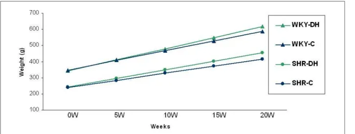
Table 1 - Linear regression models of body weight oscillations in time
Groups Linear Regression Model
Coeficient of
Determination (%)
C-WKY Weight = 346.816 + 12.102 w 67.7 *
HFD-WKY Weight = 341.336 + 13.791 w 77.1 *
C-SHR Weight = 239.900 + 8.752 w 77.3 *
HFD-SHR Weight = 243.827 + 10.477 w 85.2 *
* - p<0.001 for comparative analysis between different weeks (W).
Figure 1 - Linear regression models of body weight values in different weeks; 0W: initial moment; 5W: 5th week; 10W: 10th week; 15W: 15th week; 20W: 20th week.
water content was analyzed with the formula: [(WW-DW) / WW] x100 (%) where WW: wet weight; DW: dry weight.
A histological evaluation of the myocardium, involving the myocyte transverse area and the interstitial collagen fraction, was performed on samples of the LV. After being fixed in a solution of 10% formol saline22,24, the fragments
were embedded in paraffin blocks. 4 µm sections, collected in histology slides, were stained with Hematoxylin-Eosin, to evaluate the sectional area of the myocyte. In each animal, 50-70 cells were evaluated. As a principle of analysis, these cells should come from the ventricular subendocardial layer, have an elliptical shape, and contain a visible and centralized core22,24. The collagen content was evaluated in tissue sections
stained in Picro-sirius red22. At least 20 quarters were used,
and perivascular regions were not taken into account. The histological sections were projected at a magnification of 40 times, with the aid of a microscope (Leica DM LS) coupled to a video camera that projected the images on a IBM PC, which was equipped with the image analyzer software Image Pro-plus (Media Cybernetics, Silver Spring, Maryland, USA).
Statistical analysis
The results were expressed in descriptive measures. Weight change against time, measured in weeks, was analyzed by linear regression models. The comparison between models was performed by slope test and linear regression constant. The nutritional and cardiovascular variables were evaluated by the two-factor ANOVA, followed, respectively, by the Tukey test, for parametric distributions, and the Dunn test, for non-parametric data. The findings were discussed at a statistical significance level of 5%.
Results
Nutrition Proile
Weight change against time, evaluated by linear regression models, is shown in Figure 1. The regression model constructed for the HFD-SHR group had the best coefficient prediction:
85.2% (Table 1). In all groups, there was a significant weight increase in relation to the initial time (p <0.001). In both treatments, weight changes in SHR differed from WKY in every week of the study. In both strains, no significant weight differences were found between the two diets.
The results of the nutritional profile are shown in Table 2. The SHR group showed lower caloric intake than the C-WKY group, whereas the HFD-SHR group showed less energy efficiency than the HFD-WKY group. The WKY showed higher BW, adiposity, glucose, total cholesterol (TC), triacylglycerol (TG), leptin and insulin levels, compared to the SHR, in both treatments. In the WKY, efficiency, adiposity and leptin increased in the HFD-WKY; the diet reduced blood glucose, as compared to the C-WKY.
Cardiovascular proile
Table 2 - Evaluation of nutritional and biochemical proiles, according to lineage and treatment
Variables Lineage
Treatment
Control (C) D. Hypercaloric (HFD)
Caloric intake (Kcal) WKY 82.3 ± 8.4 83.2 ± 10.8 SHR 71.5 ± 2.3 ## 78.8 ± 4.5 *
Energy eficiency (g/ Kcal) WKY 0.15 ± 0.03 0.17 ± 0.02 *
SHR 0.13 ± 0.01 0.15 ± 0.03 #
Body weight (g) WKY 539 ± 69 566 ± 60 SHR 370 ± 16 ## 409 ± 10 ##
Adiposity Ind. (%) WKY 8.55 ± 2.96 11.27 ± 3.45 * SHR 3.65 ± 0.30 ## 5.54 ± 0.93 ##
Glucose (mg/dL) WKY 144 ± 13 128 ± 13 * SHR 98 ± 12 ## 95 ± 18 ##
Total Cholesterol (mg/dL) WKY 123.2 ± 21.3 119.1 ± 17.7 SHR 49.7 ± 5.0 # 38.1 ± 7.4 #
TG (mg/dL) WKY 105.2 ± 48.4 94.1 ± 48.6 SHR 34.7 ± 4.9 # 39.8 ± 3.8 #
Leptin (ng/ dL) WKY 8.28 ± 3.36 11.06 ± 4.00 * SHR 1.89 ± 0.58 # 3.36 ± 1.08 #
Insulin (ng/ dL) WKY 2.21 ± 2.47 1.64 ± 0.57 SHR 0.63 ± 0.62 ## 0.51 ± 0.93 #
Values in mean ± standard deviation; TG: triacylglycerol; * - p <0.05 vs C; # - p <0.05; ## - p <0.01 vs WKY; ANOVA and Tukey test. Insulin values expressed in median ± semi-amplitude: ANOVA and Dunn test.
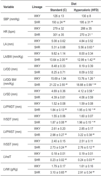
Table 3 - Evaluation of systolic blood pressure and heart structure echocardiography, according to lineage and treatment
Variable Lineage Diet
Standard (C) Hypercaloric (HFD)
SBP (mmHg) WKY 126 ± 13 130 ± 8
SHR 193 ± 24 ## 195 ± 31 ##
HR (bpm) WKY 279 ± 32 288 ± 35
SHR 301 ± 35 270 ± 21 *
LA (mm) WKY 5.08 ± 0.62 4.84 ± 0.52 SHR 5.31 ± 0.68 5.56 ± 0.63 #
LA/BW (mm/Kg) WKY 9.82 ± 1.14 8.00 ± 0.34 SHR 13.64 ± 2.05 ## 12.99 ± 1.42 ##
LVDD (mm) WKY 8.48 ± 0.33 8.19 ± 0.39 SHR 8.25 ± 0.71 8.09 ± 0.52
LVDD/ BW (mm/Kg)
WKY 15.69 ± 1.94 13.76 ± 1.26 *
SHR 21.22 ± 2.55 ## 18.88 ± 0.99 * ##
LVSD (mm) WKY 4.69 ± 0.36 4.12 ± 0.58 * SHR 4.39 ± 0.61 4.06 ± 0.59
LVPWDT (mm) WKY 1.52 ± 0.08 1.59 ± 0.08 SHR 1.84 ± 0.13 ## 1.95 ± 0.16 * ##
IVSDT (mm) WKY 1.55 ± 0.06 1.60 ± 0.07 SHR 1.87 ± 0.08 ## 1.96 ± 0.15 * ##
LVPWST (mm) WKY 2.61 ± 0.20 2.85 ± 0.17 SHR 2.98 ± 0.27 ## 3.22 ± 0.38 ##
IVSST (mm) WKY 2.40 ± 0.15 2.51 ± 0.11 SHR 2.73 ± 0.24 ## 2.76 ± 0.12 ##
LVrelT WKY 0.18 ± 0.01 0.19 ± 0.01 SHR 0.23 ± 0.02 ## 0.24 ± 0.03 ##
LVMI (g/Kg) WKY 1.79 ± 0.17 1.61 ± 0.16 SHR 3.10 ± 0.65 ## 2.91 ± 0.34 ##
Values in mean ± standard deviation; SBP: systolic blood pressure, HR:
heart rate; LA: diameter of the left atrium, LA/BW: LA and inal body weight
ratio; LVDD: LV diastolic diameter; LVDD/BW: body weight and LVDD ratio; LVSD: systolic diameter of LV; LVPWDT: posterior wall diastolic thickness; IVSDT: diastolic thickness of the interventricular septum; LVPWST: posterior wall systolic thickness; IVSST: interventricular septum systolic thickness; LVRelT: relative thickness on the LV wall; LVMI: left ventricular mass index; * - p <0.05; ** - p <0.01 vs C; # - p <0.05; ## - p <0.01 vs WKY, ANOVA and Tukey test.
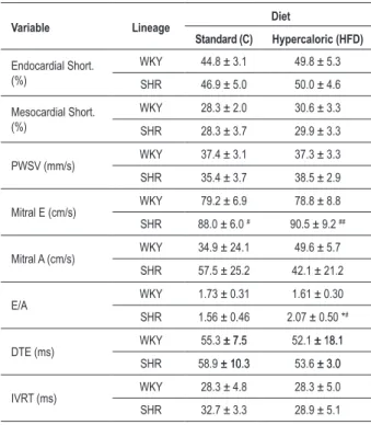
results of the echocardiographic evaluation of the ventricular function. E wave differences, larger in the C-SHR than in the C-WKY (p <0.05), were more pronounced among the HFD groups (p <0.01). The E/A ratio, similar in the C groups, was higher in the HFD-SHR than in the HFD-WKY; the diet increased the E/A ratio in the SHR, indicating the existence of an interaction between the strain and the treatment.
Table 5 shows cardiopulmonary morphology and heart histology findings. In both treatments, pulmonary moisture, VE weight, RV/BW and LV/BW ratios, morphometry and collagen were higher in the SHR than in the WKY. In SHR, the HFD increased A and VE weight, as well as the collagen content.
Discussion
Hypercaloric diets, rich in lipids, are frequently used in experimental trials to induce metabolic disorders commonly found in humans1,2,19. Despite hundreds of investigations
employing high-fat diets, there is no consensus on the content and composition of the fatty acids, saturated or unsaturated, used in these interventions2. In this research, the high-fat
diet was obtained by adding a combination of industrialized products to a standard diet17. Similar treatments are very
common in experiments with rodents3,5,17.
In this study, the SHR had weight lower than the WKY, at the
Table 4 - Echocardiographic assessment of systolic and diastolic function of the left ventricle, according to lineage and treatment
Variable Lineage Diet
Standard (C) Hypercaloric (HFD)
Endocardial Short. (%)
WKY 44.8 ± 3.1 49.8 ± 5.3
SHR 46.9 ± 5.0 50.0 ± 4.6
Mesocardial Short. (%)
WKY 28.3 ± 2.0 30.6 ± 3.3
SHR 28.3 ± 3.7 29.9 ± 3.3
PWSV (mm/s) WKY 37.4 ± 3.1 37.3 ± 3.3 SHR 35.4 ± 3.7 38.5 ± 2.9
Mitral E (cm/s) WKY 79.2 ± 6.9 78.8 ± 8.8 SHR 88.0 ± 6.0 # 90.5 ± 9.2 ##
Mitral A (cm/s) WKY 34.9 ± 24.1 49.6 ± 5.7 SHR 57.5 ± 25.2 42.1 ± 21.2
E/A WKY 1.73 ± 0.31 1.61 ± 0.30
SHR 1.56 ± 0.46 2.07 ± 0.50 *#
DTE (ms) WKY 55.3 ± 7.5± 7.5 52.1 ± 18.1± 18.1 SHR 58.9 ± 10.3± 10.3 53.6 ± 3.0± 3.0
IVRT (ms) WKY 28.3 ± 4.8 28.3 ± 5.0 SHR 32.7 ± 3.3 28.9 ± 5.1
Values in mean ± standard deviation; Endocardial Short.: relative endocardial shortening fraction; Mesocardial Short.: relative mesocardial shortening fraction; PWSV: velocity of shortening of the posterior wall; Mitral E: mitral E wave; Mitral A: mitral A wave; E/A: ratio of E and A waves; DTE: deceleration time of E wave; IVRT: isovolumetric relaxation time of the LV; * - p <0.05; ** - p <0.01 versus C; # - p <0.05; ## - p <0.01 vs. WKY, ANOVA and Tukey test.
Table 5 - Evaluation of lung moisture, cardiac morphology, according to lineage and treatment
Variable Lineage
Diet
Standard (C) Hypercaloric (HFD)
Lung
WW/DW WKY 4,37 ± 0,21 4,27 ± 0,47 SHR 4,90 ± 0,25 # 4,77 ± 0,19 #
Moisture (%)
WKY 77,1 ± 1,2 76,4 ± 2,5
SHR 79,5 ± 1,0 # 79,0 ± 0,8 #
Atria (g) WKY 0,092 ± 0,013 0,110 ± 0,015 SHR 0,086 ± 0,012 0,120 ± 0,071 *
A/FBW (mg/g) WKY 0,17 ± 0,02 0,20 ± 0,03 SHR 0,24 ± 0,04 0,30 ± 0,02 #
RV (g) WKY 0,21 ± 0,03 0,22 ± 0,03
SHR 0,22 ± 0,05 0,24 ± 0,08
RV/FBW (mg/g) WKY 0,39 ± 0,03 0,40 ± 0,03 SHR 0,60 ± 0,15 # 0,59 ± 0,22 #
LV (g) WKY 0,83 ± 0,10 0,85 ± 0,09
SHR 0,94 ± 0,10 ## 1,06 ± 0,05 ** ##
LV/FBW (mg/g) WKY 1,53 ± 0,11 1,48 ± 0,63 SHR 2,54 ± 0,37 ## 2,58 ± 0,15 ##
Fibrosis (%) WKY 3,70 ± 0,65 5,35 ± 0,65 SHR 7,70 ± 0,65 ## 11,31 ± 0,69 ** ##
CSA (µm2) WKY 197,1 ± 7,0 201,3 ± 7,0 SHR 272,0 ± 7,0 ## 276,5 ± 7,3 ##
Variables expressed as mean ± standard deviation: A/FBW: ratio between
atria and inal body weight; RV: right ventricle weight; RV/FBW: ratio between RV and inal body weight; LV: left ventricle weight; LV/FBW: ratio between LV weight and inal body weight. WW/DW - wet weight/dried weight ratio. Variables
expressed as mean ± standard error: Fibrosis and CSA: cardiomyocyte cross-sectional area; * - p <0.05; ** - p <0.01, versus C; # - p <0.05; ## - p <0.01, versus WKY; ANOVA and Tukey test.
data agree with the authors who analyzed these responses in other strains of rats5,25,26. In the Wistar5,25 and
Sprague-Dawley26 studies, HFD treatments promoted increased body
adiposity, but did not change body weight. For the SHR, the diet did not promote changes in adiposity, although the calorie intake was higher in the HFD-SHR. Compared to other strains, the SHR strain is resistant to weight gain through high-fat interventions13; when there are weight changes due
to the treatment, these variations are mild, compared to those observed among normotensive rats12. However, other authors
reported an increase in body weight14-16 and adiposity16 in the
SHR on high-fat diet, during briefer interventions of eight15 or
twelve weeks14,16. Probably, these divergent results are mainly
due to the dietary compositions used in these studies, which were characterized by a high intake of saturated fatty acids. In our study, the HFD was mostly made up of unsaturated fatty acids from plant origin, such as corn and peanut oils17.
As to the biochemical profile, the C-SHR had lower levels of blood glucose and insulin than the C-WKY group (Table 2). The SHR have a higher genetic predisposition to hyperglycemia and insulin resistance, which arise especially with the maturation11,27. Due to the young age of the SHR in
this study (7 to 8 months), it is possible that this predisposition has not yet been observed. In literature, research on blood glucose and insulin in WKY and SHR rats had conflicting
results. Whereas some authors28 found hyperglycemia and
insulin resistance in the SHR, others27 reported no differences
between the two strains. These discrepancies may result from many factors, including the age of the animals27. The
HFD-WKY and the HFD-SHR groups showed similar results to those found in the control groups, although the diet only reduced the blood glucose in the WKY, thus confirming the interaction between the strain and the treatment. In the postprandial state, the hyperinsulinemia that results from pancreatic β cells stimulation enhances tissue glucose uptake and, as the fasting state is maintained, reduces glycemia27.
This mechanism explains the glucose decrease observed in the HFD-WKY. However, in both lineages, it was not possible to verify insulin level changes due to the treatment, although the difference between the strains, most evident on standard diet (p<0.01), was milder among the HFD groups (p<0.05). Additional studies are needed to clarify this finding.
in the C-SHR than in the WKY. Leptin, which is synthesized by the adipose tissue, is associated with body adiposity29,
which was higher in the C-WKY. This hormone has a lipolytic influence and, during prolonged fasting periods, reduces the uptake of TG in adipocytes, contributing to increased serum TG levels29. Therefore, the increase of leptin, due to increased
adiposity, favors the TG increase in the C-WKY as compared to the C-SHR. Similar behavior was observed among HFD groups, although the diet only increased leptin in the HFD-WKY. Triacylglycerol was not changed by the treatment in both strains (Table 2). This finding may be possibly due to the action of insulin, which remained unchanged by the diet in both strains. Insulin stimulates the uptake of TG in adipocytes, contributing to adipogenesis, and inhibits lipolysis, therefore preventing the increase of serum triacylglycerols30.
Total cholesterol was also lower in the SHR, in both treatments. Due to an increased excretion of cholesterol, combined with deficiencies in enteric capture and molecular transport, the SHR are hypocholesterolemic compared to the WKY31. In both strains, the HFD did not promote changes
in cholesterol. Unsaturated fatty acids, the components of the HFD used in this investigation17, stimulate liver uptake
of cholesterol18.
As to the cardiovascular assessment, echocardiography is an alternative for the evaluation of ventricular function, and can provide important information on the heart structure and performance in small animals32. It is a versatile, safe, painless,
noninvasive and valid method for in vivo serial analyses23. The
echocardiographic evaluation in animal experiments is usually performed under the action of anesthetic agents that can cause physiological changes in the cardiovascular and respiratory systems21-24. Ketamine hydrochloride has minimal influence
on the cardiopulmonar system32. This drug may cause muscle
rigidity, which is minimized by the combination with xylazine hydrochloride, a drug with sedative and analgesic action. This association is indicated for many animal species because it induces analgesia and reduces the muscle tone23,32.
The overall cardiovascular results indicate that the C-SHR group shows cardiac remodeling, characterized by enlargement of the left atrium, concentric left ventricular hypertrophy, right ventricular hypertrophy and myocardial interstitial fibrosis, as compared to the C-WKY. The functional assessment showed preserved systolic function with an increase in mitral E-wave in the C-SHR. These results, associated with a higher pulmonary moisture, indicate a diastolic dysfunction. In the SHR, the neurohormonal activation via the renin-angiotensin-aldosterone system (RAAS), endothelins and catecholamines associated with sympathetic nerve activity may cause water retention and promote structural changes of the myocardium and vascular wall, causing ventricular remodeling10,21. This combination of
responses may enhance myocardial stiffness, with reduction of ventricular compliance and filling, which cause water retention and venocapillar pulmonary hypertension10,33. In
the SHR, this event is common in older age groups (18 to 24 months), and is associated with other clinical signs of ventricular dysfunction and atrial remodeling10,33. Accordingly,
although the increase in pulmonary moisture found in the C-SHR (Table 5) is an unexpected finding because of the young
age of the animals (7-8 months), we can not discard that this result indicates a change in functional performance. In parallel with this assumption, other authors found evidence of diastolic dysfunction in the SHR at the age of 12-14 months34,35. The
results indicate that the remodeling observed in the C-SHR maintained normal ventricular systolic performance, with a possible change in the diastolic function.
Cardiac structural differences between the strains on standard diet remained unaltered on HFD, except the diameter of the left atrium (Table 3) and the A/BW ratio (Table 5), which increased in the HFD-SHR as compared to the WKY-HFD. The echocardiographic functional assessment (Table 4) showed that the E wave and the E/A ratio were increased in the HFD-SHR. The E/A ratio increase, modified by relaxation and ventricular compliance disturbances, coupled with the increase in the diameter of the left atrium and the interstitial collagen fraction, indicated the presence of a ventricular diastolic dysfunction10,33. The preservation of systolic function
and other indicators of diastolic performance, as the IVRT and DTE, which were not altered by the treatment, suggesting the occurrence of a pseudonormal pattern36. However, the heart
rate decrease in the HFD-SHR, as compared to the C-SHR, is noteworthy. This change increases the mitral blood inflow during the passive relaxation, which magnifies the values of the E wave, and, therefore, of the E/A ratio, explaining the results found in the HFD-SHR group. The heart rate decrease is an unexpected finding, because hypercaloric and high-fat diet promotes sympathetic nervous system hyperactivity in the SHR14, causing tachycardia. Additional research could
help clarify the hemodynamic change found.
One important result is the interaction between the diet and the strain in determining the interstitial collagen fraction (Table 5). The treatment increased the myocardial interstitial fibrosis in the HFD-SHR group by approximately 50%, as compared to the C-SHR group. The interstitial remodeling stems from the balance between stimulating and inhibiting molecular agents that affect the extracellular matrix proliferation37. The
stimulating factors comprise bioactive molecules, including RAAS components, endothelins, catecholamines and cytokines, which are exacerbated in the SHR33,37. Probably,
these factors contributed to the additional diet-induced interstitial remodeling. Folder et al38 observed that saturated
or unsaturated fat-rich diets induced the activation of peptides that are involved in cardiac remodeling: activator protein-1 (AP-1) and mitogen-activated protein kinases (MAPKs). These agents are also activated by the RAAS and their effects include the increase in interstitial fibrosis through the transformation and growth factor (TGF-β)37. Although it was not possible
to detect differences in adiposity between the C-SHR and the SHR-HFD (p <0.10), the influence of fat on interstitial remodeling should not be dismissed. All RAAS components and cytokines, such as interleukin and tumor necrosis factor (TNF)-α, are secreted and regulated by the adipose tissue39.
Considering the collagen content increase in the HFD-SHR group, compared to the C-SHR, and other evidence of cardiac remodeling and pulmonary moisture in the WKY-HFD, it is very likely that the HFD-SHR group developed left ventricle diastolic dysfunction.
References
1. Sharma N, Okere IC, Duda MK, Chess DJ, O’Shea KM, Stanley WC. Potential impact of carbohydrate and fat intake on pathological left ventricular hypertrophy. Cardiovasc Res. 2007; 73 (2): 257-68.
2. Buettner R, Schölmerich J, Bollheimer LC. High-fat diets: modeling the metabolic disorders of human obesity in rodents. Obesity. 2007; 15: 798-808. 3. Du Toit EF, Nabben M, Lochner A. A potential role for angiotensin II in obesity
induced cardiac hypertrophy and ischaemic/reperfusion injury. Basic Res Cardiol. 2005; 100 (4): 346-54.
4. Akiyama T, Tachibana I, Shirohara H, Watanabe N, Otsuki M. High-fat hypercaloric diet induces obesity, glucose intolerance and hyperlipidemia in normal adult male Wistar rat. Diabetes Res Clin Pract. 1996; 31 (1): 27-35. 5. Naderali EK, Fatani S, Williams G. Chronic withdrawal of a high-palatable
obesity-inducing diet completely reverses metabolic and vascular abnormalities associated with dietary-obesity in the rat. Atherosclerosis. 2004; 172: 63-9. 6. Carroll JF, Zenebe WJ, Strange TB. Cardiovascular function in a rat model of
diet-induced obesity. Hypertension. 2006; 48 (1): 65-72.
7. Girard A, Madani S, Boustani ESE, Belleville J, Prost J. Changes in lipid metabolism and antioxidant defense status in spontaneously hypertensive rats and Wistar rats fed a diet enriched with fructose and saturated fatty acids. Nutrition. 2005; 21: 240-8.
8. Ricci E, Smallwood S, Chouabe C, Mertani HC, Raccurt M, Morel G, et al. Electrophysiological characterization of left ventricular myocytes from obese Sprague-Dawley rat. Obesity. 2006; 14: 778-86.
9. Wilson CR, Tran MK, Salazar KL, Young ME, Taegtmeyer H. Western diet, but not high fat diet, causes derangements of fatty acid metabolism and contractile dysfunction in the heart of Wistar rats. Biochem J. 2007; 406 (3): 457-67. 10. Bing OH, Brooks WW, Robinson KG, Slawsky MT, Hayes JA, Litwin SE, et
al. The spontaneously hypertensive rat as a model of the transition from compensated left ventricular hypertrophy to failure. J Mol Cell Cardiol. 1995; 27 (1): 383-96.
11. Aitman TJ, Gotoda T, Evans AL, Imrie H, Heath KE, Trembling PM, et al. Quantitative trait loci for cellular defects in glucose and fatty acid metabolism in hypertensive rats. Nature Genetics. 1997; 16 (2): 197-201.
12. Contreras RJ, King S. High fat/sucrose feeding attenuates the hypertension of spontaneously hypertensive rats. Physiol Behav. 1989; 46 (2): 285-91. 13. Zhang T, Reid K, Acuff CG, Jin CB, Rockhold RW. Cardiovascular and analgesic
effects of a highly palatable diet in spontaneously hypertensive and Wistar-Kyoto rats. Pharmacol Biochem Behav. 1994; 48 (1): 57-61.
14. Sedová L, Berubé J, Gaudet D, Dumont M, Tremblay J, Hamet P et al. Diet-induced obesity delays cardiovascular recovery from stress in spontaneously hypertensive rats. Obes Res. 2004; 12: 1951-8.
15. Sato T, Nara Y, Kato Y, Yamori Y. Effects of high-calorie diet on blood pressure and sodium retention in spontaneously hypertensive rats and normotensive Wistar-Kyoto rats. J Diabet Complications. 1995; 9: 220-3.
16. Pausova Z, Sedová L, Berube J, Hamet P, Tremblay J, Dumont M, et al. Segment of rat chromosome 20 regulates diet-induced augmentations in adiposity, glucose intolerance, and blood pressure. Hypertension. 2003; 41 (5): 1047-55.
17. Nascimento AF, Sugizaki MM, Leopoldo AS, Lima-Leopoldo AP, Luvizotto RAM, Nogueira CR, et al. Ciclo de dietas hipercalóricas induz obesidade e co-morbidades em ratos Wistar. Arq Bras Endocrinol Metab. 2008; 52 (6): 968-74.
18. Diniz YS, Cicogna AC, Padovani CR, Santana LS, Faine LA, Novelli ELB. Diets rich in saturated and polyunsaturated fatty acids: metabolic shifting and cardiac health. Nutrition. 2004; 20: 230-4.
19. Dourmashkim JT, Chang GQ, Gayles EC, Hill JO, Fried SK, Julien C, et al. Different forms of obesity as a function of diet composition. Int J Obes. 2005; 29: 1368-78.
20. Dobrian AD, Davies MJ, Schriver SD, Lauterio TJ, Prewitt RL. Oxidative stress in a rat model of obesity-induced hypertension. Hypertension. 2001; 37: 554-60.
and LVPWDT, indicators of myocardial hypertrophy in the SHR-HFD. However, these findings were not associated with morphometry, which showed that the myocyte sectional area was unchanged by the HFD. Gerdes40 points out four
factors that restrict the technique precision and may lead to erroneous morphometrical interpretations: variability of the tissue cutting angle, heterogeneous contractility of cardiac fibers, considering only the measurements performed in the nucleus and the isolated sectional area which does not accurately reflect the degree of cellular hypertrophy. Other procedures are needed for a more accurate assessment of myocardial hypertrophy.
Although the WKY had increased treatment-induced adiposity (p <0.001), we observed a slight increase in the collagen content, with no statistical significance (p <0.10). In the functional assessment of the WKY, the HFD reduced the LVSD. While this finding may indicate better ventricular emptying, this was not consistent with the shortening rate, which was similar in the C-WKY and the HFD-WKY (Table 4). Further studies may contribute to the clarification of this decoupling of variables.
In conclusion, in the SHR, although it did not significantly
affect the nutritional profile, the high-fat diet increased cardiac remodeling and precipitated the emergence of ventricular diastolic dysfunction. In the WKY, the treatment promoted moderate nutritional changes, characterized by increased adiposity and leptinemia, accompanied by non-significant cardiovascular changes. These findings validate the hypothesis of an increased susceptibility of the SHR to diet-induced cardiac remodeling.
Potential Conflict of Interest
No potential conflict of interest relevant to this article was reported.
Sources of Funding
This study was funded by FAPESP.
Study Association
21. Cicogna AC, Padovani CR, Georgette JC, Aragon FF, Okoshi MP. Effects of protein-calorie restriction on mechanical function of hypertrophied cardiac muscle. Arq Bras Cardiol. 1999; 72 (4): 436-40.
22. Matsubara LS, Narikawa S, Ferreira ALA, Paiva SAR, Zornoff LAM, Matsubara BB. Remodelação miocárdica na sobrecarga crônica de pressão ou de volume no coração de ratos. Arq Bras Cardiol. 2006; 86 (2): 126-30.
23. Bregagnollo EA, Mestrinel MA, Okoshi K, Carvalho FC, Bregagnollo IF, Padovani CR, et al. Relative role of left ventricular geometric remodeling and of morphological and functional myocardial remodeling in the transition from compensated hypertrophy to heart failure in rats with supravalvar aortic stenosis. Arq Bras Cardiol. 2007; 88 (2): 225-33.
24. Zornoff LAM, Matsubara BB, Matsubara LS, Minicucci MF, Azevedo PS, Campana AO, et al. Cigarette smoke exposure intensifies ventricular remodeling process following myocardial infarction. Arq Bras Cardiol. 2006; 86 (4): 276-81.
25. Estadella D, Oyama LM, Dâmaso AR, Ribeiro EB, Nascimento CMO. Effect of palatable hyperlipidic diet on lipid metabolism of sedentary and exercised rats. Nutrition. 2004; 20: 218-24.
26. Smith BK, Kelly LA, Piña R, York DA, Bray GA. Preferential fat intake increases adiposity but not body weight in Sprague-Dawley rats. Appetite. 1998; 31 (2): 127-39.
27. Natalucci S, Ruggeri P, Cogo CE, Picchio V, Brunori A, Burattini R. Age-related analysis of glucose metabolism in spontaneously hypertensive and normotensive rats. Exp Physiol. 2003; 88 (3): 399-404.
28. Umeda M, Kanda T, Murakami M. Effects of angiotensin II receptor antagonists on insulin resistance syndrome and leptin in sucrose-fed spontaneously hypertensive rats. Hypertens Res. 2003; 26 (6): 485-92.
29. Ainslie DA, Proietto J, Fam BC, Thorburn AW. Short-term, high-fat diets lower circulating leptin concentrations in rats. Am J Clin Nutr. 2000; 71: 438-42. 30. Saltiel AR, Kahn CR. Insulin signalling and the regulation of glucose and lipid
metabolism. Nature. 2001; 414: 799-806.
31. Yuan YV, Kitts DD. Dietary fat source and cholesterol interactions alter plasma lipids and tissue susceptibility to oxidation in spontaneously hypertensive (SHR) and normotensive Wistar Kyoto (WKY) rats. Mol Cell Biochem. 2002; 232 (1-2): 33-47.
32. Schwarz ER, Pollick C, Meehan WP, Kloner RA. Evaluation of cardiac structures and function in small experimental animals: transthoracic, transesophageal, and intraventricular echocardiography to assess contractile function in rat heart. Basic Res Cardiol. 1998; 93 (6): 477-86.
33. Bing OH, Conrad CH, Boluyt MO, Robinson KG, Brooks WW. Studies of prevention, treatment and mechanisms of heart failure in the aging spontaneously hypertensive rat. Heart Fail Rev. 2002; 7 (1): 71-88. 34. Radin MJ, Holycross BJ, Sharkey LC, Shiry L, McCune SA. Gender modulates
activation of renin-angiotensin and endothelin systems in hypertension and heart failure. J Appl Physiol. 2002; 92 (3): 935-40.
35. Emter CA, McCune SA, Sparagna GC, Radin MJ, Moore RL. Low-intensity exercise training delays onset of decompensated heart failure in spontaneously hypertensive heart failure rats. Am J Physiol Heart Circ Physiol. 2005; 289 (5): 2030-8.
36. Zile MR, Brutsaert DL. New concepts in diastolic dysfunction and diastolic heart failure: part II. Causal mechanisms and treatment. Circulation. 2002; 105: 1503-8.
37. Boluyt MO, Bing OH. Matrix gene expression and decompensated heart failure: the aged SHR model. Cardiovasc Res. 2000; 46 (2): 239-49.
38. Földes G, Vajda S, Lakó-Futó Z, Sármán B, Skoumal R, Ilves M, et al. Distinct modulation of angiotensin II-induced early left ventricular hypertrophic gene programming by dietary fat type. J Lipid Res. 2006; 47 (6): 1219-26. 39. Engeli S, Schling P, Gorzelniak K, Boschmann M, Janke E, Ailhaud G, et al. The
adipose-tissue renin-angiotensin-aldosterone system: role in the metabolic syndrome? Int J Biochem Cell Biol. 2003; 35 (6): 807-25.