Instituto Dante Pazzanese de Cardiologia/CARPE
Mailing address: Carlos A. C. Pedra - Instituto Dante Pazzanese de Cardiologia Av. Dr. Dante Pazzanese, 500 - Cep 04012-180 - São Paulo, SP, Brazil E-mail: [email protected]
Received 7/1/02 Accepted 4/14/03
Arq Bras Cardiol, volume 81 (nº 5), 444-52, 2003
Carlos A.C. Pedra, Simone F. Pedra, César A. Esteves, Francisco Chamiê, Sérgio Ramos,
Sérgio C. Pontes Jr, João Carlos Tress, Sérgio L. N. Braga, Larry A. Latson, Valmir F. Fontes
São Paulo, SP - Rio de Janeiro, RJ - Brazil
Initial Experience in Brazil with the Helex Septal Occluder
for Percutaneous Occlusion of Atrial Septal Defects
In the last 10-15 years, major advances in the percuta-neous treatment of atrial septal defects with the use of seve-ral occluding devices has occurred with progressive impro-vement in the results 1-14. Because this approach currently
provides excellent occlusive results with minimum morbidity and mortality in short and medium follow-up with obvious advantages regarding the surgical approach 8-17, it is
conside-red the method of choice for the treatment of selected patients in several medical centers around the world. However, some devices have not been widely accepted by interventionists because they possess characteristics far from those considered ideal 1-7. The most commonly used, including in
our group, are the Amplatzer Septal Occluder (AGA Medical Corporation, Golden Valley, MN) 10-16 the CardioSEAL septal
occluder and the STARFlex (Nitinol Medical Technologies, Boston, MA)8,9. Recently, a new device, the Helex septal
occluder (W.L. Gore and Associates, Flagstaff, AZ) was introduced for clinical use after encouraging results from ex-perimental studies 18. We report herein the initial experience at
2 Brazilian centers with the new Helex device.
Methods
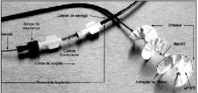
The Helex septal occluder is composed of a single fle-xible nitinol wire frame tridimensionally helically shaped, with a 0.012-inch diameter, covered with a thin polytetrafluo-roethylene membrane (PTFE better known as Gore-Tex) (fig.1). For implantation, the frame is sealed in a central man-drel and pulled inside a delivery catheter (9F) (fig 1). As the device is extruded from the catheter, it assumes a helicoidal configuration (therefore the name Helex). It is composed of 2 flexible disks with round borders that are positioned through the hole within the septum. The Helex septal occlu-der is available on the market in 5 diameters, 15 to 35mm, in 5mm increments. Three small metal loops also called “eye-lets” are found in the center of the disks, one more distal (outside the disk on the left side), one between the 2 disks, and another proximal, (outside the disk on the right side). Within the proximal waist is a retrieval cord made of po-lytetrafluoroethylene 18,19.
Objective - To evaluate the initial clinical experience with the Helex septal occluder for percutaneous closure of atrial septal defects.
Methods - Ten patients underwent the procedure, 7 patients with ostium secundum atrial septal defects (ASD) with hemodynamic repercussions and 3 patients with per-vious foramen ovale (PFO) and a history of stroke. Mean age was 33.8 years and mean weight was 55.4 kg. Mean diameter by transesophageal echocardiography and mean stretched ASD diameter were 11.33 ± 3.3mm, and 15.2 ± 3.8mm, respectively. The Qp/Qs ratio was 1.9 ± 0.3 in patients with ASD.
Results - Eleven occluders were placed because a pa-tient with 2 holes needed 2 devices. It was necessary to retrieve and replace 4 devices in 3 patients. We observed im-mediate residual shunt (< 2mm) in 4 patients with ASD, and in those with patent foramen ovale total occlusion of the defect occurred. No complications were noted, and all pa-tients were discharged on the following day. After 1 month, 2 patients with ASD experienced trivial residual shunts (1mm). In 1 patient, we observed mild prolapse in the pro-ximal disk in the right atrium, without consequences.
Conclusion - The Helex septal occluder was safe and effective for occluding small to moderate atrial septal de-fects. Because the implantation technique is demanding, it requires specific training of the operator. Even so, small technical failures may occur in the beginning of the learning curve, but they do not involve patient safety.
The Helex septal occluder comes preshaped out of the 9Fr delivery catheter, connected to its components the man-drel, control or implant catheter, and the retrieval cord that runs parallel to the mandrel within the control catheter (fig.1), and this within the delivery catheter, forming a tri-coaxial implantation system. For device loading, the central mandrel must be sprung progressively to stretch the nitinol frame, enabling the device to be pulled by the controlling catheter inside the delivery catheter under the water seal. Saline solution injections are performed through a Y system connected in the proximal end of the catheter to carefully withdraw the air bubbles from the system. An extremely flexible 9Fr delivery catheter is supplied with radiopaque markers at its distal tip, and it is advanced within the short sheath, inserted in the femoral vein, reaching the right atrium, and maneuvered toward the left atrium through the atrial septal defect, without the need for using long sheaths and guidewires in the left atrium 18,19.
Once positioned in the left atrium, incremental advan-cement of the control catheter and withdrawal of the man-drel results in configuration of the left atrial disk (fig. 2). When the left atrial portion of the device is completely formed, it is possible to observe a small central eyelet outside the tip of the delivery catheter. At this time, the left disk is positioned within the left atrium. The whole system is tractioned until the left disk reaches the interatrial septum (fig. 2). At this point, the operator feels a little resistance in the catheter and observes, on fluoroscopy the device mo-ving during a heartbeat. Correct positioning must be confir-med through transesophageal echocardiography. If device prolapse occurs through the atrial septal defect, the distal
disk may be recaptured, performing movements contrary to those described regarding the mandrel and the control ca-theter. The configuration of the right atrium disk is perfor-med as follows: the position of the device near the septum is maintained through mild and constant traction in the man-drel. The right atrial portion of the device is exposed when the delivery catheter is pulled to the inferior vena cava, and the control catheter is advanced for reconfiguration of the right disk, documented by fluoroscopy (fig. 2). Positioning of the device, which must “straddle” the septum, is confir-med through echocardiography. If the positioning is not adequate, the device may be withdrawn inside the delivery catheter, pushing the mandrel and tractioning the control catheter within the delivery catheter, just as the device is loaded before its entrance into the body. If the positioning is satisfactory, the device will be released from the compo-nents of the system after it is locked into place. To do this, the delivery catheter is advanced again over the control ca-theter to stabilize the device next to the septum. A red retrie-val cord cap (which tightens the retrieretrie-val cord) is removed from the proximal part (operator) and the control catheter is withdrawn around 1-2 cm inside the delivery catheter and advanced again to give some slack in the retrieval cord bet-ween the control catheter and the device. The mandrel is pulled definitively outside the system until a click is heard, which demands greater strength to finish the maneuver. This action results in the release of the tip of the nitinol fra-me, which leaves the control catheter and captures the pro-ximal metallic eyelet of the right disk, locking the disks and the device in the interatrial septum. The delivery catheter is again withdrawn to the inferior cava to allow the occluder to be completely free in the septum, without any traction from the catheters (fig. 3). However, the device is still connected to the system through the retrieval cord. At this moment, fi-nal positioning and the presence and size of residual shunts are assessed by echocardiography. If the final posi-tion is not satisfactory, the device can still be removed from the septum. The red retrieval cord cap of the device is con-nected to the system to immobilize the retrieval cord. The control catheter is smoothly and constantly tractioned pul-ling the cord, which pulls the device inside the delivery ca-theter (fig. 4). The operator may see and feel the thin polyte-trafluoroethylene membrane to be perforated by the hook
Fig. 1 - Helex device connected to the implant system with all its components.
Fig. 2 - Implant sequence in plastic model documented by fluoroscopy; (A and B) distal disk set after the delivery catheter reached the left atrium; C) left atrium disk withdrawn towards the interatrial septum with satisfactory position; (D and E) proximal disk set in the right atrium. The device is still connected to the implant system through the mandrel and the retrieval cord.
that blocks the disks. The tactile sensation is similar to that of a tissue removal, previously sewn to another with the stitches falling apart. After the total device is withdrawn within the guide catheter, the device loses its special confi-guration, and it can no longer be used. Such a rescue ma-neuver from the device through the retrieval cord must be carefully performed to avoid resistance that could lead to a fracture in the cord and loss of connection with the device, and consequent embolization. If the device is well
positio-ned, the retrieval cord should be released, advancing the delivery catheter to stabilize the device next to the septum and the control catheter should be carefully pulled taking with it the retrieval cord that is completely released from the proximal wire. Make sure the free end of the cord in the pro-ximal end (from the operator) of the system is moving freely during this maneuver. When the control catheter and the re-trieval cord are outside the delivery catheter, the device is fi-nally free from any connection with the system 18,19. As a
ge-neral rule, the diameter of the occluder should be around 1.7 to 2 times the stretched diameter. Although the determina-tion of the stretched diameter is important for selecting the appropriate Helex, it is not as crucial as it is with the Am-platzer10,14, where its occlusive mechanism and its stability
in the septum are dependent on its central portion, which is effectively inside the atrial septal defect and which must have a similar diameter to that stretched. The correlation between the size of the atrial septal defect in transesopha-geal echocardiography and the size of its stretched diameter depends on distensibility of the atrial septum, which may vary. Defects with similar diameters seen on echocardiogra-phy may have varied dimensions after stretching. Because the Helex has mechanisms different from those of the Amplatzer for fixation and stabilization in the septum, the diameter measured through echocardiography is as impor-tant as the stretched diameter. For example, a 12-mm central defect with a stretched diameter of 22 mm, can generally be occluded with a 30- or 35-mm Helex and should not be ex-cluded from selection. In cases of patent foramen ovale without an atrial septum aneurysm, 20- or 25-mm prostheses are selected. In the presence of an aneurysm, prostheses of 30- and 35-mm are those most recommended.
Diagnosis of ostium secundum atrial septal defects (ASD) was performed based on clinical, radiographic, elec-trocardiographic, and echocardiographic findings by using transthoracic echocardiography. Detailed anatomic charac-teristics of the defects were determined by transesopha-geal echocardiography performed after transthoracic echo-cardiography according to already described protocols 20.
As the Helex device was used only for mild to moderate atrial septal defect occlusion in previous experimental and clinical studies 18,19, we tried to select for this initial
expe-rience patients with ASDs 15- to 18-mm in diameter (at its longer axis), located in the center of the septum and with thick, broad rims. In adult patients with a history of tran-sient stroke, the transesophageal echocardiographic study with injection of microbubbles and use of the Valsalva ma-neuver demonstrated significant passage of the microbub-bles from right to left through foramen ovale. No other ap-parent cause for stroke was found after extensive neurolo-gical, cardiac, blood, and peripheral vascular investigation in these patients.
After venous puncture and introduction of small 9Fr sheaths, patients received heparin (100UI/kg, maximum 10,000UI). A routine cardiac catheterization with angiogra-phic studies and collection of samples for oximetry was per-formed before the occlusion period. In patients with patent
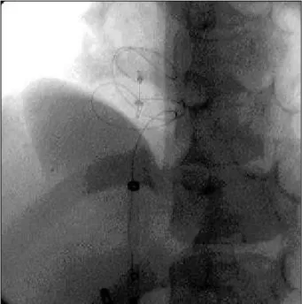
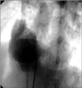
Fig. 3 - In-vivo device implanted documented by fluoroscopy. The mandrel was withdrawn and the device remains connected to the system through the retrieval cord. No tension exists on the device, enabling a better assessment of the final posi-tion in the septum.
foramen ovale, angiography and collection of samples for oxi-metry were not performed. The stretched diameter was deter-mined only in patients with atrial septal defects, using sizing balloons (AGA Medical Corporation) (24mm) with techni-ques similar to those already described8-14. After determining
the stretched diameter, the devices were selected and placed, following the steps described above. After final release of the device and angiographic, and echocardiographic evalua-tions, sheaths were withdrawn and hemostasis was obtained through manual compression. Patients recovered from anesthesia, were extubated in the catheterization laboratory, and then transferred to a recovery room.
The follow-up protocol required clinical evaluation with a chest X-ray and transthoracic echocardiography be-fore hospital discharge. Patients were advised to take aspi-rin (dosage 3-5mg/kg/day; maximum 100mg) and to perform prophylaxis for infectious endocarditis for at least 6 months, according to the routine protocol after percu-taneous closure of an atrial septal defect 8-14. Follow-up was
scheduled for 1, 3, 6, and 12 months after the procedure, with performance of transthoracic echocardiography at 1 and 6 months, and transesophageal echocardiography at 12 months. Fluoroscopy was routinely scheduled at 6 months to detect eventual fractures in the nitinol frame, or per-formed according to the individual need of each patient.
Due to the complexity of maneuvering the tri-coaxial system septal occluder device, 3 of the authors received spe-cific practical training in animals at W. Gore and Associates in Flagstaff, AZ, USA, before the experience in human subjects started; this training was sponsored by the company.
This study was approved by the ethical committees at both institutions. Although the Helex septal occluder is le-gal in Europe, it was going through aproval procedures by the Brazilian legislation when this study took place, and it is predicted to be approved in the 2nd quarter of 2002. All pa-tients or guardians gave written consent after a detailed
ex-planation of the procedure and of the features of this new device was provided.
Regarding the statistical analysis, values were expres-sed as mean standard deviations or median with minimum and maximum values. Fisher’s exact test was used to com-pare septal movement before and after the procedure. The ANOVA test was used to assess progressive decreases in right ventricle dimensions. The statistical significance level was determined as <0.05.
Results
Ten patients were selected for percutaneous occlu-sion of atrial septal defects with the Helex septal occluder under general anesthesia and transesophageal echocardio-graph guidance, following already described protocols 20,21.
Seven patients had ostium secundum atrial septal defects with hemodynamic repercussions, and 3 patients had patent foramen ovale and a history of strokes. The mean age was 12.3±4.8 years old (5-17; median 14 years), and weight was 41.7±19.1 kg (20-63; median 45 kg) in patients with atrial septal defects (tab. I). In patients with patent foramen ovale, the mean age was 41 years old (median 40 years, and weight was 73.3 kg (median 72 kg) (tab. II). All patients with atrial septal defects were asymptomatic and were not taking medication. Patients with patent foramen ovale had expe-rienced transient stroke episodes some months before the procedure and were taking aspirin or clopidogrel.
The mean diameter of the septal atrial defects in our patients was 11.3±3.3 mm (7.5-17 mm; median 10 mm) (tab. I). One patient with 2 ASDs (3.6 and 5mm) with a 12-mm distance between the holes was selected for the possible implantation of 2 devices, according to previous reports 22,23. The smaller
hole was in a postero-inferior position in the septum, near the inferior vena cava, and the other was in the antero-superior
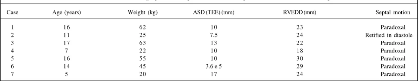
Table I - Clinical an echocardiographic data of patients with atrial septal defects before the occlusion procedure
Case Age (years) Weight (kg) ASD (TEE) (mm) RVEDD (mm) Septal motion
1 16 62 10 23 Paradoxal
2 11 25 7.5 24 Retified in diastole
3 17 63 13 22 Paradoxal
4 7 22 10 18 Paradoxal
5 16 55 10 30 Paradoxal
6 14 45 3.6 e 5 29 Paradoxal
7 5 20 17 24 Paradoxal
ASD - atrial septal defects; ETE - transesophageal echocardiography; RVEED - right ventricular end-diastolic diameter.
Table II - Clinical, hemodynamic and procedural data of patients with patent forame ovale
Case Age (years) Weight (kg) MPA (mmHg) Device (mm) Time (h) Technical problems Immediate RS
1 60 72 24/13 20 1 -
-2 23 70 20/8 20 0.7 -
-3 40 75 25/15 20 1 -
position behind the aortic torus. The mean of the stretched diameter was 15.2±3.8 mm, ranging from 12 to 20 mm in single atrial septal defects. In the patient with 2 holes, the stretched diameter from both ASDs was around 11-12 mm (fig. 5).
In all patients with atrial septal defects, the right ven-tricle was shown to be dilated on the transthoracic echocar-diography, 2 standard deviations higher than normal, and the interventricular septum had abnormalities in its move-ments, demonstrating hemodynamic repercussions (tab. I). All patients had normal pressure in the pulmonary ar-tery (tab. II and III). In patients with atrial septal defects, the ratio Qp/Qs was 1.9±0.3, according to the Fick method, and angiographic studies confirmed the presence of defects and a normal pulmonary vein connection.
Implantation was successful in all cases. However, 3 patients required withdrawal of 4 devices before the final re-lease due to technical problems related to implantation. In the first patient, a mandrel fracture occurred during opening of the left atrium disk due to inappropriate maneuvering of
the system by the operator (tab. III). The device was with-drawn within the delivery catheter and discarted. A new de-vice with the same diameter was successfully placed une-ventfully. In the 3rd patient, the retrieval cord was inadver-tently tractioned together with the withdrawal of the man-drel after proper locking of the disks. There was traction of the device that was already well positioned in the interatrial septum leading to erasure of its configuration (tab. III). With traction movements in the retrieval cord and rotation of the delivery catheter, the device was withdrawn toward the catheter inside the inferior vena cava and withdrawn outside the body without problems (fig. 4). A new device with the same diameter was successfully implanted. In the 4th patient, the operator did not maintain enough traction in the mandrel during the locking of the disks, leading them far from the interatrial septum, with an unsatisfactory final appearance. Using the retrieval cord, the device was with-drawn from the septum with erasure of is configuration and was withdrawn within the delivery catheter as already des-cribed. In a second attempt, accentuated tension in the man-drel was applied during locking of the disks, causing incom-plete locking with the “eyelet” of the proximal disk excluded from the locking hook. Still connected in the retrieval cord, the device was dismantled, and retrieved from the body as previously described. A third device, with the same diame-ter (25 mm), was successfully implanted (tab. III). In the other patients, the devices were implanted at the first attempt, without technical problems. In the patient with 2 ASDs, both devices were successfully implanted at the first attempt without problems. The inferior atrial septal defect was initially approached followed by the superior. The device used in the anterior-superior hole was greater than desirable because a smaller device was not available for implantation at that time. Thus, the superior part of the distal disk was in contact with the roof of the left atrium leading to mild prolapse towards its interior, but with satisfactory final results (fig. 6). In the 3 patients with patent foramen ovale, immediate echocardiography with microbubbles and Valsalva’s maneuver (hyperinflation with ambu ± abdominal compression) was negative without passage of contrast from the right to the left side (tab. II). Immediate residual shunt was present in 4 patients with atrial septal defects in the catheterization laboratory (tab. IV). In these patients, the
Fig. 5 - Stretched diameter determination in the patient with 2 separated holes. Two balloon catheters (AGA Medical Corporation, Golden Valley, MN) were positio-ned, one at each defect.
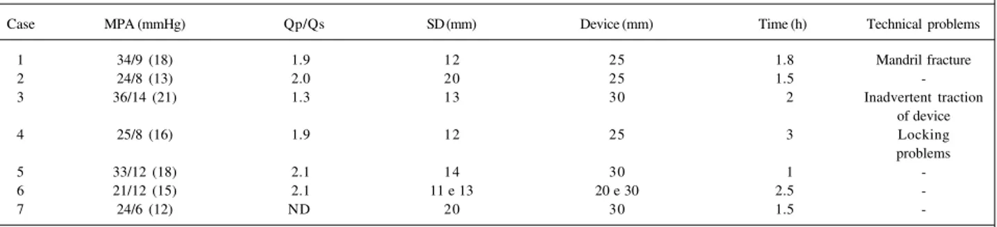
Table III - Hemodynamic and procedural data of patients with atrial septal defects
Case MPA (mmHg) Qp/Qs SD (mm) Device (mm) Time (h) Technical problems
1 34/9 (18) 1.9 12 25 1.8 Mandril fracture
2 24/8 (13) 2.0 20 25 1.5
-3 36/14 (21) 1.3 13 30 2 Inadvertent traction
of device
4 25/8 (16) 1.9 12 25 3 Locking
problems
5 33/12 (18) 2.1 14 30 1
-6 21/12 (15) 2.1 11 e 13 20 e 30 2.5
-7 24/6 (12) ND 20 30 1.5
duration of the procedure varied from 1 to 3 hours, with a mean of 1.9±0.7 hours (tab. IV). In patients with patent foramen ovale, the mean duration of the procedure was even shorter (0.9h) (tab. II). All patients were extubated in the catheterization laboratory, transferred to the anesthesia recovery room, and then to the ward. Intensive care was not necessary for any of the patients. Complications due to the procedure did not occur during hospitalization. Transthora-cic echocardiography demonstrated normalization of the septal movement in all patients with atrial septal defects the next morning. All patients were discharged the next day and returned for follow-up 1 month after the procedure.
A significant reduction in right ventricle diameter did not occur in the group of patients with atrial septal defects (final right ventricle diameter before the procedure, 24.3±4.5mm; at 24 hours, 22.5±3.6mm; at 1 month, 20 ± 3.6mm; P=0.17) (tab. IV). However, 5 patients had right ven-tricle dimensions within normal values for body surface at 1-month follow-up. In 1 patient (case 5), echocardiography revealed mild prolapse of the proximal disk into the right atrium without alterations in the local flow or adjacent struc-tures (cava, coronary sinus, tricuspid valve), and without residual shunt, confirmed at fluoroscopy. In the patient where 2 devices were implanted (case 6), a 1-mm residual shunt was present in the superior portion of the inferior de-vice and, the distal disk in the left atrium maintained the same initial configuration and positioning investigated by fluo-roscopy. Another patient (case 2) had a 1-mm residual shunt (tab. IV). All patients were asymptomatic and had no evi-dence of arrhythmias on the electrocardiogram.
Discussion
Although this experience with the Helex septal occlu-der is limited, we noted of some features of this device that were close to the ideal ones 19, and its advantages and
disad-vantages over other devices. Because it does not require long sheaths for implantation, it minimizes the risk of an unintentional air embolism 19. Its profile is extremely low,
fa-cilitating rapid in-growth of new tissue, which occurs at around 1 month, according to experimental studies 18.
Because the device is made of polytetrafluoroethylene, the long-term biocompatibility was not questioned 19. Its
flexi-ble nitinol frame and its round nonabrasive borders consi-derably minimize the risk of cardiac perforations 18,19,23. The
implantation system is controlled, and it may be recaptured and repositioned before its final deployment. Even after lo-cking of the disks, the device is still attached to the system through a thin retrieval cord made of Gore-Tex. It is possible to assess the device’s position in the atrial septum, without any tension on the delivery systems. This feature is only found in this device. Because it is very flexible and has an atraumatic contour, its rescue, in case of embolism, is safer, and with low risk of damage to the cardiac or vascular struc-tures 18,19,23. However, some disadvantages make its use
more limited: the implantation technique is not so simple, re-quiring special training of the operator. Even so, experienced operators who are familiar with percutaneous occlusion of atrial septal defects may make technical mistakes during the initial learning curve, as occurred during our experience. However, because of the features of the device and the
im-Table IV - Evolution of residual shunting, right ventricular dimensions and ventricular septal motion in patients with atrial septal defects
Case Immediate RS (mm) RS (mm) 24 hours RVEDD (mm) Septal motion RS (mm) (TTE) RVEDD (mm) Septal motion
(TEE) (TTE) 24 hours 24 hours 1 mounth 1 month 1 month
1 - - 23 Normal - 20 Normal
2 1 - NA Normal 1 19 Normal
3 - - 21 Normal - 19 Normal
4 2 2 17 Normal - 16 Normal
5 - - 28 Normal - 27 Normal
6 1 1 24 Normal 1 22 Normal
7 2 2 22 Normal - 17 Normal
RS - residual shunt; RVEDD - right ventricular end-diastole diameter; TEE - transesophageal echocardiography; TTE - transthoracic echocardiography; NA - non available.
plantation system, the problems resulting from these mista-kes were easily dealt with, putting neither the well-being of patients nor the success of the procedure in jeopardy. A current technical limitation of this device is the locking sys-tem between the 2 disks, which still requires simplification and greater operator experience to operate, as seen in 2 of our patients. Because its maximum diameter is 35mm, only small to moderate defects can be approached with such a device 19. Although the device is not self-centered during
implantation, this feature may be advantageous in cases of multiple close atrial septal defects or of a fenestrated sep-tum that may be occluded with a single device8,9,19,23.
Dis-tant defects may need 2 devices as was seen in our study and in other experiences 23. In these cases, the Helex’s low
profile is an advantage because it results in less intracardiac material. Patients with atrial septal defects associated with septal aneurysms with thin, highly flexible borders, also be-nefit from the stability mechanism in the septum, which does not depend on the determination of the stretched diameter, also observed with the CardioSEAL and STARFlex 8,9,18,19.
As polytetrafluoroethylene is a hydrophobic material and is not filled with water, echocardiographic views of the devi-ce are not very accurate, but this does not hinder the im-plantation procedure. Its cost is similar to that of other devi-ces available on the market (Amplantzer, CardioSEAL, STARFlex), which is still a limiting factor in our country.
Although this is still an initial experience, we consider the results obtained satisfactory regarding the safety and efficiency of this device. International experience based on a greater number of cases also corroborates this impression. As the clinical use was only started in December 2000 in Glasgow, Scotland, no articles are available reporting the results of parge series of patients undergoing implantation with this device, only abstracts of congresses 24-27. Thus far,
the Helex has been used in Europe, which has a less strict legislation for the use of such devices. A phase I study has been conducted in the USA to determine the safety and effi-ciency of its use in human beings. Data below were obtained from the international record controlled by the company that makes the device, and refer to ostium secundum atrial septal defects (Dr. Larry A. Latson, personal communication, São Paulo, Brazil, March 2002). Of 266 implantation attempts, in 21 cases (7.9%) the procedure was abandoned with removal of all devices in the catheterization laboratory. Of the 245 suc-cessful cases (92.1%), 68% were performed in females, me-dian age was 9 years old (1 to 74 years old) and the meme-dian weight was 33 kg (7.6 to 131kg). The mean diameter (not stretched) determined by transesophageal echocardiography was 13mm, ranging from 4.5 to 21mm. The occlusion rate was 95% after a 1-year follow-up, with no patient experiencing significant shunting, requiring reintervention. No significant immediate or late mortality or morbidity occurred related to the procedure. No clinical episodes suggestive of stroke or systemic thrombosis were observed. Late arrhythmias did not occur. However, fractures in the nitinol frame were noti-ced in 8.5% of the cases, with all patients being asymptoma-tic, without clinical complications due to this occurrence. In
only 1 patient was late withdrawal of the device the option. Embolism of the device occurred in 6 cases (2.5%), all retrieved in the catheterization laboratory, with no need for surgery and clinical complications. As we have seen in our initial experience and in the international cohorts, the use of the Helex septal occluder is safe and efficient for the closure of mild to moderate atrial septal defects.
Regarding patent foramen ovale occlusion in patients with a history of strokes, some considerations are necessary. Foramen ovale is not a atrial septal defect, but a valve that may open in special circumstances, with an increase in pressure in the right cavities, enabling flow from the right to the left 28-30 atrium. Epidemiologic studies demonstrate that
the occurrence of foramen ovale is more common in patients with stroke without a definitive cause (cryptogenic stroke) than in the general population (60% vs 10-25%) 31-33. This
as-sociation suggests that patent foramen ovale acts as a car-diac anatomic substrate for a probable paradoxical emboli-zation, although this is not strong enough to define a causal correlation. When an atrial septum aneurysm is present, this association is even stronger 33,34. The treatment of this
di-sease is based on secondary prophylaxis of recurrent episo-des of stroke and anti-coagulant and/or platelet antiaggre-gants are usually administered, with no consensus regar-ding the best scheme 35-37. Surgical occlusion has been
spo-radically reported in the literature 38,39, with recurrence rates
similar to those found with clinical treatment (3-5%) Howe-ver, it has intrinsic disadvantages because it is an invasive procedure with significant morbidity, especially regarding thoracotomy and extracorporeal circulation. With the ad-vent and improvement of devices for occlusion of atrial sep-tal defects, these patients have also been successfully trea-ted in the catheterization laboratory 40-44. Despite the
absen-ce of prospective and randomized studies in the literature, comparing clinical treatment with anticoagulants and per-cutaneous closure of atrial septal defects, longitudinal stu-dies from series of patients suggest that percutaneous treatment is, at least, as effective as clinical treatment, de-creasing the recurrence rate to around 2-4% per year 41-45.
Furthermore, taking into account the intention to treat it is probably more effective because it is permanent, since ma-ny patients discontinue anticoagulant treatment after some time. Morbidity from the procedure also seems to be smaller when compared with the chances of significant episodes of bleeding with the chronic use of anticoagulants 36,37. The
Amplatzer and CardioSEAL are the devices that have been more commonly used for the percutaneous occlusion of patent foramen ovale 43-45. Recent studies 43,44 have
demons-trated the use of the Helex septal occluder for this purpose with excellent results and lower rates of failure or complica-tions. The advantages of its properties are the basis for its use in the percutaneous treatment of ASD, demonstrated in our study. Closure rates above 95% are observed with the most commonly used devices 41-45. The absence of residual
shunt, observed in the 3 patients with patent foramen ovale in our study, is a predictive factor of lower rates of annual recurrences, about 1% 45.
ex-1. King TD, Mills NL. Nonoperative closure of atrial septal defects. Surgery 1974; 75: 383-8.
2. King TD, Sandra L, Thompson RN, Steiner C, Mills NL. Secundum atrial septal defect. Nonoperative closure during cardiac catheterization. JAMA 1976; 235: 2506-9.
3. Rashkind WJ. Transcatheter treatment of congenital heart disease. Circulation 1983; 67: 711-6.
4. Lock JE, Rome JJ, Davis R, et al. Transcatheter closure of atrial septal defects: ex-perimental studies. Circulation 1989; 79: 1091-9.
5. Sideris EB, Sideris SE, Fowlkes JP, Ehly RL, Smith JE, Gulde RE. Transvenous atrial septal occlusion in piglets using a Buttoned double disc device. Circula-tion 1990; 81: 312-8.
6. Babic UU. Double umbrella device for transvenous closure of patent ductus arterio-sus and atrial septal defects: first experience. J Interv Cardiol 1991; 4: 283-94. 7. DAS GS, Voss G, Jarvis G, Wyche K, Gunther R, Wilson RF. Experimental atrial
septal defect closure with a new catheter, self-centering device. Circulation 1993; 88: 1754-64.
8. Pedra CAC, Pihkala J, Lee KJ, et al. Transcatheter closure of atrial septal defects using the CardioSEAL implant. Heart 2000; 84: 320-6.
9. Pedra CAC, McLaughlin P, Benson LN. The role of CardioSEAL and Star-flex de-vices in atrial defect occlusion. Curr Interv Cardiol Rep 2000; 2: 274-82. 10. Sharafudin MI, Gu X, Titus JL, et al. transvenous closure of secundum atrial
sep-tal defects: preliminary results with new self-expanding nitinol prosthesis in a swine model. Circulation 1997; 95: 2162-8.
11. Fontes VF, Pedra CAC, Pedra SRF, et al. Experiência inicial no fechamento percu-tâneo da comunicação interatrial com a prótese de Amplatzer. Arq Bras Cardiol 1998; 70: 147-53.
12. Berger F, Ewert P, Bjornstad PG, et al. Transcatheter closure as standard treatment for most interatrial defects: experience in 200 patients treated with the Amplatzer Septal Occluder. Cardiol Young 1999; 9: 468-73
13. Hijazi ZM, Cao Q, Patel HT, Rhodes J, Hanlon KM. Transesophageal echocar-diographic results of catheter closure of atrial septal defect in children and adults using the Amplatzer device. Am J Cardiol 2001; 85: 1387-90.
14. Fontes VF, Pedra SRF, Braga SLN, Pedra CAC. Fechamento percutâneo da comu-nicação interatrial. Rev Soc Cardiol Estado de São Paulo 2002; 12: 293-305. 15. Berger F, Vogel M, Alexi-Meskishvili V, Lange PE. Comparison of results and
complications of surgical and Amplatzer device closure of atrial septal defects. J Thorac Cardiovasc Surg 1999; 118: 674-8.
16. Cowley CG, Lloyd TR, Bove EL, Gaffney D, Dietrich M, Rocchini AP. Compari-son of results of closure of secundum atrial septal defect by surgery versus Am-platzer septal occluder. Am J Cardiol 2001; 88: 589-91.
17. Pedra CAC, Pedra SRF, Fontes VF. Comunicação interatrial do tipo ostium se-cundum. Do tratamento cirúrgico ao percutâneo e os dinossauros do futuro. Arq Bras Cardiol 2003;80:650-5.
18. Zahn EM, Wilson N, Cutright W, Latson LA. Development and testing of the He-lex Septal Occluder, a new expanded polytetrfluoroethylene atrial septal defect occlusion system. Circulation 2001; 104: 711-6.
19. Latson LA, Zahn EM, Wilson N. Helex septal occluder for closure of atrial septal defects. Current Interv Cardiol Reports 2000; 2: 268-73
20. Pedra SRFF, Pedra CAC, Assef JE, et al. Fechamento percutâneo da comunicação interatrial: o papel da ecocardiografia. Arq Bras Cardiol 1999; 72: 59-64. 21. Bjornstad PG. Transcatheter closure of atrial septal defects demands co-operation
between the interventionist and the echocardiographer. Cardiol Young 2000; 10: 462-4.
22. Pedra CAC, Fontes-Pedra SRF, Esteves CA, Assef J, Fontes VF, Hijazi ZM. Multi-ple atrial septal defects and patent ductus arteriosus: successful outcome using two Amplatzer septal occluders and Gianturco coils. Cathet Cardiovasc Diagn 1998; 45: 257-9.
23. Dobrolet NC, Iskowits S, Lopez L, Whalen R, Zahn EM. Sequential implanta-tion of two Helex septal occluder devices in a patient with complex atrial septal anatomy. Cathet Cardiovasc Interv 2001; 54: 242-6.
24. Latson LA, Wilson N. A new transcatheter ASD-closure device. J Am Coll Cardiol 1999; 33 (Suppl.2): 520A
25. Wilson N, Latson L, Zahan E. A new low profile Nitinol – ePTFE flexible double disk occlusion device. Cardiol Young 1999; 9 (Suppl. 2): 14.
26. Zahn EM, Cheatham J, Latson LA, Wilson N. Results of in vivo testing of a new Ni-tinol ePTFE septal occlusion device. Cathet Cardiovasc Diagn 1999; 47: 124. 27. Zahn EM, Latson LA, Wilson N. Acute and long term follow up results with the
He-lex septal occluder in an animal model J Am Coll Cardiol 2000; 35 (Suppl. 2): 498A. 28. Chambers JB, de Belder MA, Moore D. Echocardiography in stroke and transient
ischemic attack. Heart 1997; 78 (Suppl. 1): 2-6.
29. Nagelhoult DA, Pearson AC, Labovitz AJ. Diagnosis of paradoxical embolism by echocardiography. Am Heart J 1991;121: 1552-4.
30. Di Tullio M, Sacco RL, Venketasubramanian N, et al. Comparison of diagnostic technique for the detection of patent foramen ovale in stroke patients. Stroke 1993; 24: 1020-4.
31. Lechat P, Mas JL, Lascault G, et al. Prevalence of patent forame ovale in patients with stroke. N Engl J Med 1988; 318: 1148-52.
32. Webster MW, Chancellor AM, Smith HJ, et al. Patent foramen ovale in young stroke patients. Lancet 1988; 2: 11-2.
33. Di Tullio M, Sacco RL, Gopal A. Patent foramen ovale as a risk factor for cryptoge-nic stroke. Ann Intern Med 1992; 117: 461-5.
34. Cabanes L, Mas JL, Cohen A, et al. Atrial sepral aneurysm and patent forame ovale as risk factors for cryptogenic stroke in patients less tha 55 years of age: a study using transesophageal echocardiography. Stroke 1993; 24: 1865-73. 35. Mas JL. Patent forame ovale, atrial septal aneurysm, and ischemic stroke in young
adults. Eur Heart J 1994: 15: 446-9.
36. Sacco RL, Di Tullio MR, Homma S. Treatment of patent forame ovale and stroke: to close or not to close, that is not yet the question. Eur Neurol 1997; 37: 205-6. 37. Nendaz M, Sarasin F, Bogousslavsky J. How to prevent stroke recurrence in
pa-tients with patent forame ovale: anticoagulants, antiaggregants, forame closure or nothing? Eur Neurol 1997; 37: 199-204.
38. Dearani JA, Ugurlu BS, Danielson GK, et al, Surgical patent foramen ovale clo-sure for prevention of paradoxical embolism-related cerebrovascular ischemic events. Circulation 1999; 100 (Suppl. 1): II-171-5.
39. Homma S, Di Tullio MR, Sacco RL, et al. Surgical closure of patent forame ovale in cryptogenic stroke patients. Stroke 1997; 28: 2376-81.
References
perience with the Helex device for percutaneous occlusion of atrial septal defect. Although some adjustments in its lo-cking mechanism and special training for the operator are re-quired, its features make it extremely useful for the treatment of select patients with mild to moderate atrial septal defects. Studies with a larger number of patients and with longer fol-low-up are necessary to better define the role of the Helex in the percutaneous closure of atrial septal defects and of patent foramen ovale.
Addendum - After this article was accepted for publication, another 2 procedures were performed for patent foramen ovale occlusion in patients with a history of stroke. In both cases, the defect was successfully occluded in the catheterization laboratory. In 1 case, device
withdra-wal was required before its final release for the placement of another with a larger diameter that could be better positio-ned in the atrial septum. None of the patients with patent fo-ramen ovale has experienced new episodes of stroke after implantation in an 8-month follow-up. In the 2 patients with atrial septal defect who had residual shunts at the initial cli-nical visits, complete occlusion was demonstrated by tran-sesophageal echocardiography performed during the clinical visit 6 months after implantation.
Acknowledgments
40. Bridges ND, Hellenbrand W, Latson L, et al. Transcatheter closure of patent fora-men ovale after presumed paradoxical embolism. Circulation 1992; 86: 1902-8. 41. Windecker S, Whal A, Chaterjee T, et al. Percutaneous closure of patent foramen ovale in patients with paradoxical embolism: long-term risk of recurrent throm-boembolic events. Circulation 2000; 101: 893-8.
42. Hung J, Landzenberg MJ, Jenkins KJ, et al. Closure of patent foramen ovale for pa-radoxical emboli: intermediate term. J Am Coll Cardiol 200; 35: 1311-6.
43. Sievert H, Horvath K, Zadan E, et al. Patent foramen ovale closure in patients with transient ischemia attack/stroke. J Interv Cardiol 2001; 14: 261-6. 44. Krumsdorf U, Keppeler P, Horvath K, Zadan E, Schrader R, Sievert H. Catheter
closure of atrial septal defects and patent forame ovale in patients with an atrial septal aneurysm using different devices. J Interv Cardiol 2001; 14: 49-55. 45. Meier B. Patent forame ovale - beauty spot or health threat? Cardiology Rounds