Comparison of Inflammatory Biomarkers between Diabetic and
Non-Diabetic Patients with Unstable Angina
Marçal de Oliveira Huoya
1; Rafaela Andrade Penalva
2; Sílber Rodrigues Alves
2; Gílson Soares Feitosa
1; Sandra Gadelha
3;
Ana Marice Teixeira Ladeia
2,4Divisão de Cardiologia - Hospital Santa Izabel1; Fundação Baiana Para o Desenvolvimento das Ciências2; Fundação Osvaldo Cruz3, Faculdade de Medicina da Universidade Federal da Bahia4, Salvador - Brazil
Summary
Background: Studies comparing inflammatory activity between diabetic and non-diabetic individuals with acute coronary syndrome are scarce, and none including only patients with unstable angina (UA) has been published to date.
Objective: We compared serum C-reactive protein (CRP), and interleukin-6(IL-6) between diabetic and non-diabetic patients with unstable angina (UA) to determine if difference in inflammatory activity is responsible for a worse prognosis in diabetic patients. We also evaluated the correlation between inflammatory markers and the metabolic profile in diabetic patients and the correlation between inflammatory response and in-hospital outcomes: death, acute myocardial infarction, congestive heart failure, and length of stay in hospital.
Methods: A prospective cohort study of 90 consecutive patients admitted to a chest pain unit with UA and divided into two groups, diabetic and non-diabetic. Serum CRP, IL-6, metabolic profile and leukocyte count were measured at hospital arrival.
Results: Forty-two patients (47%) were diabetic (age 62±9) vs. 48 (53%) non-diabetic (age 63±12). No differences between median C-reactive protein (1.78 vs. 2.23mg/l,p=0.74) and interleukin-6 (0 vs. 0pg/ml,p=0.31) were found between the two groups. There was a positive correlation between CRP and total cholesterol (rs = 0.21,p = 0.05), CRP and LDL-cholesterol (rs=0.22,p=0.04) and between CRP and leukocyte count (rs = 0.32, p = 0.02) in both groups. No associations were found between inflammatory markers and in-hospital outcomes.
Conclusion: We found no difference in inflammatory activity between diabetic and non-diabetic patients with UA, suggesting that this clinical condition may result in balanced inflammatory activity between the two groups and increase acute-phase proteins independently of metabolic state. (Arq Bras Cardiol 2009;92(4):269-274)
Key words: Atherosclerosis / complications; diabetes mellitus; inflammation / complications; angina pectoris.
Mailing address: Dr. Marçal de Oliveira Huoya •
Praça Almeida Couto, 500 - 40.000-000 – Salvador, BA - Brazil. E-mail: [email protected]
Manuscript received October 6, 2007; Revised manuscript received January 17, 2008; Accepted January 26, 2008.
Introduction
Atherosclerosis is responsible for 80% of all deaths in diabetic patients1. As the prevalence of diabetes is estimated
to double by the year 2025, the burden of cardiovascular disease with this condition will dramatically increase2.
Compared with non-diabetic individuals, diabetic patients have a two- to four-fold increased risk of coronary heart disease3, two fold-higher risk of short-term mortality
after acute myocardial infarction4, and poorer results when
submitted to angioplasty with increased risk of restenosis5.
Inflammatory activity is increased in individuals with type 16 and type 2 diabetes and strongly associated with risk of
atherothrombosis7.
C-reactive protein (CRP) is the principal downstream mediator of acute phase response and is primarily derived
via IL-6 dependent hepatic biosynthesis and has long been recognized as a very stable protein8. Levels of CRP increase
very rapidly in response to trauma, inflammation, and infection and decrease just as rapidly with the resolution of the condition9. Thus, measurement of CRP is widely used to
monitor various inflammatory states. The synthesis of CRP is predominantly under the control of IL-610, which in turn
has been assumed to originate from activated leukocytes, adipocytes, and endothelial cells11.
Increased levels of C-reactive protein (CRP) and interleukin-6 (IL-interleukin-6) are associated with higher incidence of adverse cardiac events in patients with acute coronary syndromes, as well as in patients undergoing coronary stenting12. Aggarwal et al13
found increased CRP levels (two fold) in diabetic than in non-diabetic subjects with acute coronary syndrome before and after coronary stenting.
interleukin-6 and leukocyte count levels between diabetic and non-diabetic patients to determine if differences in inflammatory activity could explain the worsened clinical or procedure results in diabetic patients with acute coronary syndrome.
Methods
Study design
In this observational, cross-sectional study with a comparison group, performed at the chest pain unit of the Santa Izabel Hospital from February to October of 2005, 90 patients with unstable angina pectoris were consecutively included, irrespective of age and gender. Patients were classified into two groups: diabetic and non-diabetic subjects. At admission, all patients were immediately examined by a cardiologist who measured their heart rate, blood pressure and pulse oximetry. All patients received 200 mg of aspirin and underwent 12-lead electrocardiograms. Blood samples for laboratory evaluation were drawn within 15 minutes of admission.
Inclusion criteria were age > 30 years old, both sexes, unstable angina and time from onset of chest pain to admission not superior to 12 hours.
Unstable angina was defined according to Braunwald14, and
only patients with class IIIB were included in the study. Patients with recent myocardial infarction (less than four weeks), dilated cardiomyopathy or Chagas heart disease, atrial fibrillation, valvular heart disease, raised levels of myocardial necrosis markers within 12 hours of admission (troponin T > 0.1 µg/ml and creatine kinase-MB mass > 4.94 µg/ml with a characteristic rise and fall curve), thromboembolism, surgery, PTCA in the last four weeks, inflammatory or neoplastic disease, chronic renal failure and fever in the previous week were excluded.
Patients were classified according to: age, sex, precordial pain at admission, TIMI risk score, diabetes duration, hypertension, smoking, previous coronary artery disease, percutaneous transluminal coronary angioplasty (PTCA), coronary artery bypass grafting (CABG), previous myocardial infarction, dyslipidemia, chest pain duration, time from onset of chest pain to admission (delta T in minutes), alcohol intake, family history of premature coronary artery disease and body mass index calculated from self-reported height and weight. Previous treatment used before inclusion in this study and also in-hospital treatment according to the chest pain unit routine protocol were recorded.
Diabetes was defined by occasional blood glucose � �00� �00 200 mg/dL or fasting glucose � ��� mg/dl� use of oral �ypoglycemic� ��� mg/dl� use of oral �ypoglycemic 126 mg/dl, use of oral hypoglycemic drugs or insulin treatment or self-reported history15.
Dyslipidemia was defined by high lipid levels according to the Third National Cholesterol Education Program16 or by use
of lipid-lowering drugs.
Death, acute myocardial infarction (AMI) characterized by troponin T levels � 0.� μg/ml and ck-MB-mass levels � 5.0 µg/ml with a characteristic rise and fall curve, congestive heart failure (CHF) occurring before discharge and length of stay in hospital were considered as in-hospital outcomes.
The local Bioethics Committee of our center approved the protocol study and all patients provided informed consent.
Laboratory evaluation
On admission, blood venous samples were obtained before intravenous administration of any drug (except for aspirin according to chest pain unit protocol), with a delay not superior to 15 minutes. Coded plasma samples were stored at –85o C. CRP was measured via a high-sensitivity latex
enhanced immunonephelometric assay on a BN II analyzer (Dade Bering, Newark, Del) at a Clinical Analyses Laboratory (LPC). The detection limit of C-reactive protein was 1.3 mg/l. The interassay coefficient of variation was 2.2%, and the intra-assay coefficient was 2.9%.
Plasma concentration of IL-6 was measured by a commercially available enzyme-linked immunosorbent assay (Quantikine human IL-6, R&D Systems, Minneapolis, MN) at Osvaldo Cruz Foundation (FioCruz). The lower detection limit of interleukin-6 was under 0.9 pg/ml. The interassay coefficient of variation was 6.4% and the intra-assay coefficient was 4.2%.
Plasma glucose, triglyceride, total cholesterol, and HDL-cholesterol were determinedusing enzymatic colorimetric assays (reagents obtained from Boehringer Manhein Diagnostics, Mannheim, Germany).In subjects who had plasma triglyceride levels < 400 mg/dl,LDL-cholesterol was calculated according to the Friedewald formula. When triglyceride levels were > 400 mg/dl, LDL-cholesterol was measured by the enzymatic method on a Hitachi 902 automatic analyzer (Boehringer Mannheim,Mannheim, Germany).
HbA1c levels were determined by a immunoturbidimetric immunoassay (Roche Diagnostics GmbH, D-68298 Manhein) with a normalrange of 4.5–6, %.
Statistical analyses
The study was designed with 80% power to detect a two-fold C-reactive protein value difference between two groups. To obtain this difference, a mean value of 2.8 mg/L and a standard deviation of 4.48 mg/L were used17. Considering a
type I error of 0.05, and a type II error of 0.20, 80 patients were required.
C-reactive protein and IL-6 concentrations are presented as median and interquartile range. Baseline characteristics were compared by chi-square test or Fisher’s exact test for categorical variables and Wilcoxon’s rank-sum or Student’s t test for continuous variables. Nonparametric tests were used to compare hs-CRP and IL-6 between groups (Mann-Whitney U test) and to determine correlations (Spearman’s p test). All p values are 2-tailed, and values lower than 0.05 were considered statistically significant.
SPSS statistical software (version 10.0, SPSS Inc., Chicago, Illinois, USA) was used for data analysis.
Results
Baseline characteristics
Three out of the 24 patients had diabetes. Ninety subjects who fulfilled the inclusion criteria were studied. Mean age was 62 ±9 (range 34-87) years; 42 were diabetic and 48 non-diabetic. Table 1 shows the baseline clinical and laboratory characteristics of non-diabetic and diabetic patients. There were no differences in baseline clinical characteristics between groups except for higher systolic blood pressure (158 ± 31 vs. 143 ± 31 mm/Hg, p = 0.02) and heart hate (75±15 vs. 67±12 beats/min, p=0.01) in diabetic patients at admission. The mean time from the onset of chest pain to admission in the chest pain unit was similar for diabetic and non-diabetic patients (4.3± 0.6 vs. 4.0± 0.5 h, p= 0.83). No differences were observed for previous treatment between diabetic and non-diabetic patients except for the use of hypoglycemic drugs and insulin.
Time since last meal until blood samples were drawn was also similar between groups (4.9± 3.1 vs. 5.0±3.3 h, p=0.95). There were no differences in laboratory characteristics between groups except for higher admission plasma glucose (178±78 vs. 105±27 mg/dl, p<0.001), fasting plasma glucose (130±40 vs 96±12 mg/dl, p<0.001), and HbA1c (6.3±4.2 vs. 3.4 ± 1.2 %, p < 0.001).
Inlammatory markers
In the entire population, median hs-CRP, IL-6 and leukocyte count was respectively 2 mg/L (interquartile range 0.8 – 4.7 mg/L), 0 pg/ml (interquartile range 0 – 0 pg/ml) and 7900 l/mm³ (interquartile range 6327– 9895 l/mm³).
Median hs-CRP (1.78 mg/L vs 2.23 mg/L, p = 0.74), IL-6 concentrations (0 pg/mL vs, 0 pg/mL, p = 0.31) and the leukocyte count (8245 l/mm³ vs, 7520 l/mm³, p = 0.74) were similar in diabetic and non-diabetic subjects (Table 2).
No difference was observed between groups when analyzed in accordance with three CRP ranges (<1mg/L, 1 to 3mg/L and >3mg/L): < 1mg/L (16% vs, 14%, p=0.5), 1-3 mg/L (10% vs. 18%, p=0.32) and > 3mg/L (14%vs 14%, p=0.7). The diabetic and non-diabetic groups had also similar distribution when analyzed in accordance with quartiles: <0.8 mg/L (15% vs, 12%, p=0.36), 0.81-2.04 mg/L (12% vs. 13%, p=0.95), 2.05-4.7 mg/L (8% vs, 16%, p=0.12 and >4.7 mg/L (14% vs. 13%, p=0.66).
When IL-6 levels were analyzed as a categorical variable (0 pg/mL = no and > 0 pg/mL = yes), positive results were similar in both groups, (17% vs. 8%, p=0.12).
In the entire population, C-reactive protein levels were positively correlated with total cholesterol levels, (rs = 0.21 p=0.05), LDL cholesterol levels (rs = 0.22, p=0.04) and leukocytes count (rs = 0.32, p = 0.02).
In-hospital outcomes
The overall mean hospital stay was 12 days, with a median of 7 days (range 2-125 days).
During the in-hospital period there were no differences between diabetic and non-diabetic subjects with respect to occurrence of congestive heart failure (5% vs. 0%, p=0.21), acute myocardial infarction (5% vs. 2%, p=0.60), death (25 vs. 0%, p=0.47) and median hospital stay (8 days vs. 7
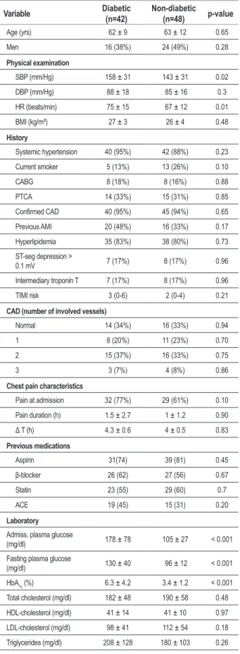
Table 1 - Baseline Characteristics
Variable Diabetic (n=42) Non-diabetic (n=48) p-value
Age (yrs) 62 ± 9 63 ± 12 0.65
Men 16 (38%) 24 (49%) 0.28
Physical examination
SBP (mm/Hg) 158 ± 31 143 ± 31 0.02
DBP (mm/Hg) 88 ± 18 85 ± 16 0.3
HR (beats/min) 75 ± 15 67 ± 12 0.01
BMI (kg/m²) 27 ± 3 26 ± 4 0.48
History
Systemic hypertension 40 (95%) 42 (88%) 0.23
Current smoker 5 (13%) 13 (26%) 0.10
CABG 8 (18%) 8 (16%) 0.88
PTCA 14 (33%) 15 (31%) 0.85
Conirmed CAD 40 (95%) 45 (94%) 0.65 Previous AMI 20 (48%) 16 (33%) 0.17
Hyperlipidemia 35 (83%) 38 (80%) 0.73
ST-seg depression >
0.1 mV 7 (17%) 8 (17%) 0.96
Intermediary troponin T 7 (17%) 8 (17%) 0.96
TIMI risk 3 (0-6) 2 (0-4) 0.21
CAD (number of involved vessels)
Normal 14 (34%) 16 (33%) 0.94
1 8 (20%) 11 (23%) 0.70
2 15 (37%) 16 (33%) 0.75
3 3 (7%) 4 (8%) 0.86
Chest pain characteristics
Pain at admission 32 (77%) 29 (61%) 0.10
Pain duration (h) 1.5 ± 2.7 1 ± 1.2 0.90
Δ T (h) 4.3 ± 0.6 4 ± 0.5 0.83 Previous medications
Aspirin 31(74) 39 (81) 0.45
β-blocker 26 (62) 27 (56) 0.67
Statin 23 (55) 29 (60) 0.7
ACE 19 (45) 15 (31) 0.20
Laboratory
Admiss. plasma glucose
(mg/dl) 178 ± 78 105 ± 27 < 0.001
Fasting plasma glucose
(mg/dl) 130 ± 40 96 ± 12 < 0.001
HbA1c (%) 6.3 ± 4.2 3.4 ± 1.2 < 0.001
Total cholesterol (mg/dl) 182 ± 48 190 ± 58 0.48
HDL-cholesterol (mg/dl) 41 ± 14 41 ± 10 0.97
LDL-cholesterol (mg/dl) 98 ± 41 112 ± 54 0.18
Table 2 - Comparison of Inlammatory Biomarkers
Variable Diabetic (n=42) Non-diabetic (n=48) p-value
CRP (mg/l) 1.78 (0.16-15) 2.23 (0.22-21) 0.74
IL-6 (pg/ml) 0 (0-40) 0 (0-17) 0.31
Categorical IL-6 7 (17%) 4 (8%) 0.12
WBC
(leukocytes/mm³) 8245 (3350-13100) 7520 (5170-16930) 0.74
Values expressed as number (%) or median (minimum-maximum).; RP - C-reactive protein; IL-6 - interleukin-6; WBC - white blood cells;
days, p=0.44). When combined outcomes were analyzed, events were also similar in both groups (12% vs. 2%, p=0.06, RR=5.7; 95% CI 0.7 to 56) (Table 3).
When subjects who had in-hospital events were compared with those without events, median hs-CRP was similar in both groups for CHF (8.1mg/L vs 2.4 mg/L, p=0.47), AMI (7.8mg/L vs 1.7 mg/L, p=0.81) and cardiovascular death (5.66 mg/L vs 2.04 mg/L, p=0.41). No difference was likewise observed for respectively median IL-6 and leukocyte count to CHF (0 pg/mL vs 0 pg/mL, p = 0.58; 9590 l/mm³ vs 7460 l/mm³, p = 0.53), AMI (0 pg/mL vs 0 pg/mL, p = 0.58; 7925 l/mm³ vs 7860 l/mm³, p = 0.5) and death (0 pg/mL vs 0 pg/mL, p = 0.70; 11500 l/mm³ vs 7900 l/mm³, p = 0.22).
There was no correlation between length of stay in hospital and hs-CRP levels (rs=0.02, p=0.86), IL-6 (rs=0.09, p=0.44) or leukocyte count (rs=0.21, p=0.85).
Discussion
There are few clinical studies comparing inflammation markers between diabetic and non-diabetic subjects with acute coronary syndrome, especially involving exclusively patients with unstable angina.
Moreno et al18 found that, in diabetic patients, coronary
tissue exhibits a larger content of lipid-rich atheroma, macrophage infiltration, and subsequent thrombosis than tissue from patients without diabetes, suggesting that there is an increased vulnerability for plaque disruption and thrombosis in patients with diabetes mellitus. The same author reviewed the pathogenesis of diabetes atherosclerosis19 and
supported the idea that patients with diabetes mellitus have
Table 3 - In-Hospital Outcomes in Diabetic and non-Diabetic Patients
Variable Diabetic (n=42) Non-diabetic (n=48) Valor de p
CHF 2 (5%) 0 (0%) 0.21
AMI 2 (5%) 1 (2%) 0.60
CV death 1 (2%) 0 (0%) 0.47
hospital STAY (day) 8 (3-125) 7 (2-44) 0.44
Combined outcomes 5 (12%) 1 (2%) 0.06
Values as expressed as number (%) or median (minimum-maximum).; HF - congestive heart failure; AMI - acute myocardial infarction; CV - cardiovascular.
more inflammatory activity than the general population with atherosclerosis.
However, in this study no differences were observed in hs-CRP and IL-6 concentrations between diabetic and non-diabetic patients with unstable angina.
Aggarwal et al13, evaluating inflammatory activity measuring
hs-CRP levels in 75 patients with acute coronary syndrome before, during and after coronary stenting in diabetic and non-diabetic individuals, concluded that diabetic patients with acute coronary syndrome had increased inflammatory activity before the procedure but after 24 hours there were no differences between diabetic and non-diabetic patients. Nevertheless the authors in their methodology did not describe fundamental information to conclude that in fact they had studied a population with acute coronary syndrome. Data on the time from onset of chest pain to admission or even the stenting procedure (first blood sample) was not informed. Moreover the author included patients with stable angina and non-ST elevation acute myocardial infarction when the necrosis factor could act as a confounding variable20 and
differently from our study, which used validated criteria to define diabetes, the authors did not even describe glucose levels.
Analyzing the CRP levels between the two groups, we observed before stenting procedure that diabetic patients had two-fold inflammatory activity than non-diabetic patients but after coronary stenting (endothelial aggression) there were no differences in hs-CRP concentrations between the two groups. Gottsauner et al21 found that the persistence of elevated
C-reactive protein plasma levels 96 h after stent implantation might reflect a prolonged inflammatory reaction to coronary stent implantation.
The role of acute endothelial aggression as a stimulus to an intense inflammatory response could be also present in unstable angina and would justify our findings and enhance the significance of our results.
It is interesting to note that in our study, we used a very careful methodology to include UA patients. All subjects were rigorously labeled by validated criteria as unstable angina to avoid interference of the necrosis factor as a confounding variable. Were admitted in a reference emergence unit to cardiologic patients which the pos-test probability is higher Continuation of Table 1 - Baseline Characteristics
Ck-mb mass (μg/ml) 2.37 ± 1.0 2.4 ± 1.5 0.86
Troponin T(μg/ml) 0.01(0.01-0.09) 0.01 (0.01-0.14) 0.58
than 80% to patients with typical chest pain and age equal or superior to 60 years. Moreover, at the end of the study 94% of the patients had the atherosclerotic disease confirmed by coronary angiography. Lastly, the homogeneity between groups was demonstrated by the lack of statistically significant differences in baseline characteristics.
Our findings with statistically significant differences (SBP and HR) may be explained by the trend towards more severe chest pain at admission in the diabetic group, contrary to the original concept22 that is currently being highly questioned23.
Contrary to our study, Sanchez et al24 observed that
diabetic patients with non-ST elevation acute coronary syndrome showed higher inflammatory activity than non- diabetic ones. Nevertheless, diabetic patients had a higher leukocyte count than non-diabetic subjects, and the authors included no instrument in their methodology to detect silent infectious process, especially in diabetic women25. In our
study, we conducted urinalysis in all the patients and explored the upper airways, including the auditory canal, to exclude silent infections. Moreover, there were no differences in leukocyte count measured at admission between diabetic and non-diabetic patients, suggesting no differences in previous inflammatory condition due to systemic infectious process.
Moreover, Sanchez did not describe admission and fasting blood glucose and lipid profile. It is important to underscore that the metabolic state of diabetes is characterized by hyperglycemia and dyslipidemia and is associated to endothelial lesion and consequently to increased inflammation markers19,26,27.
Regarding the positive correlation between hs-CRP concentrations and leukocyte count, Danesh et al28 described
a strong association between leukocyte count and coronary artery disease development and Stewart et al29 observed higher
mortality reduction associated to the use of pravastatin in those patients who showed increased leukocyte count values at admission.
In this study, upon analyzing all included patients with unstable angina, a weak correlation was observed between hs-CRP and total and LDL cholesterol, but superior to that found in previous studies30,31 that did not include patients with
acute coronary syndrome.
In our study, we did not observe higher in-hospital outcomes between diabetic and non-diabetic patients despite literature findings32, probably because of the reduced follow-up period
(median 8 days) and few patients included (small sample), to find more in-hospital outcomes such as CHF, AMI and death (reduced power) in patients with unstable angina.
Another limitation was the fact that the mean time from onset of chest pain to admission in the emergency room (4 h) until collection of blood samples to measure hs-CRP may have been insufficient to find the inflammatory process in course.
However, this occurrence would not explain the similar hs-CRP results between two groups.
Our findings suggest that events involved in an acute coronary screening, such as unstable angina, propitiate an important, yet similar, inflammatory response between diabetic and non-diabetic patients, suggesting that inflammation response resulting from an acute coronary syndrome is more intense and acute than the inflammation response resulting from diabetes where the process is more continuous and slower and endothelial aggression is lasting. Another possibility is that the use of statin (in our study similar in both diabetic and non-diabetic patients) may be more effective in diabetic patients, since more intense anti-inflammatory effect was described in individuals with enhanced inflammatory activity29.
It is important to underscore that our diabetic patients were well controlled (mean Hba1c=6.3%) and this finding may have decreased hyperglycemia impact on endothelial aggression.
Regarding everything we discussed in our study, some questions must be answered: Are the inflammation characteristics observed in diabetes different from those in acute coronary syndrome? Is there any way to evaluate this? Is the statin effect higher among diabetic than among non-diabetic patients? Could this superior effect explain the equality of inflammation activity between groups when they were admitted in the study with acute coronary syndrome? Is the inflammatory response in an acute coronary syndrome so intense that it could equilibrate inflammation activity between diabetic and non-diabetic patients?
Our findings allow us to conclude in this group of patients that inflammation activity is similar between diabetic and non-diabetic patients with unstable angina and in these patients hs-CRP showed a positive correlation with total and LDL cholesterol as well as with leukocyte count. These findings also underscore the importance of CRP as an endothelial aggression marker in acute coronary syndrome, as well as of the multifactorial aspect of the atherothrombosis process.
Potential Conflict of Interest
No potential conflict of interest relevant to this article was reported.
Sources of Funding
This study was partially funded by Fundação de Amparo a Pesquisa do estado da Bahia (FAPESB).
Study Association
References
1. Roffi M; Topol EJ. Percutaneous coronary intervention in diabetic patients with non-ST-segment elevation acute coronary syndromes. Eur Heart J. 2004; 25 (3): 190-8.
2. King H, Aubert RE, Herman WH. Global burden of diabetes, 1995-2025: prevalence numerical estimates, and projections. Diabetes Care. 1998; 21: 1414-31.
3. Wilson PWF, Kannel WB. Epidemiology of hyperglycemia and atherosclerosis. In: Ruderman N, Williamson J, Brownlee M. (eds.) Hyperglycemia, diabetes, and vascular disease. New York: Oxford University Press; 1992. p. 21-9.
4. Woodfield SL, Lundergan CF, Reiner JS, Greenhouse SW, Thompson MA, Rohrbeck SC, et al. Angiographic findings and outcome in diabetic patients treated with thrombolytic therapy for acute myocardial infarction: the GUSTO-I experience. J Am Coll Cardiol. 1996; 28: 1661-9.
5. West Ne, RuyGrok PN, Disco CM, Webster MW, Lindeboom WK, O’Neil WW, et al. Clinical and angiographic predictor of restenosis after stent deployment in diabetic patients. Circulation. 2004; 109 (7): 867-73.
6. Schalkwijk CG, Poland DC, van Dijk W, Kok A, Emeis JJ, Drager AM, et al. Plasma concentration of C-reactive protein is increased in type-I diabetic patients without clinical macroangiopathy and correlates with markers of endothelial dysfunction: evidence for chronic inflammation. Diabetologia. 1999; 42: 351-7.
7. Jager A, Van Hinsbergh VW, Kostense PJ, Emeis JJ, Yudkin JS, Nijpels G, et al. Von Willebran factor, C-reactive protein and 5-year mortality in diabetic and non-diabetic subjects: the Horn Study. Arterioscler Thromb Vasc Biol. 1999; 19: 3071-8.
8. Rifai N, Ridker PM. Population dsistributions of C-reactive protein in apparently healthy men and women in the United States: implication for clinical interpretation. Clin Chem. 2003; 49: 666-9.
9. Du Clos TW. Function of C-reactive protein. Ann Med. 2000; 32: 274-8.
10. Heinrich PC, Castell JV, Andus T. Interleukin-6 and the acute phase response. Biochem J. 1990; 265: 621-36.
11. Danesh J, Collins R, Peto R. Chronic infections and coronary heart disease: is there a link? Lancet. 1997; 350: 430-6.
12. Buffon A, Liuzzo G, Biasucci LM, Pasqualetti P, Ramazzotti V, Rebuzzi AG, et al. Preprocedural serum levels of C-reactive protein predict early complications and late restenosis after coronary angioplasty. J Am Coll Cardiol. 1999; 34: 1512-21.
13. Aggarwal A, Schneider DJ, Sobel BE, Dauerman HL. Comparison of inflammatory markers with diabetes mellitus versus those without before and after coronary arterial stenting. Am J Cardiol. 2003; 92: 924-9.
14. Braunwald E. Unstable angina: a classification. Circulation. 1989; 80: 410-4.
15. Kehoe R, Wu SY, Leske MC, Chylack LT Jr. Comparing self-reported and physician reported medical history. Am J Epidemiol. 1994; 139 (8): 813-8.
16. National Cholesterol Education Program (NCEP) Expert panel on detection, evaluation, and treatment of high blood cholesterol in adults (Adult Treatment Panel III). Third Report of the National Cholesterol Education Program (NCEP) Expert Panel on Detection, Evaluation, and Treatment of High Blood Cholesterol in Adults (Adult Treatment Panel III) final report. Circulation. 2002; 106: 3143-421.
17. Schulze MB, Rifai N, Rimm EB, Stampfer MJ, Li T, Hu FB. C-reactive protein and incident cardiovascular events among men with diabetes. Diabetes Care. 2004; 27: 889-94.
18. Moreno PR, Murcia AM, Palacios IF, Leon MN, Bernardi VH, Fuster V, et al. Coronary composition and macrophage infiltration in atherectomy specimens from patients with diabetes mellitus. Circulation. 2000; 102: 2180-4.
19. Moreno PR, Fuster V. New aspects in the pathogenesis of diabetic atherothrombosis. J Am Coll Cardiol. 2004; 44: 2293-300.
20. De Servi S, Mariani M, Mariani G, Mazzone A. C-reactive protein increase in unstable coronary disease: cause or effect? J Am Coll Cardiol. 2005; 46: 1496-502.
21. Gottsauner-Wolf M, Zasmeta G, Hornykewycz S, Nikfardjam M, Stepan E, Wexberg P, et al. Eur Heart J. 2000; 21: 1152-8.
22. Bradley RF, Schonfield A. Diminished pain in diabetic patients with acute myocardial infarction. Geriatrics. 1962; 17: 322-6.
23. Airaksinen KEJ. Silent coronary artery disease in diabetes: a feature of autonomic neuropathy or accelerated atherosclerosis? Diabetologia. 2001; 44: 259-66.
24. Sanchez PL, Morinigo JL, Pabon P, Martin F, Piedra I, Palacios IF, et al. Prognostic relations between inflammatory markers and mortality in diabetic patients with non-ST elevation acute coronary syndrome. Heart. 2004; 90: 264-9.
25. Stapleton A. Urinary tract infections in patients with diabetes. Am J Med. 2002; 113 (Suppl. 1A): 80S–84S.
26. Schram MT, Chaturvedi N, Schalkwijk C, Giorgino F, Ebeling P, Fuller JH, et al. Vascular risk factors and markers of endothelial function as determinants of inflammatory markers in type 1 diabetes. Diabetes Care. 2003; 26: 2165-73.
27. Weyer C, Yudkin JS, Stehouwer CD, Schalkwijk CG, Prateley RE, Tataranni PA. Humoral markers of inflammation and endhotelial dysfunction in relation to adiposity and in vivo insulin action in Pima Indians. Atherosclerosis. 2002; 161: 233-42.
28. Danesh J, Collins R, Appleby P, Peto R. Association of fibrinogen, C-reactive protein, albumin, or leukocyte count with coronary heart disease. JAMA. 1998; 279: 1477-82.
29. Stewart RAH, White HD, Kirby AC, Heritier SR, Simes RJ, Nestel PJ, et al. White blood cell count predicts reduction in coronary heart disease mortality with pravastatin. Circulation. 2005; 111: 1756-62.
30. Ridker PM, Stampfer MJ, Rifai N. Novel risk factors for systemic atherosclerosis: a comparison of C-reactive protein, fibrinogen, homocysteine, lipoprotein (a), and standard cholesterol screening as predictors of peripheral arterial disease. JAMA. 2001; 285: 2481-5.
31. Ridker PM, Rifai N, Rose L, Buring J, Cook NR. Comparison of C-reactive protein and low-density lipoprotein cholesterol levels in the prediction of first cardiovascular events. N Engl J Med. 2002; 347: 1557-65.