From the Laboratory of Medical Investigation 51, Clinical Emergencie Discipline, Hospital das Clínicas, Faculty of Medicine, University of São Paulo – São Paulo/SP, Brazil.
E-mail: [email protected] Received for publication on
July 31, 2003.
ORIGINAL RESEARCH
LOW CORONARY PERFUSION PRESSURE IS
ASSOCIATED WITH ENDOCARDIAL FIBROSIS IN A
RAT MODEL OF VOLUME OVERLOAD CARDIAC
HYPERTROPHY
Maria Carolina Guido, Márcia Kiyomi Koike and Clovis de Carvalho Frimm
GUIDO MC et al. Low coronary perfusion pressure is associated with endocardial fibrosis in a rat model of volume overload cardiac hypertrophy. Rev. Hosp. Clín. Fac. Med. S. Paulo 59(4):228-235, 2004.
Left ventricular hypertrophy following volume overload is regarded as an example of cardiac remodeling without increased fibrosis accumulation. However, infarction is associated with increased fibrosis within the noninfarcted, hypertrophied myocardium, particularly in the subendocardial regions. It is conceivable to suppose that, as also occurs postinfarction, low coronary driving pressure may also interfere with accumulation of myocardial fibrosis following aortocaval fistula.
PURPOSE: To investigate the role of acute hemodynamic changes in subsequent deposition of cardiac fibrosis in
response to aortocaval fistula.
METHOD: Aortocaval fistula were created in 4 groups of Wistar rats that were followed over 4 and 8 weeks: aortocaval
fistula 4 and aortocaval fistula 8 (10 rats each) and their respective controls (sham-operated controls - Sh), Sh4 and Sh8 (8 rats each). Hemodynamic measurements were performed 1 week after surgery. Hypertrophy and fibrosis were quantified by myocyte diameter and collagen volume fraction at the end of follow up.
RESULT: Compared with Sh4 and Sh8, pulse pressure, left ventricular end-diastolic pressure, and +dP/dt were higher in
aortocaval fistula 4 and aortocaval fistula 8, but –dP/dt wassimilar. Coronary driving pressure (mm Hg), used as an estimate of perfusion pressure, was lower in aortocaval fistula 8 (52.6 ± 4.1) than in Sh8 (100.8 ± 1.3), but comparable between aortocaval fistula 4 (50.0 ± 8.9) and Sh4 (84.8 ± 2.3). Myocyte diameter was greater in aortocaval fistula 8, whereas interstitial and subendocardial fibrosis were greater in aortocaval fistula 4 and aortocaval fistula 8. Coronary driving pressure correlated inversely and independently with subendocardial fibrosis (r2 = .86, P <.001), whereas left ventricular systolic pressure (r2 = 0.73,
P = .004) and end-diastolic pressure (r2 = 0.55, P = 012) correlated positively and independently with interstitial fibrosis.
CONCLUSION: Coronary driving pressure falls and ventricular pressures increase early after aortocaval fistula and are
associated with subsequent myocardial fibrosis deposition.
KEY WORDS: Myocardial fibrosis. Aortocaval fistula. Coronary driving pressure. Volume overload. Cardiac remodeling.
An increase in myocyte volume ac-companied by an expansion of the ex-tracellular matrix characterize the de-velopment of myocardial hypertrophy in response to cardiac overload.1,2,3 The
positive effect is the preservation of cardiac performance by preventing an increase in myocardial oxygen con-sumption.4,5,6
Although initially beneficial, hypertrophy may ultimately result in
cardiac failure.1,7,8 One of the potential
mechanisms involved is the advent of myocardial ischemia, which may fol-low a disproportionate growth of myocytes relative to coronary ves-sels.9,10,11 Recurrent ischemic episodes
predictably result in myocyte necrosis and scar formation.12,13,14
car-diac hypertrophy, it has been repeatedly documented that the collagen volume fraction (CVF) increases.15,16,17,18 Two
main forms of fibrillar collagen accumu-lation are currently acknowledged: re-active and reparative fibrosis.19,20,21,22
Reactive interstitial fibrosis follows the development of pressure overload hypertrophy, in particular, and is char-acterized by an increased accumulation of collagen fibers intermingled among myocytes.16,23 Reparative fibrosis results
from myocyte loss and represents wound healing scarring, such as occurs following myocardial infarction.12,15
In eccentric hypertrophic response to volume overload, myocyte growth and fibrillar collagen accumulation appear to develop in a proportional manner.24-26 However, increased
fibro-sis has recently been reported in cases of volume overload.10,11,27,28 In
infarc-tion, the development of interstitial and subendocardial (SE) fibrosis within noninfarcted myocardium has been recognized.12,29,30 In this case, SE
fibrosis appears to represent a repair process in response to persistent ischemia in this region, since the coro-nary driving pressure (CDP) has been documented to be low during the early phases after infarction12.
It is conceivable to suppose that a similar mechanism might have a role in collagen fiber accumulation in volume overload hypertrophy. If the initial hemodynamic burden affects the CDP, as already demonstrated for large infarcts,12 SE fibrosis is likely to
super-vene. Furthermore, interstitial fibrosis might also follow if, in addition to vol-ume overload, afterload were increased during hypertrophy development.
In experimental cardiac hypertro-phy, hemodynamic monitoring during surgical procedures is rarely per-formed. Consequently, the effects of acute hemodynamic changes on sub-sequent myocardial remodeling are currently undetermined.
The aim of the present study is to
investigate in a experimental model of volume overload cardiac hypertrophy: 1) whether or not there is increased car-diac collagen accumulation, 2) the dis-tribution and type of fibrosis, and 3) the potential role of acute hemodynamic changes in the subse-quent development of myocardial fi-brosis.
METHOD
Animals and groups
Wistar rats weighing 280 to 350 g were used. All procedures were carried out in accordance with the norms of the Brazilian College of Animal Ex-periments and conformed to the “Guide for the Care and Use of Labo-ratory Animals”. Our Institutional Ethi-cal Committee approved the protocol (CAPPesq, 160/02).
Four groups of animals with surgi-cally created aortocaval fistula (ACF) were constituted: ACF4 and ACF8, 10 rats each, and their respective sham-operated controls (Sh), Sh4 and Sh8, 8 rats each, followed over 4 and 8 weeks, respectively.
Experimental model
Surgical procedures were carried out using a modified technique that has been previously described31,32.
Un-der 10% chloral hydrate anesthesia (300 mg/kg, intraperitoneally)33 and
positive pressure ventilation (Harvard Rodent ventilator – Model 683 -South Natick, Massachusetts, EUA), a median laparotomy was performed, and the abdominal aorta and the infe-rior vena cava were identified and iso-lated from neighboring structures un-derneath the emergence of the renal ar-teries. Arterial and venous blood flow was briefly interrupted with the aid of vascular clamps to permit the insertion of an 18-gauge needle into the aorta.
The needle was carefully advanced, and the posterior aortic wall was punc-tured 3 to 4 times to the inside of the vena cava. After removing the needle, homeostasis was conducted with local application of super glue. The abdomi-nal wall was closed and the animals returned to their cages after full recov-ery.
The mortality rate was 8% imme-diately, 29% 48 hours after surgery, and 23% during long-term follow up. Fistula permeability was assessed daily with a pediatric stethoscope.
Hemodynamic measurements
One week after surgery, surviving rats were anesthetized again and sub-mitted to simultaneous systemic and cardiac hemodynamic measurements (hemodynamics—phase 1). Briefly, a 0.05 mm polyvinyl chloride catheter was inserted through the right carotid artery into the ascending aorta and ad-vanced into the left ventricle (LV). An-other catheter was inserted through the jugular vein into the superior vena cava and advanced into the right atrium. Also, a 0.01 mm catheter was introduced through the left femoral ar-tery and advanced into the descending abdominal aorta. The catheters were connected to pressure transducers and coupled with a calibrated pre-amplifier (General Purpose Amplifier 4 - model 2, Stemtech Inc. WI, USA). Pressure tracings were recorded and analyzed using a computerized system proces-sor (Windaq - AT/Codas, Dataq Instru-ments Inc., OH, USA).
mm Hg·s-1) were obtained as measures
of LV systolic and diastolic function, respectively.
Pulse pressure (mm Hg) was calcu-lated as the difference between sys-temic systolic arterial pressure and diastolic arterial pressure and used as a measure of fistula size.
CDP (mm Hg) calculated by the difference between systemic diastolic arterial pressure and LV end-diastolic pressure was used as an estimate of myocardial perfusion pressure.34
After 4 and 8 weeks of follow up, before sacrifice, rats were again anes-thetized, and the femoral artery was catheterized for new systemic systolic arterial pressure, diastolic arterial pres-sure, and pulse pressure determinations (hemodynamics—phase 2).
All measurements corresponded to the mean value of each hemodynamic parameter continuously recorded dur-ing a 10-minute period after stabili-zation of the pressure curves.
Anatomic and morphometric study
The animals were sacrificed using a 100 mM CdCl2 solution and were perfusion-fixed with 10% formalin un-der a perfusion pressure corresponding to the systemic diastolic arterial pres-surein vivo. The heart, lungs, and liver were removed, cleaned, and weighed. The dry weight of the lungs and liver were used for calculating the percent-age of water content in each tissue, which was used as an estimate of con-gestive heart failure. After determining heart / body weight (g·kg-1), the right
and the left atrium were carefully trimmed out and weighed separately.
A 1 to 2 mm coronal slice of the heart, including both ventricles, was cut at the equatorial plane to obtain 7 µm thick paraffin-embedded tissue sections. After being stained with hematoxylin and eosin (HE) and picrosirius red,35 these sections
under-went morphometric analysis using an
image analysis system (Leica Q500 iW, Leica Imaging Systems ltd., Cam-bridge, UK).
At first, each HE tissue section was visualized under a macro lens (28 mm, Tamron, Japan) to permit the determi-nation of LV wall thickness and LV cavity dimension, both expressed as tissue areas (µm2). Subsequently, LV
myocyte hypertrophy was estimated under a 100x microscopic objective by measuring myocyte transversal diam-eter around the nucleus (µm).
Fibrillar collagen was identified in the picrosirius red-stained sections by its red color. Under a 40x microscopic objective, an estimate of CVF was de-termined to be the mean percentage of red-stained connective tissue areas per total myocardial area in each micro-scopic field (%). The CVF was ad-dressed separately in 2 distinct re-gions: the inner third of the LV, corre-sponding to the subendocardium, and the medium third of the interventricu-lar septum. An average of 15 to 20 fields was examined in each of these 2 myocardial regions.
Perivascular collagen (%) was cal-culated separately as the ratio of picrosirius red-positive stained areas around microvessels to luminal perim-eter. Only 50 to 200 µm arterioles with major to minor diameter ratio <0.60 were computed.
Statistical Analysis
Variables are expressed as mean ± SE. One-way analysis of variance or Kruskal-Wallis on ranks were used for intergroup comparisons of data. Post hoc comparisons were performed using Bonferroni’s and Dunn’s methods. Pearson’s correlation coefficient was used to test the association between hemodynamics—phase 1 data and fi-brillar collagen measures.
Multiple linear forward stepwise regression analysis was performed to identify which among the hemody-namic variables could predict subse-quent fibrosis.
A P <.05 value was considered sta-tistically significant.
Data were computed in Excel spreadsheets (Excel, 1997 Microsoft, USA) for subsequent analysis using Sigma Stat software (Sigma Stat 1994, Jandel Scientific, USA).
RESULTS
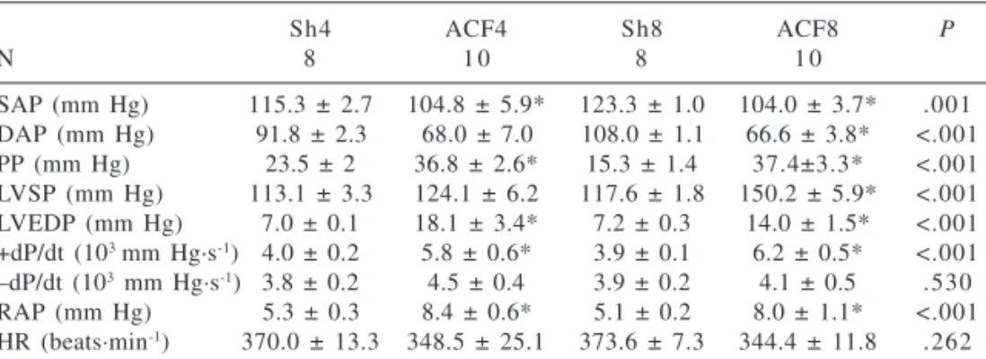
Hemodynamics – Phase 1
Table 1 summarizes the hemody-namic data obtained 1 week after the surgical experiment in the 4 groups studied: Sh4, ACF4, Sh8, and ACF8. Except for systemic diastolic arterial
Table 1 - Hemodynamic data obtained at one week following aortocaval fistula
creation (hemodynamics—phase 1) of groups Sh4, ACF4, Sh8, and ACF8.
Sh4 ACF4 Sh8 ACF8 P
N 8 1 0 8 1 0
pressure in ACF4, systemic arterial pressures were statistically lower in both ACF4 and ACF8 than in their re-spective Sh controls. Accordingly, pulse pressure was statistically greater in ACF than in Sh groups.
Intraventricular pressures showed higher LV systolic pressure in ACF8 than in Sh8 and higher LV end-diastolic pressure in ACF4 than in Sh4 and in ACF8 than in Sh8.
LV function showed greater +dP/dt in both ACF groups than in their re-spective Sh controls and comparable –dP/dt.
Right mean atrial pressure was sta-tistically higher in both ACF than in Sh, whereas heart rate did not differ.
As for LV perfusion pressure (Fig-ure 1), CDP was lower in ACF, al-though this was statistically signifi-cant only with regards to the compari-son between ACF8 and Sh8.
Hemodynamics – Phase 2
At the end of follow up (Table 2), while systemic systolic arterial pres-sure was comparable between both ACF4 and ACF8 and their respective Sh controls, systemic diastolic arterial pressure was statistically lower. Ac-cordingly, pulse pressure was signifi-cantly higher in both ACF groups than in their Sh controls. Again, heart rate was comparable among groups.
Anatomy and Morphometry
Table 3 depicts the anatomic and morphometric data. While initial weights of rats were comparable in all groups, final weights were greater in groups followed over 8 weeks than in groups followed over 4 weeks.
The percent of water in lungs and liver as well as the relative total heart and left atrium weights were not sta-tistically different among groups.
While relative left atrium weight did not differ between ACF and Sh
Table 2 - Hemodynamic data obtained at 4 and 8 weeks following aortocaval
fistula creation (hemodynamics—phase 2) of groups Sh4, ACF4, Sh8, and ACF8.
Sh4 ACF4 Sh8 ACF8 P
N 8 1 0 8 1 0
SAP (mm Hg) 125.5 ± 1.9 120.7 ± 7.9 128.8 ± 1.7 116.4 ± 6.1 .414 DAP (mm Hg) 94.3 ± 2.4 71.2 ± 5.5* 94.0 ± 2.1 67.7 ± 5.2* <.001 PP (mm Hg) 31.3 ± 1.6 49.5 ± 5.2* 34.8 ± 2.7 48.7 ± 5.0* .007 HR (beats·min-1) 373.5 ± 12.8 337.0 ± 32.3 386.8 ± 10.7 343.6 ± 20.6 .542 SAP, systemic systolic arterial pressure; DAP, systemic diastolic arterial pressure; PP, pulse pressure; HR, heart rate; *, P <.05 ACF vs.respective Sh.
Table 3 - Heart and body weights, percent of organ water weight, myocyte diameter,
and collagen volume fraction of groups Sh4, ACF4, Sh8, and ACF8.
Sh4 ACF4 Sh8 ACF8 P
N 8 1 0 8 1 0
IAW (g) 315.9 ± 7.2 309.2 ± 9.6 297.8 ± 2.6 322.0 ± 6.8 .071 FAW (g) 367.5 ± 11.4 357.0 ± 7.7 414.7 ± 15.3= 421.7 ± 6.3= <.001 % H2OLu 84.1 ± 0.8 82.0 ± 0.6 85.2 ± 1.0 82.2 ± 1.2 .061 % H2OLi 74.6 ± 0.3 72.4 ± 0.8 72.6 ± 0.8 71.2 ± 1.9 .174
HW 5.2 ± 0.2 5.5 ± 0.5 4.3 ± 0.2 5.8 ± 0.7 .07
LAW 0.09 ± 0.01 0.14 ± 0.02 0.11 ± 0.01 0.16 ± 0.02 .059
RAW 0.06 ± 0.01 0.13 ± ±0.02* 0.07 ± 0.01 0.15 ± 0.04 .001 LV WT (µm2) 42.3 ± 3.3 35.7 ± 2.2 51.9 ± 4.3 31.0 ± 2.3* .001 LV CD (µm2) 36.1 ± 6.5 25.4 ± 3.5 37.2 ± 4.2 39.8 ± 8.2 .323 MY φ (µm) 6.7 ± 0.05 9.2 ± 0.26* 6.2 ± 0.07 10.1 ± 0.19* <.001 SE CVF (%) 4.6 ± 0.47 26.7 ± 5.5* 4.0 ± 0.79 25.6 ± 3.5* <.001 IVS CVF (%) 5.5 ± 0.61 13.7 ± 1.4* 4.7 ± 0.61 13.4 ± 1.3* <.001
PVC (%) 13.7 ± 1.3 15.4 ± 1.5 14.2 ± 0.7 16.5 ± 2.9 .834
IAW, initial animal weight; FAW, final animal weight; % H2OLu, percentage of water in lungs; % H2OLi, percentage of water in liver; HW, relative heart weight; RAW, relative right atrium weight; LAW, relative left atrium weight; LV WT, left ventricular wall thickness; LV CD, left ventricular cavity dimension; MY φ, myocyte diameter; SE CVF, subendocardial collagen volume fraction; IVS CVF, interventricular septum collagen volume fraction; PVC, perivascular collagen; *, P <.05 ACF vs respective Sh; =, P <.05 8 vs respective 4.
groups, relative right atrium weight was statistically greater in ACF4 than in Sh4.
While LV wall thickness did not statistically differ between ACF4 and Sh4 groups, it was lower in ACF8 than in Sh8. The cavity dimension of the LV did not differ between ACF and Sh groups.
At the microscopic level, myocyte diameter was statistically greater in both ACF4 and ACF8 than in their re-spective Sh controls.
Thus, excentric left ventricular hypertrophy was manifested in ACF8 by larger myocyte diameter and smaller wall thickness than in Sh8, with comparable cavity dimension.
As for the extracellular matrix com-partment, CVF measured within each of the two regions examined, interven-tricular septum and SE, was statisti-cally greater in both ACF groups than in their respective Sh controls. Perivas-cular collagen did not differ among groups.
In Figure 2 is portrayed the hall-mark of the histological findings found in ACF rats, represented by LV SE fi-brosis.
Hemodynamic determinants of left ventricular fibrosis
Subendocardial fibrosis
The SE fibrosis of hearts examined 8 weeks after experimental surgery correlated positively with pulse pres-sure (r = 0.85, P <.001), LV systolic pressure (r = 0.67, P = .038), and LV end-diastolic pressure (r = 0.74, P = .001) measured 1 week after surgery. In addition, SE fibrosis at 8 weeks corre-lated inversely with systemic systolic arterial pressure (r = –0.69, P = .003), systemic diastolic arterial pressure (r = –0.91, P <.001), and CDP (r = –0.93,
P <.001).
Among all, only CDP independ-ently predicted SE fibrosis (r2 = 0.86,
SE = 0.0536, P <.001) at 8 weeks.
Interventricular septum fibrosis
Interstitial fibrosis within interven-tricular septum of hearts examined 8 weeks after experimental surgery cor-related positively with LV systolic pressure (r = 0.74, P = .001), LV end-diastolic pressure (r = 0.68, P = .004), and pulse pressure (r = 0.58, P = .018) measured 1 week after surgery. In ad-dition, septal fibrosis at 8 weeks cor-related inversely with systemic systolic arterial pressure (r = –0.60, P = .014), systemic diastolic arterial pressure (r = –0.69, P = .003), and CDP (r = –0.72,
P = .002).
Among all, LV systolic pressure (r2
= 0.73, SE = 0.0377, P = .004) and LV end-diastolic pressure (r2 = 0.55, SE =
0.1613, P = .012) were predicted inde-pendently of septal fibrosis.
DISCUSSION
The present study showed dissimi-lar reductions in systemic systolic and diastolic arterial pressures, which re-sulted in an increase in pulse pressure 1 week following experimental aortocaval fistula in rats. The de-creased systemic diastolic pressure corresponds to the characteristic hemodynamic change found in arterio-venous shunts and stands for a decrease in systemic vascular resistance17,18.
Ad-ditionally, LV hemodynamics also changed. Both LV systolic and end-diastolic pressures increased, whereas CDP decreased. Cardiac systolic func-tion, represented by LV +dP/dt was im-proved, whereas cardiac diastolic func-tion, represented by LV –dP/dt was un-altered.
The small gradient observed be-tween systemic and LV pressures must be regarded as a consequence of meas-uring systolic arterial pressure distally from the vascular shunt in the present study.
In contrast with our findings, pre-vious studies reported reductions of Figure 2 - Photomicrographs of picrosirius red-stained tissue sections under a 40x magnification
LV systolic pressure,17,18,36 indicating
the presence of cardiac dysfunction. It has to be emphasized that hemodynamic monitoring was per-formed before sacrifice in most former studies, when cardiac dysfunction has had enough time to supervene. Our 1-week hemodynamic results demon-strate that diastolic function was pre-served despite the elevation of LV end-diastolic pressure. This may be ex-plained by an enlarged LV dimension and thus increased preload, which may have in part contributed to the hypercontractility found, through the Starling mechanism. Since morphom-etry was performed only 4 and 8 weeks after the hemodynamic measurements, this premise cannot be definitely af-firmed based on the present findings.
At the end of the experiment, while systolic pressure in rats with aortocaval shunts decreased to values comparable with their respective controls, diastolic pressure remained lower as did pulse pressure. This hemodynamic pattern indicates that the vascular shunt re-mained pervious in most ACF rats over the entire period of observation.
The decrease in CDP may have contributed to ongoing myocardial ischemia and necrosis with subsequent fibrosis repair, ultimately resulting in cardiac dysfunction. To the best of our knowledge, this is the first study to demonstrate in an experimental animal model of eccentric hypertrophy that CDP actually falls. It is likely that the CDP decrease persisted for longer than 1 week, since systemic diastolic pres-sure remained low until morphometric studies were performed. It remains to be demonstrated whether left ventricular end-diastolic pressure remains elevated. The advent of cardiac hypertrophy was best demonstrated by myocyte en-largement occurring as early as 4 weeks following aortocaval fistula. Although the water content of lungs and liver was not increased, indicating that that overt heart failure did not occur in these
ani-mals, this does not necessarily mean that cardiac function was not impaired. The absence of LV hemodynamic data before sacrifice precluded an accurate estimation of cardiac function at the end of experiment.
Cardiac remodeling at the nonmyocyte compartment was illus-trated by a relative increase in fibril-lar collagen deposition within the in-terstitial space as well as within the SE region. Accordingly, 2 types of cardiac fibrosis deposition were found in hearts of rats with aortocaval fistulas: reac-tive interstitial fibrosis and reparareac-tive SE fibrosis.19,37,38 Perivascular fibrosis
was absent.
Although hemodynamics—phase 1 data were not significantly associ-ated with the presence of myocardial fibrosis 4 weeks after aortocaval fis-tula, they correlated significantly with fibrosis found at 8 weeks. Among all hemodynamic variables, it was CDP that independently predicted SE fibro-sis, whereas LV systolic and end-diastolic pressures independently pre-dicted interstitial septal fibrosis. The association of decreased CDP during the early phases of hemodynamic de-rangement following aortocaval fistula creation with subsequent SE fibrosis has not been reported before. This fact suggests that SE fibrosis may conceiv-ably be the consequence of impaired coronary perfusion. On the other hand, interstitial fibrosis appears to be con-sequence of LV overload. In this re-gard, the increase in interstitial fibril-lar collagen presently observed does not differ from that reported to occur in different experimental models of cardiac hypertrophy in response to afterload increase.16,20
The increase in interstitial myocar-dial fibrosis in this model of aortocaval fistula is actually controversial.24,27,28 It
is conceivable to speculate that while LV hypertrophy is physiological and associated with preserved ventricular function, interstitial collagen remains
relatively unchanged. In advanced stages characterized by cardiac dys-function, it may be that collagen accu-mulates within the interstitial space. To confirm this hypothesis, hemodynamic measurements are to be repeated before the sacrifice of the animals. This inves-tigation is presently under way.
In summary, hearts of rats submit-ted to volume overload consequent to aortocaval fistula creation show early reduction of LV coronary perfusion pressure and increased LV systolic and end-diastolic pressure. Subsequent LV remodeling is characterized by inter-stitial and SE fibrosis deposition. Sub-endocardial fibrosis was associated with coronary perfusion pressure and interstitial fibrosis with LV pressure.
CONCLUSIONS
Early hemodynamic changes ap-pear to interfere with cardiac remodeling after aortocaval fistula creation. Impaired coronary perfusion pressure may contribute to SE fibrosis, and pressure overload to interstitial fi-brosis.
ACKNOWLEDGEMENTS
RESUMO
GUIDO MC e col. A redução da pres-são de perfupres-são coronariana está associada com a fibrose endocár-dica no modelo de hipertrofia por sobrecarga de volume em ratos.
Rev. Hosp. Clín. Fac. Med. S.
Pau-lo 59(4):228-235, 2004.
No remodelamento que se segue às sobrecargas de volume não é descrito o aumento de fibrose miocárdica. Após o infarto, entretanto, há hipertrofia do miocárdio remoto com acúmulo de fibrose, particularmente no subendo-cárdio. Na fístula aorto-cava, tal como no infarto, é possível que a queda da pressão de perfusão coronariana inter-fira com a fibrose cardíaca.
OBJETIVO: Investigar o papel das
mudanças hemodinâmicas agudas sobre a fibrose cardíaca na fístula aorto-cava.
MÉTODO: Ratos Wistar
submeti-dos a fístula aorto-cava, seguisubmeti-dos por 4 e 8 semanas, constituíram 4 grupos, fístula aorto-cava 4 e fístula aorto-cava 8 (10 ratos cada) e seus respectivos controles (shamoperated controls -Sh), Sh4 e Sh8 (8 ratos cada). A hemodinâmica foi realizada 1 semana após a cirurgia. A hipertrofia e a fibrose foram quantificadas ao final do segui-mento pelo diâmetro dos miócitos e pela fração de volume do colágeno.
RESULTADOS: Comparados com
Sh4 e Sh8, a pressão de pulso, a prsão diastólica final do ventrículo es-querdo e a +dP/dtforam maiores em fístula aorto-cava 4 e fístula aorto-cava 8, enquanto a -dP/dt foi similar. A pres-são estimada da perfupres-são coronariana (mmHg) foi menor em fístula aorto-cava 8 (52,6±4,1) do que em Sh8 (100,8±1,3), mas comparável entre fístula aorto-cava 4 (50,0±8,9) e Sh4
(84,8±2,3). O diâmetro dos miócitos foi maior em fístula aorto-cava 8 e a fibrose intersticial e subendocárdica maior em fístula aorto-cava 4 e fístula aorto-cava 8. Houve correlação inver-sa e independente da pressão de perfusão coronariana com a fibrose subendocárdica (r2=0,86; p<0,0001) e
das pressões sistólica (r2=0,73;
p=0,0035) e diastólica final do ventrículo esquerdo (r2=0,55;
p=0.0124) com a fibrose intersticial.
CONCLUSÃO: A queda precoce
da pressão de perfusão coronariana e o aumento das pressões ventriculares após a fístula aorto-cava associam-se com fibrose miocárdica subseqüente.
UNITERMOS: Fibrose miocár-dica. Fístula aorto-cava. Pressão de perfusão coronariana. Sobrecarga de volume. Remodelamento cardíaco.
REFERENCES
1 . Olivetti G, Cigola E, Maestri R, Lagrasta C, Corradi D, Quaini F. Recent advances in cardiac hypertrophy. Cardiovasc Res 2000;45:68-75.
2 . Cohn JN, Ferrari R, Sharpe N. Cardiac remodeling - Concepts and clinical implications: a consensus paper from an international forum on cardiac remodeling. J Am Coll Cardiol 2000;35:569-82.
3 . Doggrell SA, Brown L. Rat models of hypertension, cardiac hypertrophy and failure. Cardiovasc Res 1998;39:89-105. 4 . Tomanek RJ. Response of coronary vasculature to myocardial
hypertrophy. JACC 1990;15:528-33.
5 . Bregagnollo EA, Rodrigues MA, Montenegro MR . Evolução temporal de medidas estruturais e funcionais da hipertrofia cardíaca desencadeada em ratos Wistar pela constrição da aorta abdominal. Arq Bras Cardiol 1986;46:9-17.
6 . Strauer BE. Myocardial oxygen consumption in chronic heart disease: role of wall stress, hypertrophy and coronary reserve. Am J Cardiol 1979;44:730-40.
7 . Colucci WS. Molecular and cellular mechanisms of myocardial failure. Am J Cardiol 1997;80(11A):15L-25L.
8 . Anversa P, Ricci R, Olivetti G. Quantitative structural analysis of myocardium during growth and induced cardiac hypertrophy: a review. J Am Coll Cardiol 1986;7:1140-9.
9 . Pereira V, de C, Rodrigues A, Tsutsui JM, Curi M, Mady C, Ramires J. Coronary flow velocity reserve in hypertensive patients with left ventricular systolic dysfunction. Clin Cardiol 2002; 25:95-102.
10. Brower GL, Janicki JS – Contribution of ventricular remodeling to pathogenesis of heart failure in rats. Am J Physiol Heart Circ Physiol 2001; 280:H674-H683.
11. Namba T, Tsutsui H, Tagawa H, Takahashi M, Saito K, Kozai T, et al. Regulation of fibrillar collagen gene expression and protein accumulation in volume-overloaded cardiac hypertrophy. Circulation 1997;95:2448-54.
12. Frimm CC, Koike MK, Cúri M. Subendocardial fibrosis in remote myocardium results from reduction of coronary driving pressure during acute infarction in rats. Arq Bras Cardiol 2003;80:515-20.
13. Norton GR, Woodiwiss AJ, Gaasch WH, Mela T, Chung ES, Aurigemma GP, et al. Heart failure in pressure hypertrophy— The relative roles of ventricular remodeling and myocardial dysfunction. J Am Coll Cardiol 2002;39:664-71.
14. Verdouw PD, van den Doel MA, de Zeeuw S, Duncker DJ. Animal models in the study of myocardial ischaemia and ischaemic syndromes. Cardiovasc Res 1998;39:121-35.
16. Stilli D, Berni R, Sgoifo A, Costoli T, Bocchi L, Cacciani F, et al. Social stress, myocardial damage and arrhythmias in rats with cardiac hypertrophy. Physiology & Behavior 2001;73:351-58.
17. Liu Z, Hilbelink DR, Crockett WB, Gerdes AM. Regional changes in hemodynamics and cardiac myocyte size in rats with aortocaval fistulas. 1. Developing and established hypertrophy. Circ Res 1991;69:52-8.
18. Liu Z, Hilbelink DB, Crockett WB. Regional changes in hemodynamics and cardiac myocyte size in rats with aortocaval fistulas. 2. Long-term effects. Circ Res 1991;69:59-65. 19. Weber KT, Brilla CG, Janicki JS. Myocardial fibrosis: functional
significance and regulatory factors. Cardiovasc Res 1993;27:341-8.
20. Brilla CG, Weber KT. Reactive and reparative myocardial fibrosis in arterial hypertension in the rat. Cardiovasc Res 1992;26:671-7 .
21. Weber KT, Brilla CG, Campbell SE, Reddy HK. Myocardial fibrosis and the concepts of cardioprotection and cardioreparation. J Hypertens 1992;10:S87-S94.
22. Weber KT, Brilla CG. Pathological hypertrophy and cardiac interstitium. Fibrosis and renin-angiotensin-aldosterone system. Circ Res 1991;83:1849-65.
23. Kamogawa Y, Biro S, Maeda M, Setoguchi M, Hirakawa T, Yoshida H, et al. Dystrophin-deficient myocardium is vulnerable to pressure overload in vivo. Cardiovasc Res 2001;50:509-15. 24. Emery JL, Omens JH. Mechanical regulation of myocardial growth
during volume-overload hypertrophy in the rat. Am J Physiol Heart Circ Physiol 1997;273:H1198-H1204.
25. Michel JB, Salzmann JL, Ossondo Nlom M, Bruneval P, Barres D, Camilleri JP. Morphometric analysis of collagen network and plasma perfused capillary bed in the myocardium of rats during evolution of cardiac hypertrophy. Basic Res Cardiol 1986;91:142-54.
26. Salzmann JL, Michel JB, Bruneval P, Ossondo Nlom M, Barres DR, Camilleri JP. Automated image analysis of myocardial collagen pattern in pressure and volume overload in rat cardiac hypertrophy. Anal Quant Cytol Histol 1986;8:326-32. 27. Narikawa S, Stefano LM, Georgetti JC . Quantificação da
hipertrofia cardíaca e fibrose intersticial em corações de ratos submetidos a sobrecarga de pressão ou de volume. Rev Soc Cardiol Estado de São Paulo 2002;12(2):46.
28. Brower GL, Henegar JR, Janicki JS. Temporal evaluation of left ventricular remodeling and function in rats with chronic volume overload. Am J Physio Heart Circ Physiol 1996;40:H2071-H2078.
29. Frimm CC, Sun Y, Weber KT. Wound healing following myocardial infarction in the rat: role for bradykinin and prostaglandins. J Mol Cell Cardiol 1996;28:1279-85. 30. Frimm CC, Moraes AV, Medeiros C . Papel da pressão arterial
casual e do exercício e a influência de fatores clínicos sobre a hipertrofia ventricular esquerda na hipertensão arterial. Arq Bras Cardiol 1994;63:21-6.
31. Perry GJ, Mori T, Wei CC, Xu XY, Chen YF, Oparil S, et al. Genetic variation in angiotensin-converting enzyme does not prevent development of cardiac hypertrophy or upregulation of angiotensin II in response to aortocaval fistula. Circulation 2001;103:1012-6.
32. Garcia R, Diebold S. Simple. Rapid, and effective method of producing aortocaval shunts in the rat. Cardiovasc Res 1990;24:430-2.
33. Capasso JM, Li P, Zhang X, Anversa P. Heterogeneity of ventricular remodeling after acute myocardial infarction in rats. Am J Physiol Heart Circ Physiol 1992;262:H486-H495.
34. Cross C, Riechen P, Salisbury P. Coronary driving pressure and vasomotor tonus as determinants of coronary blood flow. Circ Res 1961;9:589-600.
35. Junqueira LC, Bignolas G, Brentani RR. Picrosirius staining plus polarization microscopy, a specific method for collagen in tissue sections. Histochem J 1979;11:447-55.
36. Wang X, Ren B, Liu S, Sentex E, Tappia PS, Dhalla NS. Characterization of cardiac hypertrophy and heart failure due to volume overload in the rat. J Appl Physiol 2003;94:752-63.