147
ABSTRACT
BACKGROUND AND OBJECTIVES: Surface electromy-ography (SE) provides a non invasive evaluation of the bio-electric phenomenon of the evaluated muscle at rest, as well as the comparison with its activity during muscle contrac-tion. his study aimed at evaluating the efectiveness of SE in patients with temporomandibular disorders according to Re-search Diagnostic Criteria for Temporomandibular Disorders (RDC/TMD) axis I criteria.
CONTENTS: Literature was reviewed as from LILACS, Med-line and Scielo databases in the period from January 1987 to February 2012. Randomized controlled clinical trials, clini-cal trials and cliniclini-cal tests evaluating signs and symptoms of temporomandibular disorders (TMD) diagnosed according to RDC/TMD were included. Search strategy has resulted in 182 articles of which eight have fulilled inclusion criteria, being one randomized clinical trial and seven longitudinal studies without randomization criteria. In all studies, SE was the method used to detect and evaluate electric activity of masticatory muscles (body of the masseter and anterior tem-poral bundle), being somewhat easily applied and following test standards. However, diferent experimental models and
Surface electromyography for temporomandibular disorders: systematic
review*
Eletromiografia de superfície em disfunção temporomandibular: revisão sistemática
Andersen Ieger Celinski1, Rafael Schlogel Cunali2, Daniel Bonotto3,Aguinaldo Coelho de Farias4,Paulo Afonso Cunali5
*Received from the Federal University of Paraná. Curitiba, PR.
1. Student of the Specialization Course in Temporomandibular Disorders and Orofacial Pain, Federal University of Paraná (UFPr); Doctor in Prosthesis and Dental Material, Eb-erhard KarlsUniversitat – Universitat Klinikum Tubingen, UKT, Germany; Professor of the Federal Institute of Paraná (IFPR). Curitiba, PR, Brazil.
2. Specialist in Temporomandibular Disorder and Orofacial Pain; Professor of the Special-ization Course on Temporomandibular Disorder and Orofacial Pain, Federal University of Paraná (UFPR). Curitiba, PR, Brazil.
3. Professor of the Positivo University; Master in Sciences, Catholic University of Paraná (PUCPR); Professor of the Specialization Course on Temporomandibular Disorder and Orofacial Pain, Federal University of Paraná (UFPr). Curitiba, PR, Brazil.
4. Professor of the Federal University of Paraná; Doctor in Orthodontics, Paulista State Uni-versity (UNESP); Professor of the Specialization Course on Temporomandibular Disorder and Orofacial Pain, Federal University Paraná (UFPR). Curitiba, PR, Brazil.
5. Professor of the Federal University of Paraná; Doctor in Sciences, Federal University of São Paulo (UNIFESP); Coordinator of the Specialization Course on Temporoman-dibular Disorder and Orofacial Pain, Federal University of Paraná (UFPR). Curitiba, PR, Brazil.
Submitted in February 27, 2013. Accepted for publication in May 03, 2013.
Correspondence to: Paulo Afonso Cunali, M.D.
Rua Cel. Napoleão Marcondes França, 360 80040-270 Curitiba, PR.
Phone: +55 (41) 3322-1234 E-mail: [email protected]
sample selections were used, making diicult the comparison of results.
CONCLUSION: In spite of the limitations of this study, it was possible to observe that although SE should not be used to diagnose TMD, it may help the follow up of TMD treat-ment evolution.
Keywords: Electromyography, Masseter, Masticatory mus-cles, Research Diagnostic Criteria, Surface electromyography, Temporal.
RESUMO
JUSTIFICATIVA E OBJETIVOS: A eletromiograia de superfície (ES) permite uma avaliação não invasiva do fenô-meno bioelétrico durante o estado de repouso do músculo avaliado bem como a comparação com sua atividade durante a contração muscular. O objetivo deste estudo foi avaliar a efetividade do uso de ES em pacientes diagnosticados com disfunção temporomandibular segundo os critérios Research Diagnostic Criteria for Temporomandiublar Disorders (RDC/ TMD) eixo I.
CONTEÚDO: A revisão de literatura foi realizada a partir das bases de dados LILACS, Medline e Scielo, cobrindo o período de janeiro de 1987 a fevereiro de 2012. Ensaios cos randomizados e controlados, ensaios clínicos e testes clíni-cos que avaliaram ES, sinais e sintomas de desordens tem-poromandibulares (DTM) diagnosticados pelo critério RDC/ TMD foram incluídos. A estratégia de busca resultou em 182 artigos, dos quais oito preencheram os critérios de inclusão, sendo que um caracterizava um estudo clínico randomizado e sete eram estudos longitudinais sem critérios de randomiza-ção. Em todos os estudos, o método utilizado para detectar e analisar a atividade elétrica dos músculos da mastigação (cor-po do masseter e feixe anterior do tem(cor-poral) foi a ES, sendo empregada com certa facilidade e seguindo os padrões para o exame. No entanto, foram utilizados diferentes modelos ex-perimentais e seleção das amostras, causando diiculdades na comparação dos resultados.
CONCLUSÃO: Dentro das limitações deste estudo, foi possível constatar que embora a ES em DTM não deva ser utilizada para diagnóstico, ela pode auxiliar no acompanha-mento da evolução dos trataacompanha-mentos de DTM.
Descritores: Disfunção temporomandibular, Eletromiogra-ia, Eletromiograia de superfície, Masseter, Músculos da mas-tigação, Research Diagnostic Criteria, Temporal.
Rev Dor. São Paulo, 2013 abr-jun;14(2):147-50
© Sociedade Brasileira para o Estudo da Dor
148
Celinski AI, Cunali RS, Bonotto D et al.
Rev Dor. São Paulo, 2013 abr-jun;14(2):147-50
INTRODUCTION
Temporomandibular disorder (TMD) is a generic term used for a set of musculoskeletal disorders which may af-fect the masticatory system1. The prevalence of TMD signs and symptoms in general population is considered high2. Females are more affected by the disease in 5:1 ratio, and between 20 and 50 years of age2,3. Current understanding points to TMDs as clinical conditions with multifactorial etiology because one or more factors may contribute for its triggering or perpetuation. Among these factors there are anatomic changes, macrotrauma, microtrauma, occlusal unbalances, parafunctional habits and systemic conditions, such as emotional stress1,3.
Surface electromyography (SE) provides the non-invasive evaluation of the bioelectric phenomenon with the evalu-ated muscle at rest, and then compares it to its activity dur-ing muscle contraction. This procedure is carried out with electrodes placed on patients’ skin, in general bilaterally. Its relatively technical simplicity allows its use in Dentistry and in clinical research4.
TMDs investigation and evaluation should include be-havioral, emotional and psycho-social factors, in addition to normally observed physical changes5. The idea of put-ting together these data to get a standardization of the di-agnosis, aiming at further reliability and reproducibility was developed by Dworkin and LeResche6 by means of a set of diagnostic criteria to investigate TMD. This set was called Research Diagnostic Criteria for Temporomandibu-lar Disorders (RDC/TMD), translated (history, evaluation questionnaire and clinical evaluation form) and culturally adapted to the Portuguese language (history and evaluation questionnaire) by Pereira et al.7 and Kominsky et al.8, re-spectively.
This study aimed at evaluating, through systematic literature review, the effectiveness of SE for patients with temporo-mandibular disorders according RDC/TMD axis I criteria6.
METHOD
The strategy was based on the computerized query of the lit-erature applying keywords to Medline, LILACS and Scielo databases, covering the period from January 1987 to Febru-ary 2012. Keywords used for the query were crossed in dif-ferent combinations and were: “surface electromyography”, “electromyography”, “temporomandibular disorder”, “emg”, “tmd” and “RDC”. Relevant articles were also reviewed with regard clinical SE efficacy as from sensitivity and specific-ity. Selected articles were submitted to evaluation by two reviewers, respecting inclusion criteria to determine final articles sample, according to their titles and abstracts. Inclu-sion criteria were:
• Studies with humans were masseter muscle and anterior temporal muscle bundle were evaluated by surface electro-myography (SE);
• Randomized clinical trials, controlled clinical trials and longitudinal prospective non randomized studies;
• Studies using the RDC/TMD questionnaire as diagnostic criteria;
• Studies in English, Portuguese, Italian, German and Span-ish, published within the determined period. So, case re-ports, case reports follow-up and literature reviews, simple opinions and authors’ opinions were excluded.
RESULTS
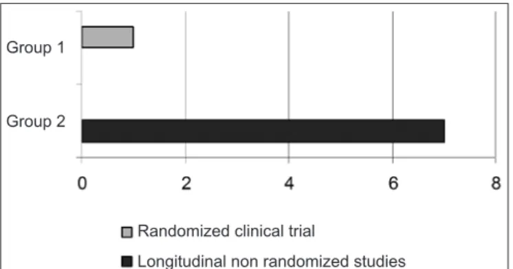
Query strategy has resulted in 182 articles. After applying inclusion/exclusion criteria, eight articles were qualified for final analysis, being the Kappa agreement index between reviewers equal to 1.00. From these studies, one was a ran-domized clinical trial and seven were longitudinal trials without randomization criteria (Graph 1).
Among selected studies, only one has not analyzed, in com-bination, muscle electric activity of masseter and temporal muscles. The remaining seven selected studies have evalu-ated the anterior temporal muscle bundle and the body of the masseter muscle (Graph 2).
Table 1 shows selected studies according to established methodological criteria.
Group 1
Group 1 Group 2
Group 2
Randomized clinical trial
Longitudinal non randomized studies
Temporal and masseter muscles Masseter muscle
Graph 1 – Studies design.
149
Surface electromyography for temporomandibulardisorders: systematic review
Rev Dor. São Paulo, 2013 abr-jun;14(2):147-50
DISCUSSION
In the search for auxiliary methods to provide better under-standing of mechanisms involved with TMD, and to establish a more objective patients’ evaluation, the authors decided for the electric evaluation of muscle electric activity, using surface electromyography, aiming at creating reference models and at comparing an asymptomatic healthy function with those situations of system disharmony or dysfunction9. SE is an ad-ditional evaluation method which allows the observation and quantiication of muscle balance, through the electric activity, both in pairs of muscles and between muscles on both sides of the body10,11.
It is known that the primary parameter to identify TMD pa-tients with regard to pain is its ratio with regard to decreased muscle strength, which may be observed by electromyo-graphic activity, especially during tooth clenching activity12. Such indings are in line with the pain adaptation model and its further integration, since pain leads to individual muscle activity changes aiming at limiting movements and at pro-tecting the system against new injuries, by decreasing agonist muscles activity13,14.
he literature suggests that SEto diagnose TMD has a much lower accuracy than was is proposed by the manufacturers of such devices15,16. In addition, recent systematic literature reviews argue that selected studies corresponded to low rele-vance and low impact trials, in addition to having conlicting inal results, possibly due to the summation of many variables such as: inadequate sample and control group selection, insuf-icient clinical conditions and incorrect use of equipment17,18. However, if due precautions are taken and a strict and stan-dardized protocol is used, electromyography may be consid-ered an eicient method to analyze the stomatognathic sys-tem, with good reproducibility and additional reference value only during clinical evaluation11,12,19-22.
he use of RDC/TMD diagnostic criteria is a major factor for standardization and comparison of studies6. Our study has found eight articles meeting such criteria. None of them were double-blind. All studies used SE to detect and analyze the electric activity of masticatory muscles (body of masseter and anterior temporal muscle bundle), being relatively easy to use and following the standards of the test. However, diferent experimental models and sample selections were used, mak-ing diicult the comparison of results.
According to some authors, SE evaluation of masticatory muscles allows the objective separation of diferent TMD subgroups diagnosed according to RDC/TMD criteria. Sig-niicant diferences are always observed in electromyographic activities and symmetry of activities at rest and during tooth clenching, between TMD and healthy patients12,18,19,23-28. he analysis of muscle electromyographic activity has also been used to evaluate TMD treatment eicacy by conven-tional methods associated or not to support therapies9,24,25,29. Low-intensity laser is an example of support therapy for TMD, which may also be relieved with electromyography. Although not promoting changes in electromyographic activ-ity of evaluated muscles, this therapy has decreased observed painful symptoms24.
Still in line with data found in selected studies, it should be taken into consideration that dentists should not use elec-tromyography or similar tools to diagnose patients who may have masticatory muscles myofascial pain. In addition, such devices should not be used in situations where the aim is an isolated evaluation, or as a complement for decision making and clinical approaches, since such tools do not meet the reli-ability and validity standards needed for such use27. However, it is observed that surface electromyographic evaluation may supply useful information for TMD diagnosis and for the therapeutic planning of the clinical case28.
It is observed that SE is, in principle, an adequate tool to evaluate neuromuscular function in Dentistry; if used accord-ing to speciic recommendations and together with patients’ history and accurate clinical and physical evaluation, EMG readings may supply objective information which may be well documented, in addition to valid and reproducible data about the functional condition of masticatory muscles of a given pa-tient. Such data may also be compared to a healthy situation and may help the follow up of the treatment through patient’s biofeedback9.
So, the primary parameter to identify patients with TMD-related pain is decreased muscle action, especially during tooth clenching12. he literature reports studies which are in line with the pain adaptation model and its further integra-tion, since pain leads to changes in muscle activity aiming at limiting movement and at protecting the system against new injuries by decreasing agonist muscles activity13,14. his way, when a sensory stimulation is received, relex protection mechanisms are activated, triggering a modulation of muscle
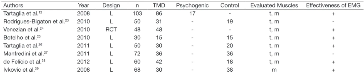
Table 1 – Studies based on the evaluation by electomyographic analysis of the activity of masticatory muscles.
Authors Year Design n TMD Psychogenic Control Evaluated Muscles Effectiveness of EMG
Tartaglia et al.12 2008 L 103 86 17 - t, m +
Rodrigues-Bigaton et al.23 2010 L 50 31 - 19 t, m
-Venezian et al.24 2010 RCT 48 48 - - t, m +
Botelho et al.25 2010 L 30 15 - 15 t, m +
Tartaglia et al.26 2011 L 50 30 - 20 t, m +
Manfredini et al.27 2011 L 72 36 - 36 t, m
-de Felício et al.28 2012 L 60 42 - 18 t, m +
Ivkovic et al.29 2008 L 68 30 - 38 m +
150
Celinski AI, Cunali RS, Bonotto D et al.
Rev Dor. São Paulo, 2013 abr-jun;14(2):147-50
activity in the stimulated area which, associated to speciic emotional situations, generates further muscle tension and, if associated to parafunctions such as tooth clenching and bruxism, lead to further muscle activity increase, which generates more pain and, consequently, more tension, and so on30.
CONCLUSION
Considering technological advances in the areas of equipment and techniques, as well as in research resources and research projects about the critical evaluation of the use of SE in cases of temporomandibular disorder, one may conclude, within the limitations of this study, that:
1. SE may be indicated for the follow-up of the efectiveness of a support therapy used for a certain clinical situation; 2. Its efectiveness could have some value as additional re-search tool to study muscle TMD features;
3. It is a procedure which should not be used as the single diagnostic tool, since it has low speciicity and sensitivity; 4. he clinical use of this method to diagnose temporoman-dibular disorders is uncertain and is currently not recom-mended.
REFERENCES
1. Okeson JP, De Leeuw R. Diferential diagnosis of temporomandibular disorders and other orofacial pain disorders. Dent Clin North Am. 2011;55(2):105-20.
2. Cooper BC, Kleinberg I. Examination of a large patient population for the presence of symptoms and sings of Temporomandibular Disorders. Cranio. 2007;25(2):114-26. 3. Oliveira AS, Dias EM, Contato RG, et al. Prevalence study of sings and
symp-toms of temporomandibular disorders in Brazilian college students. Braz Oral Res. 2006;20(1):3-7.
4. Castrolorio T, Farina D, Bottin A, et al. Surface EMG of jaw elevator muscles: efect of electrode location and inter-electrode distance. J Oral Rehabil. 2005;32(6):411-17. 5. List T, Dworkin SF. Comparing TMD diagnoses and clinical indings at Swedish and
US TMD Center Using Research Diagnostic Criteria for Temporomandibular Disor-ders. J Orofac Pain. 1996;10(3):240-53.
6. Dworkin SF, LeResche L. Research diagnostic criteria for temporomandibular disor-ders: review, criteria, examinations and speciications, critique. J Craniomandib Di-sord. 1992;6(4):301-55.
7. Pereira Jr FJ, Huggins KH, Dworkin SF, et al. Critérios de diagnóstico para pesquisa das desordens temporomandibulares RDC/TMD. Tradução oicial para a língua por-tuguesa. J Bras Clin Odontol Int. 2004;8:384-95.
8. Komisnky M, Lucena LBS, Siqueira JTT, et al. Adaptação cultural do questionário “Research diagnostic criteria for temporomandibular disorders” axis II para o portu-guês. Jl Bras Clin Odontol Int. 2004;4(1):51-61.
9. Emshorf R, Bösch R, Pümpel E, et al. Low-level laser therapy for treatment of
tempo-romandibular joint pain: a double-blind and placebo-controlled trial. Oral Surg Oral Med Oral Pathol Oral Radiol Endod. 2008;105(4):452-6.
10. Ferario VF, Sforza C, Serrao G. he inluence of crossbite on the coordinated elec-tromyographic activity of human masticatory muscles during mastication. J Oral Rehabil. 1999;26(7):575-81.
11. Ferrario VF, Sforza C, Tartaglia GM, et al. Immediate efect of a stabilization splint on masticatory muscle activity in temporomandibular disorder patients. J Oral Rehabil. 2002;29(9):810-5.
12. Tartaglia GM, Moreira Rodrigues da Silva MA, Bottini S, et al. Masticatory muscle activity during maximum voluntary clench in diferent research diagnostic criteria for temporomandibular disorders (RDC/TMD) groups. Man her. 2008;13(5):434-40. 13. Lund JP, Donga R, Widmer CG, et al. he pain-adaptation model: a discussion of the relationship between chronic musculoskeletal pain and motor activity. Can J Physiol Pharmacol. 1991;69(5):683-94.
14. Murray GM, Peck CC. Orofacial pain and jaw muscle activity: a new model. J Orofac Pain. 2007;21(4):263-88.
15. Lund JP, Widmer CG, Feine JS. Validity of diagnostic and monitoring tests used for temporomandibular disorders. J Dent Res. 1995;74(4):1133-43.
16. Greene CS. he role of biotechnology in TMD diagnosis. In: Laskin DM, Greene CS, Hylander WL, editores. TMDs: An evidence-based approach to diagnosis and treatment. Chicago, 1st ed. Quintessence Publishing; 2006. p. 193-202.
17. Klasser GD, Okeson JP. he clinical usefulness of surface electromyography in the diagnosis and treatment of temporomandibular disorders. J Am Dent Assoc. 2006;137(6) 763-71.
18. Suvinen TI, Kemppainen P. Review of clinical EMG studies related to muscle and occlusal factors in healthy and TMD subjects. J Oral Rehabil. 2007;34(9):631-44. 19. Bodéré C, Téa SH, Giroux-Metges MA, et al. Activity of masticatory muscles in
sub-jects with diferent orofacial pain conditions. Pain. 2005;116(1):33-41.
20. Bevilaqua-Grosso D, Monteiro-Pedro V, Guirro RR, et al. A physiotherapeu-tic approach to craniomandibular disorders: a case report. J Oral Rehabil. 2002;29(3):268-73.
21. Landulpho AB, E Silva WA, E Silva FA, et al. Electromyographic evaluation of mas-seter and anterior temporalis muscles in patients with temporomandibular disorders following interocclusal appliance treatment. J Oral Rehabil. 2004;31(2):95-8. 22. Ceneviz C, Mehta NR, Forgione A, et al. he immediate efect of changing
mandi-bular position on the EMG activity of the masseter, temporalis, sternocleidomastoid, and trapezius muscles. Cranio. 2006;24(4):237-44.
23. Rodrigues-Bigaton D, Berni KC, Almeida AF, et al. Activity and asymmetry index of masticatory muscles in women with and without dysfunction temporomandibular. Electromyogr Clin Neurophysiol. 2010;50(7-8):333-8.
24. Venezian G.C, da Silva MA, Mazzetto RG, et al. Low level laser efects on pain to pal-pation and electromyographic activity in TMD patients: a double-blind, randomized, placebo-controlled study. Cranio. 2010;28(2):84-91.
25. Botelho AL., Silva BC, Gentil FH, et al. Immediate efect of the resilient splint evaluated using surface electromyography in patients with TMD. Cranio. 2010;28(4):266-73. 26. Tartaglia GM, Lodetti G, Paiva G, et al. Surface electromyographic assessment of
patients with long lasting temporomandibular joint disorder pain. J Electromyogr Kinesiol. 2011;21(4):659-64.
27. Manfredini D, Cocilovo F, Favero L, et al. Surface electromyography of jaw muscles and kinesiographic recordings: diagnostic accuracy for myofascial pain. J Oral Reha-bil. 2011;38(11):791-9.
28. de Felicio CM, Ferreira Cl, Medeiros AP, et al. Electromyographic indices, orofacial myofunctional status and temporomandibular disorders severity: A correlation study. J Electromyogr Kinesiol. 2012;22(2):266-72.
29. Ivkovic N, Mladenovic I, Petkoci S, et al. TMD chronic pain and masseter silent period in psychiatric patients on antidepressive therapy. J Oral Rehabil. 2008;35(6):424-32. 30. Okeson JP, Falace DA. Nonodontogenic toothache. Dent Clin North Am.