ABSTRACT
Occlusal force, electromyographic activity of
subjects with different facial types
William CUSTODIO1, Simone Guimarães Farias GOMES1, Fernanda FAOT2, Renata Cunha Matheus Rodrigues GARCIA3, Altair Antoninha DEL BEL CURY3
1- MSc, Graduate student, Department of Prosthodontics and Periodontology, Piracicaba Dental School, State University of Campinas, Piracicaba, SP, Brazil. 2- DDS, MSc, PhD, Associate Professor, Department of Dental Prosthodontics, Dentistry School, Federal University of Pelotas, Pelotas, RS, Brazil. 3- DDS, MSc, PhD, Professor, Department of Prosthodontics and Periodontology, Piracicaba Dental School, State University of Campinas, Piracicaba, SP, Brazil.
Corresponding address: Dr. Altair Antoninha Del Bel Cury - Faculdade de Odontologia de Piracicaba - UNICAMP - Departamento de Prótese e Periodontia Av. Limeira, nº 901 - Piracicaba, SP - Brasil - 13414-903 - Phone: + 55-19-2106-5294 - Fax: + 55-19-2106-5211 - e-mail: [email protected]
O
maximal occlusal force (MOF), masticatory muscle electromyographic (EMG) activity, ! "#$ subjects were divided into 3 groups by Ricketts’s analysis: brachyfacial, mesofacial and dolychofacial. Maximum occlusal force in the molar region was bilaterally measured with a force transducer. The electromyographic activities of the masseter and anterior temporal $ $! calculated by subtracting the intermolar distance of maximum opening or protrusion from the distance in the rest position. The data were analyzed using ANOVA followed by Tukey’s %"&!$'*+!-& showed that shorter faces had higher occlusal forces (P<0.0001). Brachyfacial subjects presented higher levels of masseter electromyographic activity and medial mandibular /$ !2/ $' $ 456!6*! Conclusion: Within the limitations of the study, it may be concluded that maximum occlusal / vertical facial pattern.
Key words: Vertical dimension. Bite force. Electromyography. Masticatory muscles. Mandible.
INTRODUCTION
A variety of craniofacial morphologic features contribute to the determination of vertical facial types in humans19. However these facial
characteristics can hamper masticatory system functioning20. Aside from esthetic considerations,
presumed functional muscular activities are 8' in prostheses7 because muscle actions induce
$ opening and protrusion movements1,5.Different
craniofacial morphologies can lead to differences in neuromuscular activities8,24,26 and in muscle
cross-sectional area, volume and orientation4.
Previous studies assessing the relationship between mandibular muscles and facial morphology showed that subjects with wider transverse craniofacial dimensions have thicker and consequently stronger mandibular muscles8 and present anatomic features
such as small gonial angles25 and reduced lower
anterior facial height15. In contrast, dolichofacial
subjects present weaker muscles than do mesofacial and brachyfacial subjects15.
short-face subjects15. Previous studies12,17 have
shown a strong positive correlation between masseter muscle thickness and maximal EMG activity. Additionally, interactions among MOF magnitude, jaw muscle size and craniofacial morphology have been demonstrated11,18. However,
there is no agreement with regard to the role of the vertical facial craniofacial apparatus and muscular function8,11.
Muscular forces exerted by the masticatory rehabilitation. The action of jaw muscles can generate interocclusal forces responsible for elastic flexure of the mandible and significant ' lower arch during stomatognathic functioning14.
Muscular contraction without tooth contact results in narrowing of the arch during opening and protrusion, and an increase during mandibular retrusion10,27,30. In contrast, during tooth contact
the mandible can twist due to eversion of the masseteric processes, causing medial displacement of the coronoid processes13,30. As a consequence, the
occurrence of these mandibular deformations can compromise the viability of long-term conventional
' # 14.
Several attempts have been made to investigate the relationship between features of the mandibular muscles and the vertical facial pattern21. However,
an integrated analysis of the jaw muscles and their effects on functional response with regard to craniofacial morphology has never been conducted in a homogeneous population. Additionally, previous studies22,23 have evaluated the maximal occlusal
force using thicker sensor pads, which lead to a greater distance between antagonistic teeth. The results may therefore not be reliable. Thus, this blinded controlled study aimed to evaluate the effects of different facial patterns on the maximal occlusion force, jaw muscle EMG activity during maximum voluntary clenches and the narrowing of the dental arch (MMF) in a sample of fully dentate adults.
MATERIAL AND METHODS
Subjects
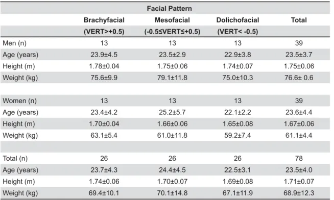
The convenience sample of this cross-sectional study included patients and students recruited from Piracicaba Dental School, State University of Campinas, Brazil. The sample included 78 subjects (39 men and 39 women) with a mean age of 23.5 years (standard deviation: 4.0). All of them were healthy and without facial deformities. They had complete dentition (except for a lack of third molars) and showed no malocclusion, no occlusal vertical dimension alteration, no history of maxillofacial surgery or mandibular injuries, no
orthodontic treatment for at least 2 years prior to the study, no periodontal disease, no caries, no temporomandibular disorders and parafunctional habits. The study protocol was approved by the Local Research and Ethics Committee, and all subjects signed written, informed consent. All subjects were submitted to anthropometric measurements, in which height was measured in centimeters (cm) with the subject in the erect position without shoes and weight was recorded in kilograms (kg) (Mechanical anthropometric scale R110; Welmy, Santa Bárbara D’Oeste, SP, Brazil).
Vertical facial type evaluation
Lateral cephalograms were used for facial type determination. The subjects were covered with a lead apron and placed in a cephalostat at a 90 degree angle to the tube head. All cephalograms were taken via standard procedures with the same radiographic unit (Elipsopantomograph Funk X-15; Macrotec Indústria e Comércio de Equipamentos Ltda., São Paulo, SP, Brazil) and processed with an automatic processor (Macrotec MX-2; Macrotec Indústria e Comércio de Equipamentos Ltda.). Facial patterns were determined by using digital cephalometric analysis (Radiocef v.4.0, Radio Memory Ltda, Belo Horizonte, MG, Brazil), which Q' the vertical direction. The vertical facial pattern was determined by the VERT index19 (VI), which is
the arithmetic mean of the difference among the ' / Y#Z to Pt-Gn), facial depth (FH to N-pog), mandibular height (FH to Go-Me), lower face height (ANS-Xi to Xi-Pm), and mandibular arch (Pm-Xi to Xi-Dc), divided by the standard deviation. The facial pattern '[ \] above +0.5), (2) mesofacial (VI between -0.49 and +0.49) or (3) dolichofacial (VI below -0.5). Each group included 26 subjects.
Maximum occlusal force measurement
Bilateral maximum occlusal force was determined using a bite force sensor based on force-sensing resistors (FSR no. 151 NF; Interlink Electronics Inc., Camarillo, California, USA)9. The bite force sensors
by analytical equipment (Spider 8; Hottinger Baldwin Messtechnik GmbH, Darmstadt, Hessen, Germany). The uses, limitations and reliability of these sensors have been previously discussed and reported9. Detailed experimental instructions
were given to subjects prior to the actual recording session and a preliminary trial was made to build ' $ $ as possible in the test procedure. Each subject performed one maximum clench in the intercuspal position with the occlusion force sensors placed ' $/ occlusal plane, for 7 s. The MOF value (N) describes the maximum bilateral occlusion force of each ' values from both sides28,29.
Electromyographic examination
The muscular activity of the masticatory muscles was recorded using a BioEMG electromyographic ' Y`{/Y ]!/Q/|]/ USA), and the BioPAC software program (BioPAC Systems Inc., Santa Barbara, CA, USA). The }#' ~6#66%/ [66 $ of 1550 μVPP. Analogue-to-digital conversion was performed at a sampling rate of 1000 samples s-1 channel-1 with a maximal resolution of 0.625 μV bit-1. In order to eliminate noise due to extraneous or cardiac artifacts, the program includes an additional 26 dB of 60 Hz noise reduction, reducing by 95% any record line-frequency interference in the EMG signals. Electromyographic activity was recorded from masseter and the anterior belly of temporal muscles bilaterally using surface bipolar, self-adhesive and pre-gelled electrodes with a contact area of 10 mm (BioPAC Systems Inc.). Skin impedance was reduced by alcohol scrub, and the electrodes were placed along the main direction ' / $ voluminous part of the muscle, as determined during maximum contraction, 25 mm apart. The '$ case, with a ground electrode placed near the 1st
thoracic vertebra3.
Data were recorded with the patients seated in a dental chair with the Frankfort horizontal plane parallel to the ground. Electromyographic activity of left and right masseter and temporal muscles was recorded during maximum voluntary clench ' Q 6!66 4 ' Structure Probe Inc., Menasha, WI, USA). Subjects were asked to clench the teeth in the intercuspal position as tightly as possible. During the endurance test, acoustic feedback on EMG activity level was provided to the subject in order to maintain isometric contraction of the masticatory muscles. Three recording tasks were performed during the
same session, on each subject, with a resting period of 2 min after each evaluation. Seven seconds of EMG activity per task were recorded for each channel (muscle), but only the median interval of 5 s (71%) was used to quantify the EMG signal. In addition, EMG data were normalized by the maximum voluntary contraction of reference (MVRC), resulting in a percentage of the maximum reference contraction based on estimated marginal means. Maximum voluntary contraction for each muscle was calculated as the mean of three maximal clenches in the intercuspal position (5-second duration). The bilaterally measured EMG activity was the sum of the data obtained from the right and left masseter as well as the right and left temporalis29.
0HGLDOPDQGLEXODUÀH[XUHDQDO\VHV
Impressions of the occlusal and incisal regions ' obtained by using a bite fork (George Gauge; Great Lakes Orthodontics Ltd., Tonawanda, NY, USA) as a tray for the vinyl polysiloxane putty material (Flexitime; Heraeus Kulzer GmbH, Hanau, Hessen, Germany). Impressions of each subject’s occlusion were carried out in three mandibular positions: relative rest position (minimum mouth opening for impression procedure), maximum opening and maximum protrusion23. After the clinical session, the
impressions obtained and a digital calipers with the measuring head set at 10-mm-width were scanned in a standardized fashion (HP ScanJet 6100 C/T; Hewlett Packard Co, Houston, TX, USA) at fashion 66+$'~66 ! images were analyzed to select two anatomical reference points located at the occlusal surface of the contralateral first molars. Analysis was performed by one previously calibrated observer. These reference points were recorded using the Adobe Photoshop 4.0 software tools (Adobe Systems Inc., San Jose, CA, USA). The width of the dental arch in each mandibular position was measured considering the intermolar linear distance set between these anatomical reference points by using the Image Tool software (University of Texas Health Science Center, San Antonio, TX, USA). The same procedure was carried out three times and a mean width of the dental arch in each position was obtained. MMF in maximum opening (MMFO) and protrusion (MMFP) were calculated by subtracting the width of the respective arch from the value obtained at the resting position22,23.
Statistical analysis
Facial Pattern
Brachyfacial Mesofacial Dolichofacial Total (VERT>+0.5) 9;<=?@<H;K (VERT< -0.5)
Men (n) 13 13 13 39
Age (years) 23.9±4.5 23.5±2.9 22.9±3.8 23.5±3.7
Height (m) 1.78±0.04 1.75±0.06 1.74±0.07 1.75±0.06
Weight (kg) 75.6±9.9 79.1±11.8 75.0±10.3 76.6± 0.6
Women (n) 13 13 13 39
Age (years) 23.4±4.2 25.2±5.7 22.1±2.2 23.6±4.4
Height (m) 1.70±0.04 1.66±0.06 1.65±0.08 1.67±0.06
Weight (kg) 63.1±5.4 61.0±11.8 59.2±7.4 61.1±4.4
Total (n) 26 26 26 78
Age (years) 23.7±4.3 24.4±4.5 22.5±3.1 23.5±4.0
Height (m) 1.74±0.06 1.70±0.07 1.69±0.08 1.71±0.07
Weight (kg) 69.4±10.1 70.1±14.8 67.1±11.9 68.9±12.3 Table 1- Anthropometric characteristics of subjects (mean ± standard deviation)
of non-constant variance were checked for each response variable using the SAS/LAB package (SAS software 8.01, SAS Institute Inc., Cary, NC, USA) and data were transformed according to Box, et al.2 (1978). Masseter EMG activity data were
transformed to the inverse (1/Ra) and temporal EMG data was exponentially transformed; MOF and MMFO data were transformed by a logarithmic function (log10) and MMFP to square root. Post-ANOVA comparisons were performed using Tukey HSD test. SAS software was used for all analysis $'*+!
The errors of measurement (Se) for the bite force magnitude, EMG activity and the also performed on repeated measurements (m1, m2) of 10 randomly selected participants (n), according to Dahlberg’s6 "[#
m2)²/2n. Percentage error was calculated using the formula: %=(Se/mean) x 100%, where Se is the result from Dahlberg’s6 (1940) formula and mean
corresponds to the mean value of the initial and second measurements.
RESULTS
Anthropometric and sample characteristics [! Z $' differences were detected among the subjects’ facial types when anthropometric measurements were considered (P>0.05).
Means and standard deviations of MOF, masticatory muscle EMG activity and MMF are presented in Table 2. One-way ANOVA showed
$' (P<0.0001) among all groups for MOF and masseter EMG activity during MVC (P<0.05). Considering the brachyfacial group, both the MOF and masseter EMG activity values were significantly higher than those of the mesofacial and dolichofacial groups, respectively. There was no significant difference (P>0.05) in the temporal EMG activity of brachyfacial and mesofacial groups, however both presented higher values than the dolichofacial group (P<0.0001).
"$' 4 were found among facial patterns (P<0.0001) (Table 2). Brachyfacial subjects showed higher values of MMFO, followed by mesofacial and dolichofacial groups. However, in the MMFP group only dolichofacial subjects differed from the other two groups (P<0.0001), showing lower values of MMF.
Error measurement - Maximum occlusal
Facial Pattern
Brachyfacial Mesofacial Dolichofacial
MOF (N)* 524.5±153.0a 389.7±162.8b 272.6±149.1c
EMG activity (%μV)
Masseter * 76.0±5.4a 75.2±5.6b 75.0±3.6c
Temporal * 85.1±4.7a 85.0±9.2a 84.7±2.9b
MMFO (mm)* 0.30±0.15a 0.21±0.13b 0.14±0.08c
MMFP (mm)* 0.23±0.09a 0.19±0.12a 0.09±0.07b
Table 2- Maximum occlusal force (MOF), combined (left and right) normalized electromyographic (EMG) activity for the PDVWLFDWRU\PXVFOHVGXULQJPD[LPXPELODWHUDOFOHQFKLQJV(0*DQGPHGLDOPDQGLEXODUÀH[XUHLQRSHQLQJ00)2DQG protrusion (MMFP) positions, according to facial patterns (mean ± standard deviation)
Different lower case letters show statistical differences among facial patterns. *Tukey test (P<0.05)
DISCUSSION
The research hypothesis was that maximum occlusion force, jaw muscle activity and medial facial morphology. Previous reports have shown the influence of particular craniomorphologic characteristics on muscular activity patterns11,18.
However, the present study assessed the effect '/ Ricketts analysis, on muscle force patterns and on !
Sample characteristics may affect occlusal force production. Variables such as gender, age, height and weight, among several others, have the potential to modulate the generated occlusal force and pattern of muscle activity24. In this
study, these variables were controlled (Table 1). Therefore, differences between subjects with distinct vertical facial patterns could be considered $ on muscular features22.
In this study, the EMG analysis allowed evaluating the activity of the masseter and temporal muscles groups at maximum effort condition. Besides, statistical differences did not predict differences in clinical terms, there were changes in the muscle activation pattern resulting from the vertical facial type. The fact that subjects with shorter face presented greater EMG activation, compared to long face subjects in the same clinical situations, can be a sign of muscular differences of the stomatognathic system caused by morphologic features of the craniomandibular complex, thus ' as demonstrated in the current research.
2 / '$ that both isolated EMG activity of the masticatory muscles and maximum occlusal force differed similarly according to facial type. All the evaluated muscular parameters showed that brachyfacial
subjects exerted more effective muscular contraction than mesofacial and dolichofacial subjects. These results are in agreement with a previous study21
that observed a tendency of subjects with shorter vertical craniofacial apparatus to present higher bite force and higher levels of muscular activity during maximum clenching. A possible explanation for this is that craniomorphologic characteristics presented by brachyfacial subjects, such as lower gonial angle and minor maxillary height, can provide mechanical advantages to the stomatognathic system by forwarding the position of the load application point, which leads to a decrease in the loading moment arm, when compared to long-faced subjects26.
Furthermore, it is well established that masseter and temporal muscles of brachyfacial subjects have larger cross-sectional areas12,13,17 and consequently
greater muscular force18. This indicated that vertical
$ load generation.
In contrast, Shinkai, et al.22 (2007) reported no
difference of MOF levels in subjects with different facial types. This may be due to methodological differences such as the sample and the thickness of the sensors used for MOF measurements. In the present study, the sample was homogeneous with regard to anthropometric characteristics and the number of volunteers in each group. In addition, the sensor assembly thickness used was around 2.25 mm, inducing a smaller mouth opening and resulting in reduced displacement of the mandibular condyle on the articular eminence of the temporomandibular joint. This condition makes the evaluation of MOF more reliable and closer to the maximal intercuspal position9.
A previous study7 showed that under functional
(P<0.05), which may be associated to differences in muscular stretching and endurance4,5. As a
consequence of muscular work, the presence of craniomorphologic characteristics related to higher muscular strength could improve the degree of MMF generation16, as shown by the present data.MMFP
values demonstrated that only the dolichofacial group presented reduced values (P<0.05), about 2.5 times inferior to those of brachyfacial subjects. The onset of lateral pterygoid activity was delayed in jaw opening as compared to jaw protrusion27.
During protrusion, both lateral and medial pterygoids are active, which results in decreased transverse arch dimensions, due to their action on the ascending mandibular ramus27. Therefore, the
medial pterygoid activity of dolichofacial subjects could be decreased similarly to the lower values observed for masseter and temporal muscles, while action of lateral pterygoid muscle seems to be shortened. Its muscular stiffness may be ' $ movement during opening movement.
From a clinical point of view, the temporary morphologic alteration caused by muscular action over the mandibular bone may generate prosthesis
' $ 1,9.
Therefore the vertical facial pattern should be taken / $ $ phenomenon of mandibular deformation, in the decision-making processes and during follow up of patients using mandibular prostheses.
The present study showed that in an adult healthy population there are wide variations in muscular functional responses according to different craniofacial morphologies. Additionally, ' presents large inter-individual and intra-individual ' $ 5,
which may also have contributed to the results observed. Our observations may help to explain why large variations in bilateral molar occlusion force, masticatory EMG activity and medial mandibular similar oral conditions. Despite the reliable results found, the surface EMG analyses only of masseter and temporal muscles could be considered a limitation of this study; the contribution of other muscular groups such as medial and lateral pterygoids was not taken into consideration. Therefore, future studies are necessary in order to verify the possible physiologic mechanisms involved in this complex relationship between functional responses of the stomatognathic system and morphologic craniofacial factors.
CONCLUSION
Based on the methodology, sample and results obtained in the present study, it was concluded that maximum occlusal force, masticatory EMG activity vertical facial patterns.
ACKNOWLEDGEMENTS
The authors would like to thank CNPq - National "'$& { }_~*66}#6 ' !
REFERENCES
1- Abdel-Latif HH, Hobkirk JA, Kelleway JP. Functional mandibular deformation in edentulous subjects treated with dental implants. Int J Prosthodont. 2000;13:513-9.
2- Box GE, Hunter WG, Hunter JS. Statistics for experimenters: an introduction to design, data analysis, and model building. New Q|"[_!!~~}#!
~# / &/Y2/4{/Y 4/ R. Surface EMG of jaw elevator muscles: effect of electrode location and inter-electrode distance. J Oral Rehabil. 2005;32:411-7. 4- Chan HJ, Woods M, Stellac D. Mandibular muscle morphology in children with different vertical facial patterns: a 3-dimensional computed tomography study. Am J Orthod Dentofacial Orthop. 2008;133:10.e1-13.
5- Chen DC, Lai YL, Chi LY, Lee SY. Contributing factors of mandibular deformation during mouth opening. J Dent. 2000;28:583-8.
6- Dahlberg G. Statistical methods for medical and biological students. New York: Interscience Publications; 1940.
7- El-Sheikh AM, Abdel-Latif HH, Howell PG, Hobkirk JA. Midline mandibular deformation during nonmasticatory functional movements in edentulous subjects with dental implants. Int J Oral Maxillofac Implants. 2007;22:243-8.
8- Farella M, Bakke M, Michelotti A, Rapuano A, Martina R. Masseter thickness, endurance and exercise-induced pain in subjects with different vertical craniofacial morphology. Eur J Oral Sci. 2003;111:183-8.
9- Fernandes CP, Glantz POJ, Svensson SA, Bergmark A. A novel sensor for bite force determinations. Dent Mater. 2003;19:118-26. [6#/Y! ! Prosthet Dent. 1990;64:483-5.
11- Garcia-Morales P, Buschang PH, Throckmorton GS, English JD. Maximum bite force, muscle efficiency and mechanical advantage in children with vertical growth patterns. Eur J Orthod. 2003;25:265-72.
12- Georgiakaki I, Tortopidis D, Garefis P, Kiliaridis S. Ultrasonographic thickness and electromyographic activity of masseter muscle of human females. J Oral Rehabil. 2007;34:121-8.
13- Hannam AG, Wood WW. Relationships between the size and spatial morphology of human masseter and medial pterygoid muscles, the craniofacial skeleton, and jaw biomechanics. Am J Phys Anthropol. 1989;80:429-45.
14- Lobbezoo F, Van Der Zaag J, Naeije M. Bruxism: its multiple causes and its effects on dental implants – an update review. J Oral Rehabil. 2006;33:293-300.
15- Pepicelli A, Woods M, Briggs C. The mandibular muscles and their importance in orthodontics: a contemporary review. Am J Orthod Dentofacial Orthop. 2005;128:774-80.
17- Raadsheer MC, Kiliaridis S, Van Eijden TM, Van Ginkel FC, Prahl-Andersen B. Masseter muscle thickness in growing individuals and its relation to facial morphology. Arch Oral Biol. 1996;41:323-32.
18- Raadsheer MC, Van Eijden TM, Van Ginkel FC, Prahl-Andersen B. Contribution of jaw muscle size and craniofacial morphology to human bite force magnitude. J Dent Res. 1999;78:31-42. 19- Ricketts RM. Perspective in the clinical application of !' ' !2$