Vania Célia Vieira de Siqueira*, Meire Alves de Sousa**, Fausto Bérzin***, Cézar Augusto Souza Casarini****
Electromyographic analysis of the orbicularis oris
muscle in youngsters with Class II, Division 1 and
normal occlusion
Aim: The purpose of this study was to make an electromyographic comparison of the
ac-tion potentials of the upper and lower segments of the orbicularis oris muscle, bilaterally, in youngsters with Class II, Division 1 malocclusion and youngsters with normal occlusion in order to verify whether or not there is a difference between the two groups with regard to the electromyographic activity that occurs. Methods: The sample consisted of 50 girls, in the age-range from 8 to 10 years, with no previous orthodontic treatment, divided into two groups: 25 with Class II, Division 1 malocclusion and 25 with normal occlusion. Elec-tromyographic signals of the orbicularis oris muscle were acquired using Ag/AgCl surface electrodes. Muscle activity was recorded in the resting position, in the isometric contraction and in the isotonic contraction and the Root Mean Square (RMS) values of each movement were determined. The data collected were submitted to statistical analyses of variance and Tukey test (α=0.05). Results: The results showed that there was a difference in electromyo-graphic activity between the young girls with Class II, Division 1 malocclusion and those with normal occlusion. Muscle activity was higher in the girls with malocclusion than in those with normal occlusion. Conclusion: This suggests lower competence of the orbicularis oris muscle in the girls with Class II, Division 1 malocclusion.
Abstract
Keywords: Electromyography. Orthodontics. Class II Division 1. Normal occlusion. Orbicularis oris muscle.
* PhD and Professor, Orthodontics Course at the Piracicaba School of Dentistry - UNICAMP, Brazil. ** PhD in Orthodontics, Piracicaba School of Dentistry - UNICAMP - Brazil;
*** Head Professor of the Anatomy Course, Department of Physiology, Piracicaba School of Dentistry - UNICAMP, Brazil. **** PhD in Anatomy, Department of Physiology, Piracicaba School of Dentistry - UNICAMP, Brazil.
How to cite this article: Siqueira VCV, Sousa MA, Bérzin F, Casarini CAS. Electromyographic analysis of the orbicularis oris muscle in youngsters with Class II, Division 1 and normal occlusion. Dental Press J Orthod. 2011 Sept-Oct;16(5):54-61.
» The authors report no commercial, proprietary, or inancial interest in the
INTRODUCTION
Electromyographic analysis of the masticatory muscles constitutes an important complementary instrument in orthodontic diagnosis, as a careful evaluation of muscle activity before and during treatment guides the professional in selecting suit-able therapy, as well as in the choice of more indi-vidualized retainers, minimizing relapses.22
Perioral musculature and lip position are de-terminant factors in the position of the teeth and shape of the dental arch because of their mod-erate yet continuous activities. The forces result-ing from the restresult-ing position of the lips help to define and maintain the occlusion. Patients with lip incompetence are unable to achieve habitual lip sealing without effort, a condition that favors dental protrusion by the reduction of lip pressure that acts on them, generating facial imbalance. The absence of contact between the lips causes a muscle imbalance that may affect various func-tions, such as breathing, swallowing and speech, besides the growth and harmonious development of the face.4,6,7,9,10,11,29
There are three swallowing patterns: Normal, with accentuated contraction of the masseter cle and limited activity of the labial and chin mus-culature; the visceral type, with little or no activity of the masseter, considerable contraction of the chin muscle and smooth activity of the lower segment of the orbicular muscle of the mouth; and the third, with marked activity of the lower segment of the orbicular and chin muscles, considerable contrac-tion of the upper segment of the orbicular muscle of the mouth and minimum activity of the masseter. Both segments of the orbicular muscle of the mouth function as separate and independent entities.14
The behavioral pattern of the upper and lower segments of the orbicular muscle of the mouth evaluated in youngsters presenting normal occlusion shows the absence of significant electromyographic activity in this muscle during mastication and de-glutition, as well as in the resting state. The lateral and medial regions, upper and lower segments may
function as independent organs between them, in spite of constituting one and the same muscle.5,18,30
The synergic patterns of muscle behavior dif-fer with regard to the anomalies of occlusion and are correlated to the existence or lack of efficien-cy of the masticatory mechanism.12,16,17
Electromyographic studies of the masseter, orbicularis oris, mentalis and anterior portion of the temporal muscles, evaluated during normal deglutition and in atypical deglutition, have re-vealed that patients with normal deglutition pre-sented accentuated contraction of the masseter muscle, little activity of the perioral muscles and absence of temporal muscle activity, whereas pa-tients with atypical deglutition presented greater activity of the perioral muscles.22,26
Electromyographic analysis of the orbicularis oris, temporal, masseter, pterygoid, mentalis and supra-hyoid muscles, during the movements of mastication, deglutition, rest, lip sealing, eleva-tion, lowering and lateral movement of the man-dible in youngsters with Class II, Division 1, when compared with a similar group of youngsters with normal occlusion, showed that all the youngsters with Class II, Division 1 presented alterations in the musculature intimately connected to the tem-poromandibular joint, and that orthodontic treat-ment could alter the action potential of some of these muscles.13,18 In mastication and deglutition, youngsters with malocclusion presented a tenden-cy towards less electromyographic activity in all the muscles.1 Lips considered incompetent before treatment became competent in approximately 25% of the evaluated cases.19 Youngsters with clin-ically normal occlusion presented more competent lips than those with Class II, Division 1.24,25,27
Electromyographic activity of the buccina-tor, mentalis, orbicularis oris, temporal, masse-ter and mandibular depressor muscles during the movements of deglutition, light contact of the teeth, forced occlusion, suction, rest and various mandibular movements did not differ among children with deciduous dentition and adults with normal occlusion.28 In patients pre-senting finger sucking habits, a predominance of activity of the orbicularis oris or mentalis muscle occurs, and during suction minimal contraction of the temporal muscle. In the presence of sucking habits, the abnormal mus-cular activity patterns become established and it is not enough to correct the habit if the mus-cular pattern persists.2
Since the shape of the dental arches and oro-facial musculature activity interact reciprocally, the present study made an electromyographic evaluation of the action potentials of the orbi-cularis oris muscle in its upper and lower seg-ments, in youngsters with Class II, Division 1 and in youngsters with clinically normal occlu-sion to verify whether or not there were dif-ferences in the electrical activity of this muscle between these groups.
MATERIAL AND METHODS
This research study was previously submitted to the Research Ethics Committee for evaluation and approval under number 147/2002 and was found to be in accordance with the Guidelines and Regulatory Rules of the National Council of Health, Resolution No. 196-1996.
The sample consisted of 50 young white girls, age-range between 8 and 10 years, who had never been submitted to orthodontic treatment before and were distributed into two groups: 25 with dental Class II, Division 1 and 25 with normal occlusion. The youngsters underwent clinical exam in which the relationship of the two per-manent maxillary and mandibular first molars, incisal relationship in the vertical and horizontal
planes (overjet and overbite) and the relationship of the permanent and/or deciduous canines were observed. Each youngster was also asked to have complementary exams performed, which con-sisted of lateral cephalometric radiographs, pan-oramic radiographs, plaster casts as well as intra and extraoral photographs.
A chart especially designed for this research was used to note the personal data and occlusal characteristics of each selected youngster. Dur-ing the clinical exam the followDur-ing characteris-tics were observed in youngsters with Class II, Division 1:
1) Maxillary permanent first molars and de-ciduous canines mesially positioned in relation to the mandibular permanent first molars and de-ciduous canines, respectively.
2) Overjet larger than 3 mm.
Cephalometricaly, the youngsters presented a vertical facial growth pattern with mean values of SN.GoGn angle of 35° and FMA angle of 27°, mean SNA angle of 80°, mean SNB angle of 75°, and mean ANB angle of 5°.
The following characteristics were considered to classify the youngsters with normal occlusion:
1) First permanent molars in key occlusion, that is, the mesio-buccal cusp of the maxillary permanent first molar occluding in the buccal sulcus of the mandibular permanent first mo-lar; permanent and/or primary canines in Class I, that is to say, the tip of the buccal cusp of the maxillary canine occluding in the buccal em-brasure between the mandibular canine and the mandibular deciduous first molar and/or man-dibular first premolar.
2) Overjet and overbite of approximately 2.5 mm; absence of malpositioned teeth and ab-sence of tooth crowding.
Electromyographic analysis
Electromyography is defined as the collection of information about muscle electrical activity by means of electrodes connected to equipment for signal amplification and recording.8,15,20,21 One of the forms of manipulating the signal occurs by determining the root mean square (Root Mean Square, RMS), representing the best method, since it considers the physiological alterations in the electromyographic signal, reflecting the num-ber, frequency and the manners of action poten-tials of the active motor units, allowing an analysis of the electromyographic signal amplitude.3
For the electromyographic recording, the
Sig-nal Conditioner Myosystem–I® (Data Hominis
Tecnologia Ltda, Brazil) was used, consisting of 12 channels with 12 bit dynamic range resolu-tion, Butterworth type filter, low pass 500 Hz and high pass 20 Hz; 100 times gain, analog-to-digital converter board (A/D) with a capacity
of 2000 Hz to 4000 Hz, 12 bits, Myosystem-I®,
version 2.12 software, for simultaneous presenta-tion of the signals from 12 channels and signal treatment (value of RMS, mean, minimum, max-imum and standard deviation), with sampling frequency of 2000 Hz.
To capture the action potentials of the orbi-cularis oris muscle, passive surface Ag/AgCl elec-trodes from Data Hominis Tecnologia Ltda. were used. A reference electrode (ground) consisting of a stainless steel metal plate was also used with the purpose of reducing noise during electromyo-graphic signal acquisition.
The volunteers remained seated on chairs, in the most comfortable manner possible with the back supported by the chair-back and the head positioned with the Frankfurt plane parallel to the ground, eyes open, feet supported on the ground and arms supported on the lower limbs. A pair of surface electrodes, previously coated with electric conductor gel, was fixed to the mid portion of the upper lip with plaster tape, 2 mm above its free edge, 1 cm distant
from one another and the same procedure was adopted for the lower lip with the electrodes fixed 2 mm below its margin. The reference electrode (ground), connected to the surface electrode and previously coated with electric conductor gel was also fixed to the right wrist of each of the volunteers, with the aid of velcro tape. Before beginning with electromyographic signal collection, all the volunteers were informed about the characteristics of the equipment and given instructions about the movements they should make, in addition to previous training.
The electromyographic recordings began with evaluation of the resting position and, after this, during isometric contraction and isotonic contrac-tion. For each of the above-mentioned situations, a collection time of 10 seconds was performed. When recording the resting position, each volun-teer kept the facial and masticatory musculature relaxed and lips in their habitual posture, with the examiner using the following command: Re-lax, reRe-lax, relax...
After this, to record isometric contraction, which was performed in maximum intercuspation, the volunteer placed parafilm material between the maxillary and mandibular teeth, bilaterally. The following verbal command was established: Force, force, force... Maintained for 10 seconds. To record isotonic contraction, non habitual mastica-tory activity, parafilm was used in the same way as in the previous movement and the masticatory cycle was determined by means of a metronome with a beat frequency of 60 bpm. The volunteer was instructed to bite the parafilm for 10 seconds whenever the sound of the metronome was heard.
60
50
50 45
40
40 35
30
30 25
RMS RMS
20
20 15
10 10
5
0 0
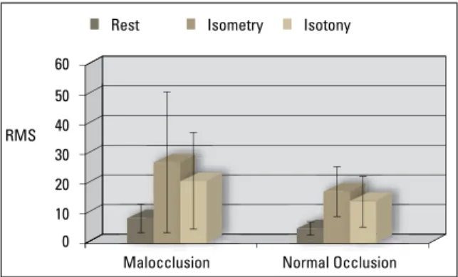
The results revealed that there was statistical-ly signifi cant difference between the two studied groups, so that youngsters with Class II, Division 1 presented a higher mean value of electrical activ-ity of the orbicularis oris muscle than youngsters with normal occlusion, in all the evaluated posi-tions. Greater electrical activity occurred during isometric contraction than in isotonic contrac-tion, however, this difference was not shown to be statistically signifi cant for the lower segment of the orbicularis oris muscle. Discrete muscu-lar activity was observed both in the group with Class II, Division 1 and in the group with normal occlusion during the resting position.
Group
Class II, Division 1 Normal Occlusion
Mean SD Mean SD
Rest 8.84 Ac 4.79 5.27 Bc 2.37
Isometry 27.66 Aa 23.64 17.83 Ba 8.41
Isotony 21.39 Ab 16.19 14.32 Bb 8.55
TABLE 1 - Mean RMS (µV) and standard deviation values with reference to the orbicularis oris muscle, upper segment (UO), of the groups with Class II, Division 1 and normal occlusion, in the resting, isometric and isotonic positions.
TABLE 2 - Mean RMS (µV) and standard deviation values with reference to the orbicularis oris muscle, lower segment (LO), of the groups with Class II, Division 1 and normal occlusion, in the resting, isometric and isotonic positions.
FIGURE 1 - Mean RMS (µV) and standard deviation values with refer-ence to the orbicularis oris muscle, upper segment (UO), of the groups with Class II, Division 1 and normal occlusion, in the resting, isometric and isotonic positions.
FIGURE 2 - Mean RMS (µV) and standard deviation values with refer-ence to the orbicularis oris muscle, lower segment (LO), of the groups with Class II, Division 1 and normal occlusion, in the resting, isometric and isotonic positions.
Means followed by different letters (capitals in the horizontal and lower case in the vertical) differ among them by ANOVA and the Tukey test (p<0.05).
Means followed by different letters (capitals in the horizontal and lower case in the vertical) differ among them by ANOVA and the Tukey test (p<0.05).
Group
Class II, Division 1 Normal Occlusion
Mean SD Mean SD
Rest 14.24 Ab 10.63 6.96 Bb 3.50
Isometry 30.63 Aa 13.24 19.90 Ba 9.43
Isotony 25.85 Aa 14.19 20.61 Ba 16.62
or diminished muscle activity at the end of the movement. In isotonic activity, one interval of the collected electromyographic tracing was selected, also avoiding the initial and fi nal intervals.
The collected data were processed by the RMS (Root Mean Square) using Myosystem–I software, and submitted to statistical analysis by the analysis of variance (ANOVA) for repeated measures and the Tukey test (α=0.05).
RESULTS
The data with reference to electromyo-graphic analysis are shown in Tables 1 and 2 and Figures 1 and 2.
Malocclusion Normal Occlusion Malocclusion Normal Occlusion
DISCUSSION
In this study, it was chosen to evaluate a group of youngsters in the age-range from 8 to 10 years, of the female gender, with dental Class II, Division 1, comparing them with a similar group with nor-mal occlusion, as a great demand for treatment for this type of malocclusion has been observed in dai-ly clinical practice. It is believed that orthodontic intervention in this age-range is more efficient be-cause of greater tissue viscoelasticity, thus contrib-uting to post-treatment stability. The choice of girls was made with the intention of avoiding variables that could interfere in the results due to inherent differences in development between the genders.
Electromyography is an extremely useful tool in the study of neuromuscular aspect of the mas-ticatory system. Nevertheless, to obtain a faithful-ly electromyographic recording it is imperative to use an adequate technique in order to minimize the interferences from the environment.
In electromyographic signal acquisition sur-face electrodes were used. The choice of elec-trodes depends on the information one wishes to collect and the location of the muscles to be studied. Surface electrodes are used in the study of muscles located immediately under the skin, as they allow the integrated electrical activity of these muscles to be estimated, whereas nee-dle electrodes enable access to deeper muscles, making them important in the study of motor units.15,16,20,21 In addition, surface electrodes have been shown to be easy to use and cause no dis-comfort to the patient.3,20
In processing the collected signal, it was cho-sen to determine the RMS. This form of analysis presents outstanding advantages, since muscle electrical activity is expressed quantitatively, with this calculation made in a simplified manner by means of specific types of software.3,21
In the present study there was evidence of a difference in electromyographic activity between youngsters with Class II, Division 1 and normal occlusion, with greater electrical activity of the
muscles in the group with malocclusion. This fact is probably related to the characterization of this malocclusion; that is, projected maxillary incisors and anteroposterior discrepancy, which makes it difficult for this musculature to perform its basic functions, generating the need for adaptations. The results obtained are in agreement with those of pre-vious studies, in which the authors observed that patients with Class II, Division 1 expend greater perioral musculature effort to make various move-ments,11,19,24,25,27 in addition to presenting lower ac-tion potentials of the orbicularis oris muscle than those patients with normal occlusion; and that re-duction in adequate dental contacts in the anterior region caused hypotonicity of the upper lip.1,12
Some studies have also verified adaptations of the perioral muscles, with an increase in their ac-tivity in the presence of alterations such as suck-ing habits, predominantly mouth breathsuck-ing and abnormal swallowing.2,6,14,22,23,26,29
of maxillary protrusion.7 However, there appears to be no doubt that the upper and lower segments of the orbicularis oris muscle function in an indepen-dent manner.5,14,18,30
In view of the results obtained in this study, it is believed that patients with Class II, Division 1 present a pattern of perioral muscle activity that differs from those with normal occlusion, with the need for greater muscle fiber participation to perform various functions.
CONCLUSIONS
Based on the literature, sample characteristics, methodology used and results obtained, it was concluded that:
1) Different electromyographic activity oc-curred between youngsters with Class II, Division 1 and those with normal occlusion.
2) This activity was shown to be greater in youngsters with Class II, Division 1, thus, suggest-ing decreased lip competence in this group.
1. Ahlgren JGA, Ingervall BF, Thilander BL. Muscle activity in normal and postnormal occlusion. Am J Orthod. 1973;64(5):445-56.
2. Baril C, Moyers RE. An electromyographic analysis of the temporalis muscles and certain facial muscles in thumb and inger sucking patients. J Dent Res. 1960;39(3):536-53. 3. Basmajian JV, De Luca CJ. Muscles alive: their function
revealed by electromyography. 5th ed. Baltimore: Williams &
Wilkins; 1985.
4. Camargo MCF, Azevedo Jr O, Briso MLG. Dispositivo indutor de vedamento labial – DIVEL. J Bras Ortodon Ortop Facial. 2001;6(33):256-61.
5. Essenfelder LRC, Vitti M. Análise eletromiográica dos músculos orbicularis oris em jovens portadores de oclusão normal. Ortodontia. 1977;10(3):180-91.
6. Gustafsson M, Ahlgren J. Mentalis and orbicularis oris activity in children with incompetent lips. Acta Odontol Scand. 1975;33(6):355-63.
7. Jung MH, Yang WS, Nahm DS. Effects of upper lip closing force on craniofacial structures. Am J Orthod Dentofacial Orthop. 2003;123(1):58-63.
8. Lehmkuhl DL, Smith KL. Cinesiologia clínica de Brunnstrom. 4ª ed. São Paulo: Manole; 1989.
9. Lowe AA, Johnston WD. Tongue and jaw muscle activity in response to mandibular rotations in a sample of normal and anterior open-bite subjects. Am J Orthod. 1979;76(5):565-76. 10. Lowe AA, Takada K. Associations between anterior temporal,
masseter, and orbicularis oris muscle activity and craniofacial morphology in children. Am J Orthod. 1984;86(4):319-30. 11. Lowe AA, Takada K, Taylor LM. Muscle activity during
function and its correlation with craniofacial morphology in a sample of subjects with class II, division 1 malocclusions. Am J Orthod. 1983;84(3):204-11.
REfERENCES
12. Marchiori SC, Vitti M. Estudo eletromiográico do músculo orbicular da boca em indivíduos com oclusão normal e maloclusões. RGO: Rev Gaúcha Odontol. 1996;44(6):331-4. 13. Moyers RE. Temporomandibular muscle contraction patterns in
Angle Class II, division I malocclusions: an electromyographic analysis. Am J Orthod. 1949;35(11):837-57.
14. Nieberg LG. An electromyographic and cephalometric radiographic investigation of the orofacial muscular complex. Am J Orthod. 1960;46(8):627-8. 15. Portney L. Eletromiografia e testes de velocidade
de condução nervosa. In: Sullivan OS, Schmitz JT. Fisioterapia: avaliação e tratamento. São Paulo: Manole; 1993. p. 183-223.
16. Pruzansky S. The application of electromyography to dental research. J Am Dent Assoc. 1952;44(1):49-68.
17. Rasheed SA, Munshi AK. Electromyographic and
ultrasonographic evaluation of the circum-oral musculature in children. J Clin Pediatr Dent. 1996;20(4):305-11. 18. Sales RD, Vitti M. Análise eletromiográica dos músculos
orbiculares oris em indivíduos portadores de maloclusão Classe I, antes e após submetidos a tratamento ortodôntico. Rev Assoc Paul Cir Dent. 1979;33(5):399-411.
19. Simpson M. An electromyographic investigation of the perioral musculature in Class II division 1 malocclusion. Br J Orthod. 1977;4(1):17-22.
20. Sodeberg GL, Cook TM. Electromyography in biomechanics. Phys Ther. 1984;64(12):1813-20.
21. Sodeberg GL, Knutson LM. A guide for use and
interpretation of kinesiologic eletromyographic data. Phys Ther. 2000;80(5):485-98.
Contact address
Vania C. V. Siqueira
Rua José Corder 87 - Jardim Modelo CEP: 13.419-325 - Piracicaba / SP, Brazil E-mail: siqueira@fop.unicamp.br
27. Vianna MS. Análise eletromiográica do músculo orbicular da boca em indivíduos com maloclusão Classe II, divisão 1 de Angle e modo respiratório predominantemente bucal ou nasal [dissertação]. Curitiba (PR): Pontifícia Universidade Católica do Paraná; 2002.
28. Vitti M, Basmajian JV. Muscles of mastication in small children: an electromyographic analysis. Am J Orthod. 1975;68(4):412-9.
29. Vitti M, Basmajian JV, Ouellette PL, Mitchell DL, Eastman WP, Seaborn RD. Electromyographic investigations of the tongue and circumoral muscular sling with ine-wire electrodes. J Dent Res. 1975;54(4):844-9.
30. Zilli AS. Estudo eletromiográico dos músculos orbiculares da boca, segmentos superior e inferior (região medial), em jovens com maloclusão Classe I de Angle [dissertação]. Piracicaba (SP): Universidade Estadual de Campinas; 1994.
Submitted: August 16, 2007 Revised and accepted: August 6, 2009 23. Tomé MC, Marchiori SC. Estudo eletromiográfico dos
músculos orbiculares superior e inferior da boca em crianças respiradoras nasais e bucais durante o repouso com e sem contato labial. J Bras Ortodon Ortop Facial. 1998;3(15):59-66.
24. Tosello DO, Vitti M, Bérzin F. EMG activity of the orbicularis oris and mentalis muscles in children with malocclusion, incompetent lips and atypical swallowing – part I. J Oral Rehabil. 1998;25(11):838-46.
25. Tosello DO, Vitti M, Bérzin F. EMG activity of the orbicularis oris and mentalis muscles in children with malocclusion, incompetent lips and atypical swallowing – part II. J Oral Rehabil. 1999;26(8):644-9.