TYMPANOMETRIC FINDINGS IN A GROUP OF STUDENTS
Achados timpanométricos em um grupo de escolares
Tâmara de Andrade Lindau (¹), Camila Ribas Delecrode (2), Ana Claúdia Vieira Cardoso (3)
(1) Philosophy and Science College of São Paulo State Univer-sity – UNESP, Marília, SP, Brasil.
(2) Studies Centre for Education and Health of the Philoso-phy and Science College of São Paulo State University – UNESP, Marília, SP, Brasil.
(3) Department of Speech Therapy of the Philosophy and Science College of São Paulo State University – UNESP, Marília, SP, Brasil.
Conlict of interest: non-existent
size), exposure to passive smoking, breastfeeding, socioeconomic level, health care and paciier use7.
These factors are not directly related to the
physio-pathology of the disease, but if present, represent an increased risk of disease, probably it inluences
one or more of the causal mechanisms3,8.
There are some behaviors that are suggestive of auditory deprivation caused by otitis media
which are: turn the head toward the sound source, frequent requests for repetition, high or low vocal
intensity, focus on the teacher´s lip, lack of attention, social isolation, learning disabilities, among others2.
These manifestations may be missed by parents
and educators, and may interfere on language development and school performance2,9.
When one considers the risk factors related to
changes of the middle ear and the impact caused
by deprivation, it becomes essential to conduct
the screening aimed at the early detection of such changes in order to minimize the losses. The
indings of such changes was reported by a study conducted with 287 students from ive to ten years
of age, it found a prevalence of 39,4% of immitance
screening failure, it showed that students between
the ages of nine and ten years have failed less than the younger ones and this difference was
statisti-cally signiicant9.
INTRODUCTION
Hearing is a pre-requisite for language acqui-sition and development. Any sensorial hearing
deprivation may result on problems in communi
-cation processes, interfering in the global cognitive
development, learning and interpersonal relation-ships, and may also impair the school and therefore the professional performance of the affected population1-3.
Recurrent otitis media is one of the most frequent causes of hearing loss3,4. Most often, approximately
70% of the cases, it would be a complication of any
upper airway infection4-6.
Some studies report that in addition to any upper
airway infection, there are other risk factors related
to otitis media, such as: care in day care centers,
seasonal variation, the presence of siblings (family
ABSTRACT
Purpose: to characterize and compare the tympanometric indings in a group of preschoolers. Method: 112 preschoolers were evaluated, both genders, aged from four years old to ive years and
eleven months old who attended a Municipal School of Early Childhood Education in the suburbs
of Marilia city– SP. Tympanometry was used as triage procedure. It was considered that the child
PASSED in the triage when it presented an A type tympanometric curve, bilaterally, being reevaluated
in case of failure. Results: it was observed a high failure index (63.4%) in the studied population. The
older female preschoolers presented a higher index of A type tympanograms, in both ears. There
was a higher incidence of failure in male preschoolers aged from four years to four years and eleven months. Conclusion: in this sample, there was a high index of tympanometric alteration. There was
a tendency among younger male students to present a higher index of tympanometric alteration when compared to older female preschoolers, this difference was not statistically signiicant.
resulting consequences, especially the learning process of reading and writing.
So the present study aimed to characterize and
to compare the tympanometric indings in a group of
preschoolers.
METHOD
This study was conducted in a Municipal
Pre-School of Education located in a suburban area
of Marilia, whose population has a low socioeco-nomic level.
At the school we develop an Extension Project
aiming to perform hearing screening, tympanometry and auditory processing screening in preschoolers. In this study, especially, the focus was the
tympano-metric indings.
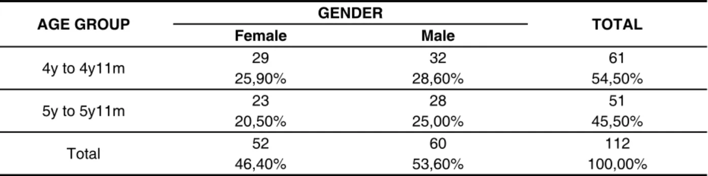
It was evaluated 112 preschoolers, from both genders, aged from four to ive years and eleven
months. The preschoolers were divided according
to gender and age as in Table 1.
It was considered as inclusion criteria: the parents or guardian sign the informed consent term and the child remaining at school in integral period. The American Speech and Hearing Association
(ASHA) screening guidelines include the following procedures to screen for outer and middle ear
disorders in children: case history obtained from the
child´s parents or guardian; otoscopic inspection
for obvious structural anomalies or obstructions of the ear canal and tympanic membrane; and
low frequency (220 or 226 Hz) tympanometry.
Pure-tone screening is not employed by contem -porary protocols for identifying outer and middle ear disorders10.
Similar to the ASHA guidelines, the American Academy of Audiology (AAA) recommends immediate medical referrals if any of the following
conditions are identiied: otalgia, otorrhea, external ear disease, and lat tympanogram with ear volume
> 1.0 cm3 (suggesting eardrum perforation) unless
there is a tympanostomy tube10,11.
Thus the hearing screening is an eficient way to determine the appropriate approach to be adopted
with students regarding peripherical hearing loss
and/or hearing abilities. If necessary, it should be
adopted medical and speech therapy approaches associated with hearing screening to minimize the
Table 1 – Preschoolers’ distribution according to the variables age group and gender
AGE GROUP GENDER TOTAL
Female Male
4y to 4y11m 29 32 61
25,90% 28,60% 54,50%
5y to 5y11m 23 28 51
20,50% 25,00% 45,50%
Total 52 60 112
46,40% 53,60% 100,00%
It is important to emphasize that in this sample there were no children with signs or symptoms which were suggestive of syndrome or craniofacial malformations.
Initially was carried out the otoscopic inspection to identify changes that would prevent the
tympa-nometry or compromise the reliability of the results. The changes observed during the inspection were considered as exclusion criteria, on these cases the
family was advised and referred to medical care. The immitanciometry was performed using a middle ear analyzer Grasson-Standler GSI-38, low frequency (226 Hz), with insert earphones and
probe system. It was inserted the probe tip into the
ear canal to seal.
To analyze the tympanometric indings were used the tympanogram classiication system proposed by Jerger (1970)12. It was considered
the PASS/FAIL criterion; the preschooler PASSED
when presented Type A tympanogram on both ears,
and FAILED when presented other tympanogram type. The preschooler who FAILED was rescreened
after six weeks11.
This research project was approved on the
Ethics Committee of the College of Philosophy
and Science – UNESP- Marília, protocol number
132/2010.
To describe the results was used the descriptive
and inferential analysis. At inferential statistical
adopted for the application of the statistical tests was 5% (0.005) and the analysis was performed
using SPSS program (Statistical Package for Social
Science) , in 19.0 version.
RESULTS
The analysis of the results showed a high failure
rate (63, 4%) in this population (Table 2 and 3). order to verify the possible differences between
genders and age groups for the variables of interest
(tympanogram type and PASS/FAIL criterion). When
analyzing the results of the association between the gender and age variables with the screening
results (PASS/FAIL criterion) it was noticed a
tendency, then it was applied the Likelihood Ratio Test adjusted by Bonferroni correction in order to identify which combinations of gender and age have tended to be different. The signiicance level
Age RIGHT EAR LEFT EAR
A Ad B C A Ad B C 4y to 4y11m
N=61
23 0 22 16 23 0 26 12
37,70% 0,00% 36,10% 26,20% 37,70% 0,00% 42,60% 19,70%
5y to 5y11m N=51
30 2 11 8 26 2 14 9
58,80% 3,90% 21,60% 15,70% 51,00% 3,90% 27,50% 17,60%
Total N=112
53 2 33 24 49 2 40 21
47,30% 1,80% 29,50% 21,40% 43,80% 1,80% 35,70% 18,80%
p = 0,026* p = 0,105
Table 2 – Tympanogram type distribution according to the age variable on right and left ear
Likelihood Ratio Test/ p-value < 0,005 = statistically signiicant*
Gender RIGHT EAR LEFT EAR
A Ad B C A Ad B C Female
N=52
33 0 13 6 25 0 16 11
63,50% 0,00% 25,00% 11,50% 48,10% 0,00% 30,80% 21,20%
Male N=60
20 2 20 18 24 2 24 10
33,30% 3,30% 33,30% 30,00% 40,00% 3,30% 40,00% 16,70%
Total N=112
53 2 33 24 49 2 40 21
47,30% 1,80% 29,50% 21,40% 43,80% 1,80% 35,70% 18,80%
p = 0,004* p = 0,275
Table 3 – Tympanogram type distribution according to the gender variable on right and left ear
Likelihood Ratio Test/ p-value < 0,005 = statistically signiicant*
Whereas the variables age (Table 2) and gender (Table 3), it was observed that the male preschoolers
with lower age presented lower rates of Type A
tympanograms in both ears when compared to
female preschoolers of higher age, this difference
was statistically signiicant on both variables in the
right ear.
As regards the pass/fail criterion for both age groups (Table 4) and genders (Table 5), the results
showed a higher fail incidence for males at the age group of four years to four years and eleven months, however, this difference was not statistically
This tendency was statistically signiicant only
when was compared the older female preschooler group and the younger male preschooler group
(Table 6). So, it can be stated that there is a tendency that more female preschoolers, at ive and ive years and eleven months, do not exhibit changes in the
middle ear when compared to male preschoolers at four and four years and eleven months of age.
When you combine the variables gender and
age (Figure 1) according to the pass/fail criterion,
it was found that preschoolers from both genders
among four years and four years and eleven
months, and males, among ive years and ive years
and eleven months, had failed more than the female preschoolers in this age group.
Age TYMPANOMETRY Total
Pass Fail
4y to 4y11m 18 43 61
29,50% 70,50% 100,00%
5y to 5y11m 23 28 51
45,10% 54,90% 100,00%
Total 41 71 112
36,60% 63,40% 100,00%
p = 0,088
Table 4 – The occurrence of pass/fail on tympanometry according to the age variable
Likelihood Ratio Test/ p-value < 0,005 = statistically signiicant*
Gender TYMPANOMETRY Total
Pass Fail
Female 24 28 52
46,20% 53,80% 100,00%
Male 17 43 60
28,30% 71,70% 100,00%
Total 41 71 112
36,60% 63,40% 100,00%
p = 0,051
Table 5 – The occurrence of pass/fail on tympanometry according to the gender variable
Legend: F- female/ M – male/ I – 4 years to 4 years and 11 months / II – 5 years to 5 years and 11 months.
Figure 1 – Scholars distribution according to gender and age and the tympanometry screening pass/ fail criterion
Pair of Categories Significance (p)
F(4y to 4y11m) x F(5y to 5y11m) 0,181
F(4y to 4y11m) x M(4y to 4y11m) 0,169
F(4y to 4y11m) x M(5y to 5y11m) 0,862
F(5y to 5y11m) x M(4y to 4y11m) 0,008*
F(5y to 5y11m) x M(5y to 5y11m) 0,136
M(4y to 4y11m) x M(5y to 5y11m) 0,235
Table 6 – Statistical analysis of the tympanometry screening pass/fail criterion matching to preschoolers’ gender and age group
Legend: F= female / M = male
Likelihood Ratio Test adjusted by Bonferroni correction/
Bonferroni alpha = 0,008512 – statistically signiicant*
DISCUSSION
Hearing screening is an eficient way to determine the appropriate approach to be adopted
with students regarding peripherical hearing loss
and/or hearing abilities. If necessary, it should be
adopted medical and speech therapy approaches associated with hearing screening to minimize the resulting consequences, especially the learning process of reading and writing9.
So, in this study, the preschoolers who failed on the tympanometric rescreening, the parents or
guardians were advised and referred to medical care.
Early detection and immediate intervention in children with auditory deprivation increases the
probability to optimize the receptive and expressive
language potential, literacy (reading and writing), academic performance, emotional and social development9.
grades3. However, a hearing screening program
developed with children from ive to ten years had found that age was not a signiicant risk factor for
middle ear disorders9.
Otitis media is a highly prevalent disease in
childhood, with a higher peak of incidence between six and 24 months of age and the second peak of incidence between four and seven years19.
The parameters race, sex and age inluence the structure of the Eusthachian tube or its function,
while the age also determines the immunological response of the host, it is evident that some factors are related, since younger children have more upper airways infections7.
Authors20 claim that sensory deprivation caused
by serous otitis media, aggravated by number and
episodes length of disease, can affect the speech perception and comprehension, especially in noise environments, and even affect the child´s language
development. The hearing luctuation caused by
middle ear disorders on early childhood can lead to auditory processing disorders, and interferes on learning.
Certain measures could be implemented in order
to reduce the incidence of middle ear disorders. Authors suggest as actions: treatment for enlarged adenoids, chronic sinusitis and allergies as some
of the necessary measures to reduce the problem
of persistent rhinorrhea, considering that regular visits to schools in order to screen children which
present persistent rhinorrhea, mouth breathing and otitis media should be part of a national program
that emphasizes the hearing health and guidelines
regarding the effect of passive smoking on the
respiratory and auditory system of children should
be emphasized in all anti-smoking campaigns15.
Considering the indings of this study, it is
necessary to implement actions to reduce the incidence of middle ear disorders in this population. Thus, it is suggested that parents and teachers
should be oriented and the implementation of
immunization programs for this population in order to reduce upper airways infections episodes.
CONCLUSION
In this sample, it was observed a high rate of
tympanometric alterations. There was a tendency of younger male preschooler group present a higher rate of tympanometric alterations when compared to the older female preschooler group, this difference
was not statistically signiicant. using antibiotics and the possible causes of these
disorders3-6,13,14.
This study veriied a high rate of tympanometric screening abnormalities. This fact can be explained by the presence of several risk factors which this population is exposed, such as: care in daycare
centers7, climatic variations7, socioeconomic level3,7,8
and persistent rhinorrhea15.
The risk factors are not involved in the physio
-pathology of otitis media, but, when present, can increase the risk of the disease, probably it can inluence one or more causal mechanisms7.
The recurrent otitis media is one of the most frequent causes of hearing loss3,4 in children at
school attendance phase2. Most of the time, around
70% of the cases, would be a complication of upper
airway infection4-6.
Some studies have reported that otitis media is a common disease in countries in development 3-15,
as occurs in Brazil, and the variability of risk factors among the countries is a relection of the
socio-cultural differences on the studied population16.
In this study the prevalence of abnormal tympa -nometry was 63,4%, in the literature was found a prevalence ranging from 39,4%9 to 74,8%3.
When analyzing qualitatively the tympano-metric was found a predominance of type B and C
tympanograms, and these indings were similar to
the literature9,17, the irst study was conducted with
287 students from ive to ten years which showed
types As, B and C tympanograms on 20% of the children9, and the second study evaluated 142
children of similar age, and found 46% of abnormal
tympanometries with the prevalence of types B,C
and As tympanograms, both studies revealed the occurrence of abnormalities in the middle ear17.
In relation to gender, there was a higher
incidence of abnormal tympanograms on males; this difference was statistically signiicant only in
the right ear. On others authors studies were found
no association between the variable gender and a
higher prevalence of otitis media in one of these genders3,9,15.
As regards the variable age, it was observed that
the youngest preschoolers failed more than the older ones, however, this difference was not statistically
signiicant. In the literature there is no consensus regarding age as a risk factor for otitis media, some
authors have associated age to a higher prevalence of otitis media3,8 but others have not9,15.
REFERENCES
1. Gatto CI, Tochetto TM. Deiciência auditiva
infantil: implicações e soluções. Rev CEFAC. 2007;9(1):110-5.
2. Vieira ABC, Macedo LR, Gonçalves DU. O diagnóstico da perda auditiva na infância. Rev. Pediatria. 2007;29(1):43-9.
3. Taha AA, Pratt SR, Farahat TM, Abdel-Rasoul GM, Albtanony MA, Elrashiedyet ALE et al. Prevalence and risk factors of hearing impairment among primary-school children in Shebin El-kom District, Egypt. Am J Audiol. 2010;19:46-60.
4. Spiro DM, King WD, Arnold DH, Johnston C,
Baldwin S. A randomized clinical trial to assess the effects of tympanometry on the diagnosis and treatment of acute otitis media. Pediatrics. 2004;114(1):177-81.
5. Winther B, Alper CM, Mandel EM, Doyle WJ, Hendley JO. Temporal relationships between colds, upper respiratory viruses detected by polymerase
chain reaction, and otitis media in young children followed through a typical cold season. Pediatrics. 2007;119(6):1069-75.
6. Revai K, Patel JA, Grady JJ, Chonmaitree T. Tympanometric indings in young children
during upper respiratory tract infections with and
without acute otitis media. Pediatr Infect Dis J.
2008;27:292-5.
7. Lubianca Neto JF, Hemb L, Silva DB. Fatores
de risco para otite média aguda recorrente: onde podemos intervir?- uma revisão sistemática da
literatura. J Pediatr. 2006;82(2):87-96.
8. Adhikari P. Otite média crônica supurada em crianças de escolas do vale katmandu. Arq. Int.
Otorrinolaringol. 2007;11(2):175-8.
9. Colella-Santos MF, Bragato GR, Martins PMF, Dias AB. Triagem auditiva em escolares de 5 a 10 anos. Rev. CEFAC. 2009;11(4):644-53.
10. American Academy of Audiology (AAA) Position
Statement: Identiication of hearing loss and
middle-ear dysfunction in preschool and school-age children. Audiology Today. 1997;9:21-3.
11. American Speech-Language-Hearing Association (ASHA). Guidelines for Audiologic Screening.
Rockville Pike.1997.
12. Jerger J. Clinical experience with impedance
audiometry. Arch Otorhinolaringol. 1970;92:311-24.
13. Meropol SB, Glick HA, Asch DA. Age
Inconsistency in the American Academy of Pediatrics Guidelines for Acute Otitis Media. Pediatrics. 2008;121(4):657-68.
14. Vergison A, Dagan R, Arguedas A, Bonhoeffer
J, Cohen R, DHooge I et al. Otitis media and its consequences: beyond the earache. Lancet Infect
Dis. 2010; 10(3):195-203
15. Sophia A, Isaac R, Rebekah G, Brahmadathan K, Rupa V. Risk factors for otitis media among preschool, rural Indian children. Int. J. Pediatric
Otorhinolaryngol. 2010;74(6):677-83.
16. Rovers MM, Kok IMCM, Schilder AGM. Risk
factors for otitis media: an international perspective.
Int J Pediatr. Otorhinolaryngol. 2006;70:1251-6. 17. Guida HL, Diniz TH. Peril audiológico em
crianças de 5 a 10 anos de idade. Arq. Int. Otorrinolaringol. 2008;12(2):224-9.
RESUMO
Objetivo: caracterizar e comparar os achados timpanométricos de um grupo de escolares. Método:
foram avaliados 112 escolares, de ambos os gêneros, na faixa etária de quatro anos a cinco anos
e onze meses que frequentavam uma Escola Municipal de Educação Infantil, localizada em um
bairro periférico da cidade de Marília. Como procedimento de triagem, utilizou-se a timpanometria.
Considerou-se que a criança PASSOU na triagem quando apresentou curva timpanométrica do tipo
A, bilateralmente, sendo reavaliada em caso de falha. Resultados: observou-se um alto índice de
falha (63,4%) na população estudada. Os escolares de maior faixa etária e do gênero feminino apre
-sentaram um índice maior de timpanogramas do tipo A, em ambas as orelhas. Houve uma maior incidência de falha nas crianças de faixa etária entre 4 anos e 4 anos e 11 meses, e do gênero mas -culino. Conclusão: nesta amostra obteve-se um alto índice de alterações timpanométricas. Houve
uma tendência de escolares do gênero masculino e de menor faixa etária apresentarem um índice maior de alterações timpanométricas quando comparados ao gênero feminino e de maior faixa etária, não sendo esta diferença estatisticamente signiicante.
In: Campos CAH, Costa HOO, editores. Tratado de Otorrinolaringologia. 1ª ed. São Paulo: Roca; 2003. Vol. 2, p. 21-7.
20. Pereira PKS, Azevedo MF, Testa JR. Alterações
condutivas em neonatos que falharam na triagem
auditiva neonatal. Braz. J. Otorhinolaryngol.
2010;76:347-54.
18. Caylan, R, Bektas, D, Atalay, C, Korkmaz, O. Prevalence and risk factors of otits media with effusion in Trabzon, a city in nrtheastern Turkey,
with an emphasis on the recommendation of OME screening. Eur. Arch. Otorhinolaryngol. 2006;263: 404-8.
19. Almeida CIR, Almeida RR. Otite media aguda.
Received on: January 31, 2012
Accepted on: May 02, 2012
Mailing Address:
Tâmara de Andrade Lindau
Rua José Chieramont, 54
Santa Rosa de Viterbo – SP – Brasil
CEP: 14270-000