RESEARCH ON FATIGUE IN FACIAL AND JAW MUSCLES:
REVIEW OF THE LITERATURE
Pesquisa da fadiga nos músculos faciais e mastigatórios:
revisão de literatura
Angela Ruviaro Busanello-Stella (1), Ana Maria Toniolo da Silva (2), Eliane Corrêa (3)
(1) Post-graduation Program in Human Communication
Disor-ders at Universidade Federal de Santa Maria – UFSM, Santa Maria, RS, Brazil.
(2) Speech Therapy Degree and the Post-graduation in Human
Communication Disorders at Universidade Federal de Santa Maria, UFSM, Santa Maria, RS, Brazil.
(3) Physical Therapy Department and Post-graduation
Pro-gram in Human Communication Disorders at Universidade Federal de Santa Maria – UFSM, Santa Maria, RS, Brazil. Fountain aid: CAPES
Conlict of interest: non-existent
2. It may be considered a natural muscle-defense process 2-5 which activates prior to the occurrence
of any damages to the organic and cellular levels 6. Its occurrence will depend on the type, length
and intensity of the exercise; on the typology of the recruited muscle ibers; on the level of training of the individual and on the environmental conditions for the performance of the exercise 7. In addition, the
study of muscle fatigue may show limitations due to its multifactorial nature and complexity 6.
The investigation about this mechanism in
facial and jaw muscles has drawn the attention of researchers in the ield of health, who carry out
studies in humans, with and without alterations, and in animals. In relation to Speech Therapy, knowing
how muscle fatigue occurs in speciic diseases
such as temporomandibular disorders (TMD) 8,9, sleep apnea 10, degenerative diseases 11 and mouth
breathing, for instance, appears to be an important
INTRODUCTION
Muscle fatigue is deined by the incapability of the musculature to maintain high levels of force in time1.
It happens due to the accumulation of substrate,
such as lactic acid, within the muscle cells, which
interferes on the concentration of intracellular pH and, consequently, in the conduction of the action potentials necessary for the activation of muscles
ABSTRACT
The purpose of this study was to deepen the knowledge about muscle fatigue of masticatory and facial muscles through analysis of scientiic literature. The search strategy was based on the statements of the Cochrane Library. Articles were selected through the PubMed database, using the descriptor “muscle fatigue” in conjunction with the following descriptors: “speech therapy”, “facial muscles”, “jaw muscles”, “mastication”, “chewing” , “lip” and “clenching”. Articles from the last ten years were included, regardless of the language. The texts were analyzed, at irst, in its abstract and those that did not it the study`s objective were excluded. Then, the full texts were analyzed, being considered: aim, study
design, control and study groups, criteria and methodological rigor, applied protocols, results and
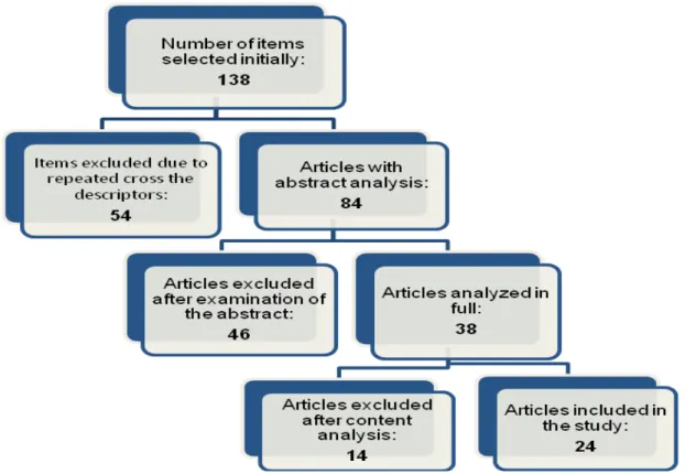
discussion about the presence of fatigue in the targeted muscles. In a last stage, studies that were not in accordance with the purpose of the study, were excluded. Thus, out of 138 articles found, 54 were excluded due to the repetition by crossing key-words and 84 were analyzed. In the abstract analysis, 46 were excluded and 38 were fully analyzed. 24 articles were maintained and 14 were excluded because they do not it the study`s criteria. Despite there are some studies regarding the analysis of fatigue, their methodology is very varied, making dificult the comparison and detailing. While some studies have found, for example, greater fatigue of the masseter and orbicularis oris muscles, others have not found the same results, which may have inluenced the target population for each study.
to locate and select the studies from the database; and the third to assess the studies found in a critical
way.
Works referring to the past ten years, regardless
the language, were selected in the Pubmed database. The descriptor “muscle fatigue” was put
together with the following descriptors: “speech
therapy”, “facial muscles”, “jaw muscles”, “
masti-cation”, “chewing”, “lip” and “clenching”.
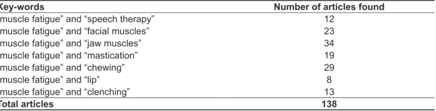
Along this step, 138 works were found, as illus -trated in Table 1.
parameter for therapeutic procedures. Nevertheless, the protocols of these studies are rather divergent,
what may hamper the comparisons among them. Thus, this study aims at deepening the knowledge
in relation to muscle fatigue of facial and masticatory muscles through the analysis of scientiic literature.
METHODS
basis to deine the steps of the study 12. The irst
step was to formulate the study problem; the second
Table 1 - Distribution of the articles found in the Pubmed database, the irst stage of the study, as the intersection of the descriptors
Key-words Number of articles found
“muscle fatigue” and “speech therapy” 12
“muscle fatigue” and “facial muscles” 23
“muscle fatigue” and “jaw muscles” 34
“muscle fatigue” and “mastication” 19
“muscle fatigue” and “chewing” 29
“muscle fatigue” and “lip” 8
“muscle fatigue” and “clenching” 13
Total articles 138
In this step the works which repeated in the
crosschecking of the descriptors were excluded. Subsequently, the analysis of abstracts was carried out and those that did not present the use
of fatigue protocols in relation to facial and/or jaw muscles were also excluded. Finally, the works selected were fully read and underwent analysis
of the following aspects: objective, study design, formation of control and study groups, criteria and
methodological strictness, applied protocols, results
found and discussion about fatigue in the muscles
concerned. In this last step, those that were not
craniofacial morphology 13. Thirty young adults at average ages between 23 and 25 years old were
divided into two groups: short faced individuals and normal-long faced individuals according to Ricketts
analysis 14. The variables analyzed were: (a)
thickness of right masseter; (b) outset and tolerance to pain and; (c) report of pain and fatigue. Thickness
was measured once through ultrasound and the
other variables were measured after gum mastication for 10 minutes at four times (immediately after, ive minutes after, ten minutes after and 24 hours after
mastication ended). The researchers observed that
individuals with short faces had greater thickness
in the masseter, whereas individuals with normal to
long faces had greater tolerance and outset of pain.
They reported the Mechanical Advantage Theory as
a plausible justiication for the differences observed, where the individuals with longer faces would have less mechanical advantage in jaw elevator muscles. Therefore, individuals with short faces would have greater occlusal force and, consequently, greater
intramuscular pressure, which may limit the
blood low through the muscles, something that is necessary for the maintenance of force.
It is believed that the indings of these researchers
match the literature bases although the gum, which
was used as food in this study, may have inluenced
the results.
REVIEW LITERATURE
From the 38 works that were fully analyzed, 14 were excluded for their content did not match
with the criteria previously described, and 24 were
included in the inal step. The textual reasons that excluded these 14 studies were:
• fatigue was not considered the objective of the
study but a symptom indirect to other diseases;
• the muscles studied were not speciied (e.g.:
masseter, temporalis, orbicularis oris, orbicularis
oculi, among others) or did not refer to the facial and jaw muscles;
• important methodological aspects did not present
a detailed description such as selection, subjects, criteria for group formation and procedure for muscle fatigue evaluation.
The studies were subsequently organized into two parts: studies developed with humans and
experimental research (with animals). Within each
session, the works were presented by the relevance
of their topics.
Research on fatigue in facial and jaw muscles in humans
A study regarding thickness, tolerance and
induction of pain of the masseter was carried out in 2003 with individuals with different types of
(b) amplitude and frequency of electromyographic
signal during isometric contraction;
(c) duration and degree of exteroceptive
suppression;
(d) pain and fatigue levels at pressure (measured
in degree scales).
The researchers concluded that exteroceptive suppression was associated with the presence of fatigue and pain (in this case, induced by the admin
-istration of glutamate), once the degree of inhibition
increased. They also emphasized that the muscle activity at rest captured by electromyography also
suffered inluence, in a distinct way, once an initial increase was observed followed by a decline.
Studies such as the ones cited above prove to be innovative and daring, once they make an attempt at reproducing, in an induced way, what would happen in natural pathological conditions, even though they analyze the electromyographic
signal mainly from the perspective of amplitude. It is believed that information regarding the reduction of median frequency (MF) as used for the analysis of muscle fatigue in the studies cited below would enrich the electromyograic indings reported so far.
A second study carried out by the same authors 17 using the same sample and methodology compared
the relationship between fatigue and muscle pain with the electrical activity of the jaw musculature in relation to gender (male and female). They did not observe differences between genders for these aspects, though they inferred that, clinically, women
could be considered more vulnerable to pain and
fatigue triggering, presumably due to the fact that the differences in their neuromuscular system
contribute to a greater propensity to developing
chronic problems of skeletal muscle pain.
Another study which considered the inluence of fatigue and muscle pain on jaw muscles 18 aimed
at investigating the effect of intense masticatory exercises on fatigue and muscle pain induction
and, consequently, in the masticatory system.
Forty men who should perform 20 ive-minute
masticatory sessions were included. Fatigue and
muscle pain scores and amplitude of mandibular stretch relex of the left masseter were measured at the end of each session, 20 minutes and 24 hours after the mastication ended, by means of the visual analogue scale (VAS). In addition, signs of TMD and threshold of pressure-muscle
pain were measured prior to, 20 minutes and 24
hours after the total exercise time, based on the tool Research Diagnostic Criteria for TMD (RDC/ TMD). Researchers observed that fatigue and pain increased, relex amplitude remained unaltered and threshold of pressure-muscle pain decreased with the performance of exercises. Despite of the fact
A study 15 aiming to examine the effects of
muscle fatigue on the short latency relex of the jaw was carried out with healthy men and women, with and without induction of muscle pain with
hypertonic saline solution (HS). According to the
researchers, the administration of intramuscular substances, especially HS, causes similar effects of pain observed in individuals with TMD and, for this reason, it becomes a useful way of studying the interaction between the dysfunction, pain and motor control. Fifteen men and thirteen women at average of 24 years old conducted a protocol of induction of muscle fatigue (mastication of chewing gum for six minutes), followed by the injection of HS and isotonic saline (IS) in the left masseter, with an interval of 45 minutes between them. Reports of pain and fatigue by degree scales as well as the amplitude of the electromyographic signal of the masseter and anterior temporalis muscles, right and left sides,
were analyzed in three situations (prior to,
immedi-ately after and 15 minutes after administration of HS). In this case, the amplitude of the electromyo
-graphic signal was used for the analysis of the jaw jerk relex. Although researchers observed that there was an increase in the amplitude of the signal after the fatigue protocol followed by HS, there was no statistical signiicance. The only difference observed was between sexes in relation to relex triggering
which, according to the authors, may have occurred
due to the difference between the muscle structure of men and women.
Another study was developed in a similar way
about the administration of intramuscular substances
to induce pain 16. The research considered the effect
of muscle fatigue induced by low-level isometric
contraction and subsequent muscle pain triggered
by the administration of glutamate on the response of exteroceptive suppression and the electromyo -graphic activity at rest in healthy humans in order to
investigate whether the glutamate would act specii
-cally on the condition of fatigue. In this study, the
masseters and anterior temporalis muscles (right and
left sides) of 23 individuals (11 women and 12 men
at ages between 25 and 23 years old, respectively)
without any signs or symptoms of TMD, headaches
or clenching were assessed. The procedure
consisted of a sustained isometric contraction at 10% of the maximum voluntary contraction (MVC) for 30 minutes in order to induce fatigue, followed by an injection of glutamate or isotonic saline solution (IS) (two different times) and subsequent rest. Considering this routine, the following aspects were analyzed at four times (outset, post-contraction, post-injection and post-rest):
observed that vibratory stimuli did not interfere in the outset of pain and fatigue.
Studies investigating muscle fatigue in speciic
occupations such as wind instrument players and
goldsmiths were also found.
Another group of Japanese researchers 21
carried out a study to investigate the inluence of tonal change of wind instruments in muscle activity, and the effects of sustained blows on fatigue.
Thirty-three individuals assessed while playing their instru-ments were selected, being analyzed the masseter,
temporal, orbicularis oris and digastric muscles of the left side. The study developed two testing steps:
Step 1 – individuals playing their instruments
for three seconds in the habitual tone and then
an octave above. Electromyographic signal was measured at rest, while the musicians played, during
maximum dental clenching and during maximum
mouth opening.
Step 2 – individuals playing their instruments for
90 minutes. Electromiographic signal was measured
prior to and after blowing activity and analyzed
through MF analysis.
Researchers observed that the electrical activity
of the masseter and temporal muscles while
playing the instrument was slightly higher than at
rest, yet extremely lower in comparison to dental clenching. As for orbicularis and digastric muscles,
the activity was relatively higher while playing the
instrument. Higher notes were followed by an increase in the electrical activity only in part of the sample. Regarding the analysis of MF there was no difference prior to and after prolonged activity. Thus, the authors concluded that the activity of jaw
muscles was low, regardless the note played, and prolonged blow activity was not enough to induce
fatigue in the studied muscles.
The study with goldsmiths was carried out in India 22 and aimed at measuring the effect of air
tubes used in their work on fatigue of facial muscles
and respiratory stress in daily work routine. One hundred male goldsmiths were included and answered a questionnaire, underwent a pulmonary
function test, peak expiratory low measurement, as well as the electromyographic evaluation of left and right buccinators and orbicularis oris, for they
are, in principle, the most engaged muscles during their work. The electromyographic evaluation was carried out throughout their workday, respec-tively in the beginning, the middle and at the end,
allowing the analysis of muscle fatigue through MF.
Results evidenced reduced lung volume and peak
expiratory low, possibly due to the great pressure needed for their activity (the use of air tubes) and the exposition to certain chemical compounds. Fatigue
was observed in all the muscles studied at the end that the study showed some limitations according to
the authors themselves, they concluded that intense
and prolonged masticatory exercises may induce fatigue, pain, and decrease of the pain threshold of pressure-masticatory muscles, without interference of the amplitude of the mandibular stretch relex.
Studies have shown that factors such as pain and fatigue are better detected in isometric contrac -tions 3,4. This is because in these situations intra-cellular substrates would accumulate more easily,
thus impairing the passage of potentials of muscular action and more rapidly yielding pain and fatigue
conditions. However, it is believed that studies
involving research on pain and fatigue in dynamic contractions are extremely relevant, once they depict what most commonly occurs in the routine of
the population.
The relation between the masseter and the sternocleidomastoid (SCM) was investigated by
Japanese researchers 19. This study aimed at
investigating the relation between the activity of the masseter and the SCM and the head and jaw movements during mastication in the protocol of muscle fatigue. The sample consisted of 12 adults
(ten men and two women) with normal occlusion
that should perform gum mastication for 30 seconds at three different times: prior to the performance of maximum clenching, immediately after it, and three minutes after clenching. The electrical activity of
masseters and right SCM was captured simultane-ously during mastication (through an
electromyo-graphic test), as well as the head and jaw movements
(through the motion capture system), being ten
masticatory cycles selected for analysis. In the electromyographic analysis, the ields of amplitude and frequency were taken into consideration. In order to ascertain the occurrence of fatigue during mastication, the median frequency of the signal was estimated. With reference to electrical activity, the masseter had lower activity after clenching, whereas the activity of the SCM increased. As for muscle fatigue, both muscles had a reduction in MF after clenching, being it restored after three minutes. For the movements, stability was observed for jaw movement and head movements increased after clenching. Therefore, the researchers concluded that there is a relation between jaw muscles and
head and neck muscles.
The inluence of vibrotactile stimulation in the masseter on the late perception of pain and fatigue
in healthy individuals has been recently investi-gated20. Twenty-ive healthy women participated in
this study, receiving vibratory stimuli for 15 seconds, sequentially performing contractions at 10%, 20% and 40% of MVC. Fatigue was investigated, as
muscles during submaximal contractions evoked. In addition, it was not possible to quantify its magnitude
or where it starts (e.g. suprabulbar motor centers,
facial motor neurons, among others).
The obstructive sleep apnea was the aim of a
study 10 in which researchers tested the use of a
treatment for apneic individuals (intra-oral device for the functional reordering of the occlusion) as a
preliminary way, in normal individuals. The sensation
of occlusal alteration caused by the use of the device
at night was assessed in 12 healthy individuals. The
variables considered were sensation of occlusal alteration, masticatory force, occlusal contact area and muscle fatigue, all measured every ifteen minutes during the irst four hours after removal of
the device. The same procedures were repeated on
another day after the individuals had slept without the device. Occlusal sensation was quantiied through VAS; masticatory force and occlusal contact
area were measured by a computer program during
three seconds of clenching; and muscle fatigue of the masseters was investigated through MF accessed by surface electromyography (sEMG). Researchers observed that the sensation of
occlusal alteration was detected by the individuals
up to 75 minutes after removal of the device and interference in masticatory force was detected right after removal. Muscle fatigue was associated to
the altered sensation in only two individuals and
with masticatory force in only one individual. Thus,
it was concluded that the most recurrent variable, alteration in occlusal perception, was probably due
to the non-physiological shift of the temporoman
-dibular joints (TMJs) that happens with the use of
the intra-oral device.
Other diseases such as Amyotrophic Lateral Sclerosis (ALS) have also been the object of studies
that investigate musculature 11. This is a progressive
disease characterized by signiicant muscle fatigue
and reduced response to repeated nerve
stimu-lation, especially in the limbs. Nevertheless, these signs may appear in facial muscles, so that, initially,
the disease may be misdiagnosed as Myasthenia
Gravis, which is, for the researchers, the reason for
this study. It aimed at investigating the occurrence
of muscle fatigue and the response to repeated nerve stimulation in facial muscles of individuals with ALS of the oropharyngeal type, in other words, with complaints about dysarthria, dysphagia and/ or hypernasal voice. The stimulation of the orbicu -laris oculi (OO), trapezoid and nasal muscles was
performed in ten patients. Six patients reported muscle fatigue while performing the tests of the
muscles concerned; three presented abnormal
reduction of stimulation in facial muscles, but not in the muscles of legs and arms; two presented of the goldsmiths’ workday. For these reasons, the
researchers concluded that the implementation of a new and more ergonomic air tube may reduce facial fatigue and respiratory stress.
With the proposal for improvements and greater ease in studies with muscle fatigue research, Italian researchers carried out a study in which fatigue was researched under conditions of continuous levels of submaximal muscle contraction 23. These
researchers stated that the use of pre-established and ixed force levels brings beneits when compared to the use of termed force of MVC of the
individuals, once they would reduce the possibility
of dental fractures, pain and discomfort. Ten healthy
individuals underwent electromyographic evaluation
of the masseter and anterior temporal, performing right unilateral clenching in a 13 kg load cell (127N) until exhaustion. In this context, MF was estimated in the outset, after one minute and at the end of the test. The resistance time observed varied from one to eight minutes, and there was a reduction of MF in the masseter and temporal, being more signiicant
in the masseter. It led the researchers to conclude
that research with ixed submaximal loads may induce muscle fatigue and would be a more acces -sible protocol, regardless the population assessed.
In a study about fatigue in the upper airway
muscles, researchers in the United States developed
a force transducer for the nasal muscle 24. They assessed the nasal dilator muscles in order to test the hypothesis that central mechanisms would
contribute to fatigue in these muscles, in condi
-tions of sustained submaximal contrac-tions. Eleven individuals were selected, ive men and six women who were able to control the mobility of the nasal
muscle and did not have any respiratory
intercur-rence. Three types of procedures were performed, with the following analyses: (a) submaximal contrac
-tions of the muscles at 20, 35 and 65% of the MVC until the force reduced in 90%, being the signal analyzed in microvolts (µV) and MF; (b) stimuli of the facial muscle and subsequent analysis of maximum potential evoked; (c) 15 nasal contractions of 10 minutes each, being analyzed the signal in µV and MF after each cycle. Despite the lack of clarity in
presenting the results, the authors observed that
the electromyographic activity in µV increased with time, the MF decreased approximately 25% in all experiments, and paradoxically the maximum
potential evoked did not alter, what according to
them indicates that fatigue (especially observed for the decrease of MF) is mainly due to the mecha
-nisms close to the neuromuscular junction. In their opinion, these data show that central fatigue may
have been present even though the central nervous
sectors were selected considering the type of headache and its absence. The analysis of MF was
carried out through electromyography throughout the
MVCs, and, a distinctive aspect of this research, as well as the following was a mold of masticatory cells speciic for each individual. The authors observed
that, among the muscles assessed, the masseter
was the most fatigable while comparing individuals
with headaches in general with the control group. In addition, women with chronic headaches showed a more rapid decrease in MF, compared to others.
TMD patients have also been the focus of other
studies8,9. One of them, carried out in Brazil8, looked
for signs of muscle fatigue in individuals with TMD,
during mastication. Twenty women participated in the study, diagnosed by the RDC as clinically normal
or with TMD of the myogenic type. The masticatory
muscles (masseter and temporal) were analyzed
EMG during 15 seconds of mastication. Fatigue was investigated through the analysis of median frequency throughout mastication, not showing signiicant alteration. According to the authors, the lack of electromyographic signals of fatigue, in all individuals, may be have happened because of the elevation of blood low due to the masticatory
movement, an aspect that may hinder the deposition
of substrates within the muscles that induce fatigue.
Another study, carried out in Italy9, focused on:
(a) assessing the use of EMGs as an objective measurement of fatigue of jaw elevator muscles; (b) comparing the EMG manifestations of fatigue of the masseter and temporal right and left; (c) assessing the recovery of the investigated muscles after testing their resistance; (d) comparing fatigue and recovery of jaw elevator muscles between
healthy individuals and TMD patients. Twenty healthy individuals and TMD patients participated in
it, performing submaximal isometric contractions at 20%, 40% and 60% of MVC for 30 seconds, and at 80% until exhaustion (resistance test) and recovery. A force transducer was used for these quantiica
-tions. According to the authors, the decrease of MF was a good indicator of fatigue in the muscles
assessed, masseter and temporal (independently
of the side) showed the same myoelectric manifes
-tations of fatigue and recovery; and the muscles
assessed showed lower values in TMD patients. Finally, they concluded that myoelectric evaluations, the way they were remarked in this study, may help
in the clinical evaluation of TMD patients.
The inluence of food texture on muscles and masticatory movements, as well as on fatigue of
this musculature was investigated by other authors 28. They analyzed, in a sample of 28 individuals,
the oxygenation of the masseter, as well as the abnormal reduction in stimulation of OO and nasal
muscles; one presented reduction only for OO. According to these indings, researchers stated that facial muscles may be affected, as one of the irst manifestations of some types of ALS, even though facial weakness is not yet evident.
Among the cervical diseases associated to the stomatognathic system there is the whip-lash
syndrome (WLS). This disease generally emerges
associated to car accidents and is due to an abrupt
movement of acceleration and deceleration in the neck, causing a series of symptoms and sequelae. Other studies also investigate muscle fatigue in jaw
muscles in this population 25,26.
The responses of the autonomic nervous system
were studied in individuals with cervical diseases associated with the stomatognathic system 25,
including muscle fatigue. The study investigated autonomic reactions theoretically more signiicant
in these individuals when compared to normal individuals in masticatory conditions. Two groups with 21 individuals each had cardiovascular aspects,
perception of fatigue and pain, and muscle fatigue through EMG evaluation, tested in a situation of
alternated unilateral mastication with chewing gum.
In fact, more intense responses were observed in the group affected by the disease, and more than half prematurely interrupted the test due to exhaustion; cardiac frequency and blood pressure were higher, pain and fatigue sensation also, although there was no perception of change in electromyographic aspects of fatigue for the masseter (amplitude and frequency of the signal). According to the authors, more intense responses of the autonomic nervous
system in individuals with cervical disease during masticatory stimulation show an overload in the mandibular motor system and may indicate central regulatory mechanisms, once the muscular
param-eters for fatigue were not observed.
Another group of researchers investigated the masticatory resistance of the same population, and
its relation with cervical lesions 26. Three groups with
50 individuals each were formed: the ones affected by cervical lesions, with TMD and/or healthy. All of them underwent a protocol of unilateral mastication of chewing gum in a ive-minute section and data referring to the report of sensation of pain and fatigue in masticatory musculature, resistance time and outset of symptoms were analyzed. The
individuals with associated cervical lesions showed
fatigue and pain more easily and earlier, evidencing
lower resistance during mastication.
A recent study 27 investigated the difference in electromyographic patterns, including muscle
fatigue, of women with sporadic and chronic
used the research on muscle fatigue to test a device for induction of eccentric contractions in masticatory
muscles and, consequently, late pain in these muscles31. Eccentric contractions happen when
the muscle performs the contraction in a contrary direction to the movement executed, what is very
hard to be reproduced in practice, especially in the masticatory muscles. The researchers remarked
that in the majority of studies about this musculature fatigue is induced by other types of contraction and, maybe for this reason, classify this musculature as
resistant. The apparatus developed by authors had
two metal axes connected by an articulating axis
which the participants should bite. The eccentric
property of contraction occurred when, by clenching the device, it was programmed to exert force against. For its testing, six individuals performed a series of eccentric contractions, 5 minutes each, with rest intervals of one minute. Sixty movements of opening and closing the mouth were performed in each series. They analyzed sensation of fatigue and pain (through VAS); maximum mouth opening without pain; sensibility to palpation; and MVC before exercise, immediately after, as well as 24, 48 hours, and one week after exercise. The researchers concluded that the device had fulilled its function, once the levels of pain and fatigue in musculature were high, maximum mouth opening had decreased after 24 and 48 hours of exercise and, within 24 hours, MVC had also been lower.
Based on the idea that repeated dental clenching would be likely to cause pain and muscle damage, even at low intensities, another study was
developed focusing on pain 32. Authors empha-sized that individuals with pain in the masticatory musculature generally maintain the same teeth
clenched for longer periods than individuals without pain, suggesting that habits such as bruxism could
emerge, even in these conditions. Thus, ten healthy women participated in the study and were supposed
to perform, on distinct days, voluntary contractions until exhaustion, at 7.5%, 10%, 15%, 25% and 40% of MVC. The variables analyzed were: (a) perception of pain and (b) fatigue, (c) actual fatigue onset, as well as (d) threshold of painful pressure for the masseters and temporal muscles in three times: prior to, immediately after and one day after exercise. The moment in which the individual would not be able to remain exercising would be considered the actual onset of fatigue. Thus, the levels of masticatory force and resistance (exercise outset until fatigue onset) widely varied among
individuals, though pain threshold in the masseter
decreased considerably immediately after and one day after exercising for the 7.5% load. For the
temporal muscle, pain threshold was still reduced
mandibular movement during mastication of chewing gum for 80 seconds with three different textures.
They concluded that the harder the chewing gum,
the greater the velocity of mandibular movements and the lower the intramuscular oxygenation, suggesting that it would lead to an increase of the
anaerobic metabolism and, consequently, to a
greater probability for emergence of muscle fatigue. There are a number of protective relexes against potential damage to oral structures. One of them is the inhibitory mandibular relex29, of which some
studies were found, relating it to muscle fatigue. It is characterized by the exteroceptive suppression of activity of jaw closing muscles, likely to be evoked by mechanical or electrical stimulation of oral
structures.
One example of these studies was one carried
out in Holland in 200929. Researchers assessed the
effect of muscle fatigue induced by exercises on the inhibitory mandibular relex in healthy individuals. Hence, eight individuals without signs of TMD and dental laws were assessed, performing two experimental sessions that aimed at investigating the reproducibility of effects. In each session the individuals were supposed to perform intense masti
-cation for 30 seconds. The subjective assessment of fatigue (VAS) and the research of inhibitory relex (through evocation of electrical stimulation caused
in the lips during mastication) were carried out in the
beginning of mastication, after and in two moments following it. As a result, it was observed that the sensation of muscle fatigue caused a reduction of 50% in the inhibitory relex, with total recovery in the irst retesting.
Another study also focused on the inhibitory relex30, used a methodology similar to the previous
one and aimed at investigating whether experimen
-tally controlled conditions, such as fatigue and pain, which simulated TMD symptoms, could interfere in the mandibular inhibitory relex. Eighteen healthy individuals participated, being previously examined with RDC to investigate the occurrence of symptoms.
Methodological procedures were based on the previous study, though pain sensation concomitantly
to fatigue was included. As well as the other authors,
these ones observed a reduction in the inhibitory
relex after induction, at 30% instead. This condition was considered a positive feedback, mainly when performed in TMD patients, once inhibition decrease
would cause masticatory muscles to be more active
in their closing, pain and fatigue would enhance and relex would consequently remain decreased,
epitomizing a cycle.
participated in the study which had the activity of
masseter, digastric and temporal muscles monitored
by means of radiotelemetry throughout one day (time quantiication). For the analysis of iber types
in muscles, a immunohistochemical staining was
performed where it was possible to analyze the iber content of heavy chains of myosin. The researchers
validated their initial hypothesis, once the muscle which had greater activation time, though at lower intensity, was the one which contained the greater
number of slow ibers (Type I and Type I + II), in the
digastric muscle in this case. In addition, all muscles showed a positive relation between activation time
and percentage of ibers type IIX, which have inter -mediary physiological property in relation to types IIA and IIB.
From the 24 studies analyzed, it was observed
that a great majority was directed to the masticatory muscles (22 studies), followed by orbicularis oris and
digastric muscles (two studies each), and orbicu-laris oculi, buccinators and nasal muscle (only one
reference each). It is understood that masticatory
muscles are the most studied once they are easily
located, contribute to most of the stomatognathic functions and, consequently, inluence on the other orofacial structures, relating to a number of pathol
-ogies of the stomatognathic system. In addition, it is possible to quantify the force in these muscles, an alternative that is widely employed in analysis of fatigue, a fact that is yet not possible for swallowing muscles, for instance.
In reference to the measurement of muscle fatigue, 12 works used EMG, 10 were based on the participants’ reports and three performed histo
-chemical analyses. All in all, the report of individuals was based on the use of VAS as a way of quanti
-ication, what implies in a great subjectivity of the indings. The use of EMG counterpoints this reality once it contributes with objective data regarding muscle fatigue, though it yet offers more than one way of analysis. In this context it is possible to perform the analysis of force peak or amplitude as well as the median frequency of the signal. In many works found in this study, researchers opted for the investigation of fatigue through analyzing the decrease of MF, a praxis that has been well accepted and defended in national and international scientiic literature.
In addition, the great variability of protocols employed in the research about muscle fatigue was
evident. Some used static contractions, either at
maximum or sub-loads, while others used dynamic contractions, aiming at researching fatigue during muscle function. The variability also refers to the difference in determining the phenomenon of fatigue. There are studies which refer to fatigue as one day after exercising at 7.5% of MVC. It led
the authors to understand that low levels of dental
clenching, in a prolonged way, induce late pain in
masticatory muscles of healthy women.
Research of fatigue in facial and masticatory muscles in studies with experimental patterns
The study of fatigue in facial and masseter
muscles with tests in animals was also developed.
A Japanese study 33 aimed at analyzing peripheral
fatigue and damage to masseter caused by prolonged low-frequency stimulation. Thirty male mice were used, divided into ive groups: S1, S2, S4, DanTr and Sham (placebo). The left masseters were chosen as experimental muscles, being the right masseters for the control group. Stimulation was performed in the left side of the neck, next to
the musculature concerned, throughout 2 hours, characterizing a session. In group S1, a session
was performed, in S2 two sessions, and in S4 four sessions (all of them with a three-minute interval
between sessions). Four sessions were also
performed in group DanTr, though Dantrolene was
administered in order to determine whether there
were artifacts of electrical current. The group Sham
(placebo) was characterized as a control group in which there was no electrical stimulation. In the other groups it occurred in the right masseter only.
The peak of maximum force during stimulation was considered in the assessment of fatigue, and for the assessment of the histological damage to the
masseter, it was dissected. The results showed that,
in each session, the maximum force of mandibular closing increased within one minute of stimulation followed by a drastic decline and became stable after it. In addition, the peak of maximum force decreased as each session developed. With reference to histo -logical analysis, it was observed that the stimulated masseter became larger and with a more irregular
arrangement of ibers, as well as the increase in the interstitial space and iniltration of mononuclear cells in the ibers, including the control groups (Sham and
DanTr). It led the researchers to the conclusion that
prolonged low-frequency stimulation in masticatory muscles may induce fatigue and damage to muscle ibers.
Another study with mice investigated the relation
of muscle ibers and the functions muscles perform34. According to the authors, skeletal muscles have a
heterogeneous structure of iber types that relects the functional demand of each muscle. This relation
has been investigated in larger animals, but not in
Due to this variability, indings referring to muscle fatigue were also divergent. The masseter, for instance, appeared to be more fatigable associated
to certain diseases such as TMD, chronic headache and cervical lesions, in some studies, while in others it was unaltered. The orbicularis oris was
more fatigable in certain occupations, as with gold-workers. In addition, the masticatory muscles of women were more fatigable than men’s.
From the data collected it is suggested that
further studies are developed in order to investigate
increasingly more diverse populations and muscles, with complete and appropriate methodological
designs for the diverse situations. the irst sign of “something different” in the muscle,
as well as the incapability of maintaining contrac
-tions. These factors have certainly inluenced in the diversity of results found in this study.
CONCLUSION
The analysis of scientiic literature about fatigue in facial and masticatory muscles has allowed the conclusion that, though there are works which focus on the analysis of fatigue, their methodology is
widely varied, either in relation to the tools used or the protocols employed, hindering a deeper analysis
of the subject and the possibility to compare.
RESUMO
O objetivo deste estudo foi aprofundar os conhecimentos acerca da fadiga muscular dos músculos faciais e mastigatórios por meio da análise da literatura cientíica. A estratégia de pesquisa baseou-se nas indicações da Biblioteca Cochrane. Os artigos foram selecionados por meio da base de dados
PubMed, utilizando-se o descritor “muscle fatigue” em conjunto com os seguintes descritores: “speech
therapy”, “facial muscles”, “jaw muscles”, “mastication”, “chewing”, “lip” e “clenching”. Foram incluídos
artigos dos últimos dez anos, independente de idioma. Os textos foram analisados inicialmente em
seu abstract, sendo excluídos os que não se adequavam ao objetivo. Em seguida foram analisados
os textos integralmente e considerou-se: objetivo, delineamento do estudo, formação dos grupos, cri
-térios e rigor metodológico, protocolos aplicados, resultados encontrados e a existência de discussão sobre a fadiga nos músculos objetivados. Nesta última etapa, aqueles que não estavam de acordo com o propósito deste estudo também foram excluídos. Assim, foram encontrados 138 artigos, dos quais 54 foram excluídos devido à repetição mediante cruzamento dos termos e 84 foram analisados. Destes, 46 foram excluídos na etapa do abstract e 38 analisados na íntegra. Destes 38, 24 foram
mantidos e 14 excluídos por não se adequarem aos critérios do estudo. Embora existam alguns tra
-balhos referentes à análise da fadiga, a metodologia dos mesmos é muito variada, diicultando a sua comparação e detalhamento. Enquanto alguns estudos observaram, por exemplo, maior fadiga dos músculos masseteres e orbiculares da boca, outros não encontraram os mesmos resultados, o que pode ter sofrido inluência da população alvo de cada estudo.
vertical craniofacial morphology. Eur J Oral Sci.
2003;111(3):183-8.
14. Ricketts RM, Roth RH, Chaconas SJ, Schulhof RJ, Engel GA. Orthodontic diagnosis and planning
their roles in preventive and rehabilitative dentistry. 1. ed. Denver: Rocky Mountain, 1982. 269 p.
15. Van Selms MK, Wang K, Lobbezoo F, Svensson P, Arendt-Nielsen L, Naeije M. Effects of masticatory muscle fatigue without and with experimental pain on jaw-stretch relexes in healthy men and women. Clin Neurophysiol. 2005;116(6):1415-23.
16. Torisu T, Wang K, Svensson P, De Laat A, Fujii H, Arendt-Nielsen L. Effect of low-level clenching and subsequent muscle pain on exteroceptive suppression and resting muscle activity in human jaw muscles. Clin Neurophysiol. 2007;118(5):999-1009. 17. Torisu T, Wang K, Svensson P, De Laat A, Fujii H, Arendt-Nielsen L. Effects of muscle fatigue induced by low-level clenching on experimental muscle pain and resting jaw muscle activity: gender differences. Exp Brain Res. 2006:174(3):566-74.
18. Koutris M, Lobbezoo F, Naeije M, Wang K, Svensson P, Arendt-Nielsen L, Farina D. Effects of intense chewing exercises on the masticatory sensory-motor system. J Dent Res.
2009;88(7):658-62.
19. Shimazaki K, Matsubara N, Hisano M, Soma K.
Functional relationships between the masseter and sternocleidomastoid muscle activities during gum chewing. Angle Orthod. 2006;76(3):452-8.
20. Dawson A, List T, Ernberg M, Svensson P. Assessment of proprioceptive
allodynia after tooth-clenching exercises. J Orofac
Pain. 2012;26(1):39-48.
21. Gotouda A, Yamaguchi T, Okada K, Matsuki T, Gotouda S, Inoue N. Inluence of playing wind instruments on activity of masticatory muscles. J
Oral Rehabil. 2007;34(9):645-51.
22. Ghosh T, Gangopadhyay S. Effect of an ergonomic intervention on muscle fatigue and respiratory stress of goldsmith’s during blowing pipe activity in India. Work. 2012 Sep 13. [Epub ahead of print]
23. Sforza C, Zanotti G, Mantovani E, Ferrario VF.
Fatigue in the masseter and temporalis muscles at constant load. Cranio. 2007;25(1):30-6.
24. Schmitt K, Dello Russo C, Fregosi RF. Force-EMG changes during sustained contractions of a human upper airway muscle. J Neurophysiol.
2009;101(2):558-68.
25. Kalezic N, Noborisaka Y, Nakata M, Crenshaw AG, Karlsson S, Lyskov E, Eriksson PO.
Cardiovascular and muscle activity during chewing
in whiplash-associated disorders (WAD). Arch Oral
Biol. 2010;55(6):447-53.
REFERENCES
1. Ascensão A, Magalhães J, Oliveira J, Duarte J, Soares J. Fisiologia da fadiga muscular. Delimitação
conceptual, modelos de estudo e mecanismos de
fadiga de origem central e periférica. Rev Portg
Cienc Despor. 2003:3(1):108-23.
2. Basmajian JV, De Luca CJ. Muscles Alive: Their
Functions Revealed by Electromyography. 5th ed.
Baltimore: Williams e Wilkins, 1985.
3. Masuda K, Masuda T, Sadoyama T, Inaki M, Katsuta S. Changes in surface EMG parametersduring static and dynamic fatiguing contractions. J Electromyogr Kinesiol. 1999:9:39-46.
4. Buzinelli RV, Bérzin F. Electromyographic analysis of fatigue in temporalis and masseter muscles during continuous chewing. J Oral Rehabil.
2001:28:1165-7.
5. Mendonça RG, Oliveira AS, Pedroni CR, Berzin F, Gastaldi AC. Electromyography assessment of chewing induced fatigue in temporomandibular disorders patients – a pilot study. Braz J Oral Sci.
2005:4(15):894-8.
6. Santos MG, Dezan VH, Sarraf TA. Bases metabólicas da fadiga muscular aguda. Rev Bras C
Mov. 2003:11(1):07-12.
7. Enoka R, Stuart D. Neurobiology of muscle fatigue. J Apll Physiol. 1992;72(5):1631-48.
8. Caria PH, Bigaton DR, de Oliveira AS, Bérzin F.
Fatigue analysis in the masseters and temporalis muscles in patients with temporomandibular
disorder during short period of mastication. Acta Odontol Latinoam. 2009;22(2):87-91.
9. Castrolorio T, Falla D, Tartaglia GM, Sforza C, Deregibus A. Myoelectric manifestations of jaw elevator muscle fatigue and recovery in healthy and TMD subjects. J Oral Rehabil. 2012;39(9):648-58. 10. Nakamura S, Sato M, Mataki S, Kurosaki N, Hasegawa M. Subjective and objective assessments of short-term adverse effects induced by oral
appliance therapy in obstructive sleep apnea: a
preliminary study. J Med Dent Sci. 2009;56(1):37-48. 11. Kim JY, Park KD, Kim SM, Sunwoo IN. Repetitive
nerve stimulation test in amyotrophic lateral sclerosis
with predominant oropharyngeal manifestations. J Clin Neurol. 2011;7(1):31-3. Epub 2011 Mar 31.
PubMed PMID: 21519524; PubMed Central PMCID: PMC3079157.
12. The Cochrane Collaboration. Cochrane
handbook for systematic reviews of interventions [Internet]. 2012 [cited 2012 10 september]. Available from: www.cochrane.org/training/
cochrane-handbook.
13. Farella M, Bakke M, Michelotti A, Rapuano A, Martina R. Masseter thickness, endurance and
30. Maillou P, Cadden SW, Lobbezoo F. The inhibitory effect of a chewing task on a human jaw relex. Muscle Nerve. 2010;41(6):845-9.
31. Türker KS, Koutris M, Sümer NC, Atiş ES, Linke IR, Lobbezoo F, Naeije M. Provocation of delayed-onset muscle soreness in the human jaw-closing
muscles. Arch Oral Biol. 2010;55(9):621-6.
32. Farella M, Soneda K, Vilmann A, Thomsen CE, Bakke M. Jaw muscle soreness after tooth-clenching depends on force level. J Dent Res.
2010;89(7):717-21.
33. Yamasaki K, Harada S, Higuchi I, Osame M, Ito G. Fatigue and damage to the masseter muscle by prolonged low-frequency stimulation in the rat. Arch
Oral Biol. 2005;50(12):1005-13.
34. Kawai N, Sano R, Korfage JA, Nakamura S, Tanaka E, van Wessel T, Langenbach GE, Tanne K. Functional characteristics of the rat jaw muscles: daily muscle activity and iber type composition. J
Anat. 2009;215(6):656-62. 26. Häggman-Henrikson B, Osterlund C, Eriksson
PO. Endurance during chewing in
whiplash-associated disorders and TMD. J Dent Res.
2004;83(12):946-50.
27. Sohn JH, Choi HC, Jun AY. Differential patterns of muscle modiication in women with episodic and
chronic tension-type headache revealed using
surface electromyographic analysis. J Electromyogr Kinesiol. 2012 Sep 1. [Epub ahead of print]
28. Yoshida T, Ishikawa H, Yoshida N, Hisanaga Y. Analysis of masseter muscle oxygenation and mandibular movement during experimental gum chewing with different hardness. Acta Odontol
Scand. 2009;67(2):113-21.
29. van der Kaaij NC, Maillou P, van der Weijden JJ, Naeije M, Lobbezoo F. Reproducible effects of subjectively assessed muscle fatigue on an inhibitory jaw relex in humans. Arch Oral Biol.
2009;54(9):879-83.
Received on: November 27, 2012
Accepted on: September 10, 2013
Mailing address:
Angela Ruviaro Busanello-Stella
Av. Presidente Vargas 2355/8º andar, sala 801- Policlínica Provedor Wilson Aita
Santa Maria/RS - Brasil
CEP: 97050-600