459
CASE REPORT
INTRODUCTION
Polyarteritis nodosa (PAN) is a systemic necrotizing vasculitis that affects mainly medium-size arteries, including renal, splenic, and temporal arteries, with the potential to affect any organ. Clinical manifestations can result from the occlusion or aneuriysmatic lesion of the affected blood vessels.1,2,3 We describe the case of a patient with polyarteritis nodosa with multisystem involvement, worsen by the rupture of renal microaneurysms, and clinical manifestation that mimicked giant cell arteritis, another systemic vasculitis.
CASE REPORT
A 48-year-old male was admitted with a 2-month history of migratory polyarthralgia and myalgia. He referred fever, nocturnal diaphoresis, anorexia and an 8 kg weight loss during this period. He also referred one episode of bilateral testicular
Received on 10/21/2008. Approved on 01/12/2009. Declaramos a inexistência de conflito de interesse. Departamento de Clínica Médica do Hospital de Clínicas da UFPR. Curitiba/PR.
Internal Medicine Department at HC-UFPR. Curitiba/PR/Brazil. 1. Internal Medicine Resident (HC-UFPR)
2. Medical Student (HC-UFPR) 3. Rheumatology Resident (HC-UFPR) 4. Attending physician (HC-UFPR) 5. Internal Medicine Professor (HC-UFPR)
Correspondence to: Lilian Schade. Rua Mariano Torres, 401, ap. 403, Centro, Curitiba, PR – Brasil. CEP: 80060-120. E-mail: [email protected]
Perirenal hematoma and involvement
of the temporal artery in a patient
with polyarteritis nodosa (PAN)
Lilian Schade1, Daniela Terumi Akish2, Salun Coelho Aragão3, Gibran Avelino Frandolo4, Eduardo dos Santos Paiva5
ABSTRACT
We report the case of a 48-year-old male with a 2-month history of migratory polyarthralgia, and myalgia associated with fever and weight loss; one episode of bilateral testicular pain; and unilateral pulsatile headache with thickening of the left temporal artery suggestive of temporal arteritis. The patient evolved with spontaneous left perirenal hematoma, splenic infarcts, and oliguric acute renal failure. Treatment included prednisone and cyclophosphamide. The left
tem-poral artery and the quadriceps muscle were biopsied. Clinical, laboratorial, and radiological indings, as well as the
intercurrences, led to the diagnosis of polyarteritis nodosa.
Keywords: polyarteritis nodosa, systemic necrotizing vasculitis, temporal arteritis, perirenal hemorrhage.
pain which subsided within two days, and severe pulsatile left temporal headache. He denied any visual symptoms. On physical examination, he presented thickening of the left temporal artery and mild hypertension, but no joint changes; neurological examination was normal.
On hospital admission, the blood cell counting showed
no signiicant abnormalities, and he had a creatinine of 1.3
mg/dL. Other tests revealed erythrosedimentation rate of 85 mm, C-reactive protein 11.3 mg/dL (reference value: up to 0.5 mg/dL), rheumatoid factor 671 IU/mL, and antineutrophil antibodies (ANCA) and ANA (antinuclear antibodies) were absent. The tuberculosis skin test (PPD skin test) was also non-reactive. Serologies for hepatitis B, C, and human
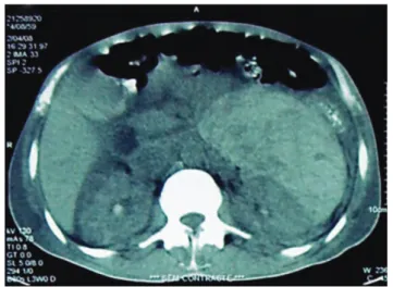
immunodeiciency virus (HIV) were negative, as well as the Venereal Disease Research Laboratories (VDRL) and blood cultures. Chest X-ray, chest CT, and echocardiogram did not show any abnormalities. Abdominal CT scan showed a cortical cyst in the left kidney (Figure 1). Echo-Doppler of the
Schade et al.
460 Bras J Rheumatol 2009;49(4):456-61
Figure 2. Right kidney: A 2.5 cm cortical cyst can be seen. A retroperitone-al mass, hyperechoic, heterogeneous, related to the left kidney, measuring 13.5 x 13.5 x 7.5 cm (approximate volume of 700 mL), suggestive of a hematoma can be seen.
Figure 1. Right kidney: Hypoechoic image with clearly deined borders, measuring 2.5 cm, in the medial third of the kidney. Wedge-shaped image with the same echoic density of renal parenchyma can be seen in the superior renal pole. Retroperitoneal lymph nodes or expansive lesions are not present.
temporal arteries showed old occlusion in the right artery and a hypoechoic halo in the left temporal artery.
The patient evolved with severe, diffuse abdominal pain, and the physical examination showed “board-like abdomen” and a reduction in the mean corpuscular volume (MCV) and haemoglobin. A new abdominal CT (Figure 2) showed a left perirenal hematoma measuring 13.5 x 13.5 x 7.5 cm (approximate volume of 700 mL) and images suggestive of splenic infarcts. Angio-MRI of the abdominal aorta showed the same hematoma, but no aneurysm. The patient developed oliguric acute renal failure requiring hemodialysis. Due to the suspicion of polyarteritis nodosa, treatment with prednisone, 60 mg/d (1 mg/kg), and pulse cyclophosphamide, 500 mg (adjusted for the renal function), was instituted. The patient improved
clinically, as well as the inlammatory activity tests. Due to the
intercurrences, biopsies were only performed when the patient was clinically stable, after four weeks of treatment. Biopsy of
the right quadriceps muscle showed a perivascular inlammatory
infiltrate, composed by mononuclear cells, suggestive of vasculitis. Biopsy of the left temporal artery did not show signs
of vasculitis. Signs and symptoms, and laboratory indings met the criteria for PAN, according to the diagnostic classiication
of the American College of Rheumatology.
DISCUSSION
The clinical manifestations of PAN frequently include
non-speciic symptoms, such as fever, fatigue, arthralgias,
and weight loss. Other symptoms depend on the organs affected. The skin is frequently involved, and it may show livedo reticularis, subcutaneous nodes, ulcers, and digital ischemia.4,5 Involvement of the nervous system is more common in mononeuritis multiplex, which occurs in 60% of the cases.4,6 Muscular involvement is also common, and it includes symptoms like myalgia and muscle weakness.7 Approximately 50% of the cases of PAN are associated with gastrointestinal manifestations, and mesenteric angina is the most common of them. Mesenteric infarction and aneurysmal rupture are rare complications, but carry a high mortality rate.4 Renal involvement, due to the presence of microaneurysms of renal and interlobar arteries, and, occasionally, of the arcuate and interlobular arteries, sparing the glomeruli, is seen in approximately 40% of the cases, and it manifests as renal failure, hypertension, and perirenal hematoma due to the rupture of microaneurysms.4,8 Although rare, acute myocardial infarction may develop as a consequence of the occlusion of a coronary aneurysm.9 There are also reports of splenic infarcts secondary to vasculitis of the splenic artery or one of its main
branches. The isolated involvement of organs, such as the skin, appendix, and gall bladder, has also been described.
The diagnosis is clinical and should, ideally, always be
conirmed by the biopsy of an affected organ, and usually
the most accessible organ is chosen. If a site for biopsy is not available, arteriography of the renal or mesenteric blood
vessels showing multiple aneurysms conirms the diagnosis.10 The American College of Rheumatology criteria for diagnostic
461
Bras J Rheumatol 2009;49(4):456-61
Perirenal hematoma and involvement of the temporal artery in a patient with polyarteritis nodosa (PAN)
89.3%.11 Henegar et al., in a study published in May 2008, emphasized the diagnostic importance of negative predictive parameters to reduce the rate of false-positive.12
We reported a case of PAN with muscular, testicular, renal, splenic, and temporal artery involvement. It should be emphasized that, occasionally, orchitis is one of its manifestations.13 Due to the multisystem involvement, a series of differential diagnosis, including neoplasias and paraneoplastic syndromes, rheumatoid arthritis, as well as other vasculitis, especially giant cell arteritis, due to the
indings in the temporal artery, was considered. The initial diagnostic dificulty allowed the development of potentially
fatal complications, such as perirenal hematoma and acute renal failure. Spontaneous perirenal hematoma is a rare complication seen more commonly as a complication of renal cell carcinoma and renal angiomyolipoma.8 Among the vasculitis, the association with PAN has been described more often.8 Due to the intercurrences, muscular and temporal artery biopsies could only be done when the patient was stable, after the institution of treatment with high doses of corticosteroids and cyclophosphamide. Muscular biopsy was suggestive of vasculitis, but not pathognomonic of PAN. Although the left temporal artery biopsy did not show signs of vasculitis, the
clinical inding of this blood vessel thickening and the
echo-Doppler showing a hypoechoic halo suggested this diagnosis. Several cases of PAN with involvement of the temporal artery have been described, giving the impression that it is a frequent finding, and also an easily accessible site for diagnostic biopsy.14,15 Manifestation is similar to that of giant cell arteritis, but the incidence of severe ocular complications is lower.
PAN treatment consists of high doses of corticosteroids,
which has a signiicant impact on survival. Oral doses of 1
mg/kg/d, or pulse therapy with 15 mg/kg IV, can be used. In severe and refractory cases, it should be associated with an immunosuppressive agent, and cyclophosphamide is the agent more commonly used. The route chosen depends on the severity of the case. Azathioprine can substitute cyclophosphamide after remission has been achieved with this drug.1,3 Clinical support for the complications of this disease is also extremely important, as well as the prevention of treatment-associated toxicity.
REFERENCES
1. Langford CA. Treatment of polyarteritis nodosa, microscopic polyangiitis, and Churg-Strauss syndrome: Where do we stand? Arthritis & Rheumatism 2001;44(3):508-12.
2. Gayraud M, Guillevin L, Toumelin P, Cohen P, Casassus FLP, Jarrousse B. Long-term followup of polyarteritis nodosa, microscopic polyangiitis, and Churg-Strauss syndrome. Arthritis & Rheumatism 2001;44(3):666-75.
3. Guillevin L, Lhote F. Treatment of polyarteritis nodosa and microscopic polyangiitis. Arthritis & Rheumatism 1998;41(12):2100-5. 4. Stone JH. Polyarteritis nodosa. Jama 2002; 288: 1632-39. 5. Gibson LE; Su WP. Cutaneous vasculitis. Rheum Dis Clin North
Am 1995;21(4):1097-113.
6. Tervaert JW, Kallenberg C. Neurologic manifestations of systemic vasculitides. Rheum Dis Clin North Am 1993;19(4):913-40. 7. PlumLey SG, Rubio R, Alasfar S, Jasin HE. Polyarteritis nodosa
presenting as polymyositis. Semin Arthritis Rheum 2002;31(6):377-83.
8. Daskalopoulos G, Karyotis I, Heretis I, Anezinis P, Mavromanolakis E, Delakas D. Spontaneous perirenal hemorrhage: a 10-year experience at our institution. International Urology and Nephrology 2004;36:15-9.
9. Kastner D, Gaffney M, Tak T. Polyarteritis nodosa and myocardial infarction. Can J Cardiol 2000;16(4):515-8.
10. Komatireddy GR, Yangco DT, Loy TS, Beale GD. Ruptured intrahepatic aneurysm as the initial manifestation of polyarteritis nodosa. J Clin Rheumatol 1998;4:333-37.
11. Lightfoot RW Jr, Michel BA, Bloch DA, Hunder GG, Zvailer NJ, McShane DJ, et al. The American College of Rheumatology 1990
criteria for the classiication of polyarteritis nodosa. Arthritis Rheum 1990;33 (8):1088-93.
12. Henegar C, Pagnoux C, Puéchal X, Zucker JD, Bar-Hen A, Le Guern V et al. A paradigm of diagnostic criteria for polyarteritis nodosa.
Arthritis & Rheumatism 2008;58(5): 1528-38.
13. Teichman JM, Mattrey RF, Demby AM, Schmidt JD. Polyarteritis nodosa presenting as acute orchitis: a case report and review of the literature. J Urol 1993;149(5):1139-40.
14. Hamidou MA, Moreau A, Toquet C, El Kouri D, de Faucal P, Grolleau JY. Temporal arteritis associated with systemic necrotizing vasculitis. J Rheumatol 2003;30(10):2165-9.