Genetic Diversity, Natural Selection and
Haplotype Grouping of
Plasmodium knowlesi
Gamma Protein Region II (PkγRII):
Comparison with the Duffy Binding Protein
(PkDBPαRII)
Mun Yik Fong*, Sarah A. A. Rashdi, Ruhani Yusof, Yee Ling Lau
Department of Parasitology, Faculty of Medicine, University of Malaya, Kuala Lumpur, Malaysia
Abstract
Background
Plasmodium knowlesiis a simian malaria parasite that has been reported to cause malaria
in humans in Southeast Asia. This parasite invades the erythrocytes of humans and of its natural host, the macaqueMacaca fascicularis, via interaction between the Duffy binding
protein region II (PkDBPαRII) and the Duffy antigen receptor on the host erythrocytes. In
contrast, theP.knowlesigamma protein region II (PkγRII) is not involved in the invasion of P.knowlesiinto humans. PkγRII, however, mediates the invasion ofP.knowlesiinto the
erythrocytes ofM.mulata, a non-natural host ofP.knowlesivia a hitherto unknown receptor.
The haplotypes of PkDBPαRII inP.knowlesiisolates from Peninsular Malaysia and North
Borneo have been shown to be genetically distinct and geographically clustered. Also, the PkDBPαRII was observed to be undergoing purifying (negative) selection. The present
study aimed to determine whether similar phenomena occur in PkγRII.
Methods
Blood samples from 78 knowlesi malaria patients were used. Forty-eight of the samples were from Peninsular Malaysia, and 30 were from Malaysia Borneo. The genomic DNA of the samples was extracted and used as template for the PCR amplification of the PkγRII.
The PCR product was cloned and sequenced. The sequences obtained were analysed for genetic diversity and natural selection using MEGA6 and DnaSP (version 5.10.00) pro-grammes. Genetic differentiation between the PkγRII of Peninsular Malaysia and North
Bor-neo isolates was estimated using the Wright’s FSTfixation index in DnaSP (version 5.10.00). Haplotype analysis was carried out using the Median-Joining approach in NET-WORK (version 4.6.1.3).
a11111
OPEN ACCESS
Citation:Fong MY, Rashdi SAA, Yusof R, Lau YL (2016) Genetic Diversity, Natural Selection and Haplotype Grouping ofPlasmodium knowlesiGamma Protein Region II (PkγRII): Comparison with the Duffy Binding Protein (PkDBPαRII). PLoS ONE 11(5): e0155627. doi:10.1371/journal.pone.0155627
Editor:João Pinto, Instituto de Higiene e Medicina Tropical, PORTUGAL
Received:December 22, 2015
Accepted:May 1, 2016
Published:May 19, 2016
Copyright:© 2016 Fong et al. This is an open access article distributed under the terms of the Creative Commons Attribution License, which permits unrestricted use, distribution, and reproduction in any medium, provided the original author and source are credited.
Data Availability Statement:All relevant data are within the paper and its Supporting Information files.
Funding:This study was funded by the University of Malaya High Impact Research Fund, UM-MOHE (UM.C/625/1/HIR/MOHE/MED/09). MYF is the recipient of the funding. The funder had no role in study design, data collection and analysis, decision to publish, or preparation of the manuscript.
Competing Interests:The authors have declared
Results
A total of 78 PkγRII sequences was obtained. Comparative analysis showed that the PkγRII
have similar range of haplotype (Hd) and nucleotide diversity (π) with that of PkDBPαRII.
Other similarities between PkγRII and PkDBPαRII include undergoing purifying (negative)
selection, geographical clustering of haplotypes, and high inter-population genetic differen-tiation (FSTindex). The main differences between PkγRII and PkDBPαRII include length
polymorphism and no departure from neutrality (as measured by Tajima’s D statistics) in the PkγRII.
Conclusion
Despite the biological difference between PkγRII and PkDBPαRII, both generally have
simi-lar genetic diversity level, natural selection, geographical haplotype clustering and inter-population genetic differentiation index.
Introduction
Malaria is caused by protozoa of the genusPlasmodium. Four species are responsible for human malaria:P.falciparum,P.vivax,P.malariaeandP.ovale. Human infections by the sim-ian malaria parasiteP.knowlesihave once been thought to be rare [1]. This was becauseP.
knowlesihad been erroneously identified under microscopy examination asP.malariae, since
both species have similar trophozoite morphology in the infected erythrocytes. This problem was eventually overcome by the use of highly specific polymerase chain reaction (PCR). Hence, a study at a district hospital in Sarawak (Malaysia Borneo) in 2004 reported PCR detection of a large number of knowlesi malaria cases that were initially diagnosed by microscopy examina-tion asP.malariaeinfections [2]. Subsequent PCR tests on archived blood smears have also detected significant number ofP.knowlesiinfections. Human knowlesi malaria has now been documented in almost all countries in Southeast Asia [3].
P.knowlesiinvades the erythrocytes of human and of its natural host, the macaqueMacaca
fascicularis, via interaction with the Duffy antigen on the erythrocytes [4–6]. The Duffy binding
protein ofP.knowlesi(PkDBP) is a large protein which is divided into seven regions (I-VII). Region II contains the critical motifs for binding to the erythrocyte Duffy antigen [5]. PkDBP is encoded by theα-gene, and is closely related to two other homologous proteins ofP. know-lesi–Pkβand Pkγ. However, unlike region II of PkDBPα(PkDBPαRII), the region II ofβ (PkβRII) andγ(PkγRII) does not binds to the Duffy antigen of human, but binds to rhesus monkey (M.mulata) erythrocytes [7]. Hence, Pkβand Pkγare responsible for the Duffy-inde-pendent pathways for invasion of rhesus erythrocytes. PkβRII binds to a sialic acid receptor on rhesus erythrocytes [8]. Although the receptor for PkγRII remains to be identified, it has recently been demonstrated that PkγRII is also a sialic acid-dependent invasion ligand [9].
PkDBPαRII haplotypes of North Borneo isolates were genetically distinct from those of Penin-sular Malaysia.
The aim of this study therefore is to determine whether PkγRII is subjected to the same selection forces as the PkDBPαRII, by measuring its genetic diversity level, selection trend (positive or negative) and geographical clustering of its haplotypes.
Materials and Methods
Ethics statement
Ethical clearance for this study was obtained from University of Malaya Medical Ethics Com-mittee (Ref No. 817.18) and the Medical Research Ethic ComCom-mittee (MREC), Ministry of Health, Malaysia (National Medical Research Register ID No.13079). Informed verbal consent was obtained from patients for use of their blood samples for diagnosis and research. Written consent was found to be unnecessary as verbal consent would be sufficient for the purpose of this study and patient details were noted down solely for personal recordkeeping. This consent procedure was approved by the ethics committees.
Blood sample collection
Seventy-eight blood samples were used in this study. The samples (0.5 ml) were collected by trained medical personnel from knowlesi malaria patients in the University of Malaya Medical Centre (UMMC), Kuala Lumpur, Peninsular Malaysia (n = 48) and several public hospitals in two Malaysian states (Sabah and Sarawak) in North Borneo (n = 30). The samples were col-lected between 2010 and 2013.
Extraction of DNA
Total DNA ofP.knowlesiwas extracted from each blood sample using the QIAGEN Blood DNA Extraction kit (QIAGEN, Hilden, Germany). In each extraction, 100μl of blood was
used. The extracted DNA was suspended in water to a final volume of 50μl.
PCR, cloning and sequencing of the Pk
γ
RII
The PkγRII was amplified by nested PCR using oligonucleotide primers F1:5'-CGCATTTTG AAGGAATCCAC-3'and R1:5'-TGCTAGACTTACCTTCACCT-3'for nest 1. The primers
for the nest 2 reaction were F:5’-TCCTCAAAAGGCGGTGACCATCC-3’and R:5’-ACTGGC TGCCTTAGATTCAACACCA-3’. Cycling conditions for nest 1 were as follows: 95°C for 4
Sequence diversity, natural selection and haplotype analyses
Multiple alignment of PkγRII sequences was performed using CLUSTAL-Omega programme [12]. Both nucleotide and the deduced amino acid sequences were aligned and analysed. DnaSP ver. 5.10.00 [13] was used in the polymorphism analysis of the PkγRII sequences. Infor-mation such as haplotype diversity (Hd) and nucleotide diversity (π) was generated. The rates
of synonymous (dS) and non-synonymous (dN) mutations were estimated and compared by the Z-test (P<0.05) in MEGA6 using the Nei and Gojobori’s method with Jukes and Cantor
correction [14]. In purifying (negative) selection, mutations are usually not advantageous, so that dN will be less than dS (dN/dS<1). However, in positive selection, non-synonymous
mutations can be advantageous and dN will exceed dS (dN/dS>1).
The Tajima’s D statistics [15] was used to test departures from the neutral theory of evolu-tion, with the assumption that the population size was constant. The Wright’s FSTfixation
index [16] was used to measure genetic differentiation between the PkγRII of Peninsular Malaysia and North Borneo isolates. Both the Tajima’s D statistics and Wright’s FSTindex
were determined using DnaSP 5.10.00. The Median-Joining method [17] in NETWORK v4.6.1.3 programme [18] was used to establish genetic relationship among the PkγRII haplotypes.
Results and Discussion
The PkγRII ofP.knowlesiclinical isolates from patients in Peninsular Malaysia and North Bor-neo were successfully amplified, cloned, and sequenced. Three clones from each isolate were used for sequencing. All clones from each isolate showed identical PkγRII sequences. A total of 78 PkγRII nucleotide sequences was obtained (GenBank Accession No. KR053974-KR054021, KU216673-KU216702). For analysis, the PkγRII sequence of the referenceP.knowlesistrain H GenBank (GenBank Accession No. M90695) was also included. Each sequence was trimmed, using the codons for amino acids at the N- and C-terminals of PkDBPαRII as reference points.
Previous studies have found no length polymorphism in PkDBPαRII [10,11]. All PkDBPαRII sequences ofP.knowlesiisolates from Peninsular Malaysia and North Borneo were 307 amino acids in length. In the present study, multiple alignment of the nucleotide sequences revealed length polymorphism in the PkγRII gene, ranging between 915 and 921 base pairs, hence encoding amino acid sequences of 305–307 in length. Full alignment of these amino acid sequences (S1 Fig) showed 54 PkγRII sequences with 307 amino acids, and 24 sequences with 306 amino acids. The PkγRII ofP.knowlesistrain H was the only sequence with 305 amino acids. Close inspection of the alignment showed two deletions in the PkγRII of
P.knowlesistrain H, N at position 55 and K at position 166. For sequences with 306 amino
acids, the deletion was K at position 166. Uniquely, no length polymorphism was seen in the North Borneo PkγRII sequences. All the North Borneo sequences were 307 amino acid in length.
these key positions may possibly explain the inability of PkγRII to bind to human andM.
fasci-culariserythrocytes.
DNA sequence analyses were conducted to determine nucleotide diversity and genetic dif-ferentiation of PkγRII (Table 1). By taking the total 79 sequences as a single population, the overall haplotype diversity (Hd) and nucleotide diversity (π) were 0.991 ± 0.005 and 0.021 ±
0.001, respectively. To determine whether natural selection contributed to the diversity, the rate of non-synonymous (dN) to synonymous mutations (dS) was determined. dN (0.019) found to be lower than dS (0.030). The dN/dS ratio was 0.633, which suggested purifying (neg-ative) selection of PkγRII. Although the overall Z-test did not show significant natural selec-tion, there was indication towards purifying selection (dN<dS,P= 0.082). Tajima’s D
statistics (-1.538,P>0.10) revealed no significant departure from neutrality.
However, when the PkγRII sequences of Peninsular Malaysia and North Borneo were ana-lysed as two separate populations, some differences were observed (Table 1). The North Borneo PkγRII was noted to have slightly higher diversity (Hd = 0.991 ± 0.011;π= 0.013 ± 0.001) than
the PkγRII of Peninsular Malaysia (Hd = 0.979 ± 0.013;π= 0.009 ± 0.002). Tajima’s D statistics
revealed significant departure of neutrality (-2.352,P<0.01) only in the Peninsular Malaysia
PkγRII. Although the dN/dS ratios showed purifying selection of PkγRII on both populations, the Z-test indicated strong purifying selection of the North Borneo PkγRII (dN<dS,P=
0.002). Comparative analysis showed that the PkDBPαRII of Peninsular Malaysia [9] and North Borneo [10] have almost similar range of haplotype (Hd = 0.986, 0.999) and nucleotide diversity (π= 0.013, 0.012) with that of PkγRII. Purifying selection on the PkDBPαRII was
equal in both population (dN<dS,P<0.05). Unlike PkγRII, the PkDBPαRII of both
Peninsu-lar Malaysia and North Borneo showed significant departure from neutrality by the Tajima’s D statistics (−2.085,P<0.05;−2.459,P<0.01 respectively) [9,10].
It is interesting that higher haplotype diversity was seen in PkDBPαRII as compared to PkγRII the despite the lesser number of samples used in the PkDBPαRII studies [10,11]. The previous and present studies used only human/clinicalP.knowlesiisolates. Therefore, the PkDBαRII of the isolates is likely to be under selection pressure to generate high polymorphism (i.e., high haplotype diversity) for the parasite to escape the host's immune defenses. PkγRII, on the other hand, does not mediate invasion ofP.knowlesiinto human erythrocyte and therefore is likely under less selection pressure to display high polymorphism for immune evasion.
Phylogenetic analysis from a previous study reported two haplotype groups of PkDBPαRII
inP.knowlesifrom Peninsular Malaysia [10]. The distribution of the haplotypes though was
uneven, because majority (83.3%) of the haplotypes clustered in a large or major group. A sub-sequent study reported distinct phylogenetic grouping of PkDBPαII haplotypes from North
Table 1. Genetic diversity and selection pressure of PkγRII ofP.knowlesiisolates in Peninsular Malaysia and North Borneo.
Origin N Hd±SD π±SD dN dS dN/dS Z-test Tajima’s D
dN = dS dN>dS dN<dS
Malaysia 79 0.991±0.005 0.021±0.001 0.019 0.030 0.633 P= 0.171# P= 1.000# P= 0.082# -1.538# Peninsular Malaysia 49 0.979±0.013 0.009±0.002 0.009 0.010 0.900 P= 0.706# P= 1.000# P= 0.375# -2.352
*
North Borneo 30 0.991±0.011 0.013±0.001 0.009 0.029 0.310 P= 0.004& P= 1.000 P= 0.002* -1.275#
N: number of sequences; Hd: haplotype diversity;π: nucleotide diversity; dN: rate of non-synonymous mutations; dS: rate of synonymous mutations
#not significant (P>0.10)
*significant (P<0.01) &
reject null hypothesis of strict neutrality, i.e., dN = dS
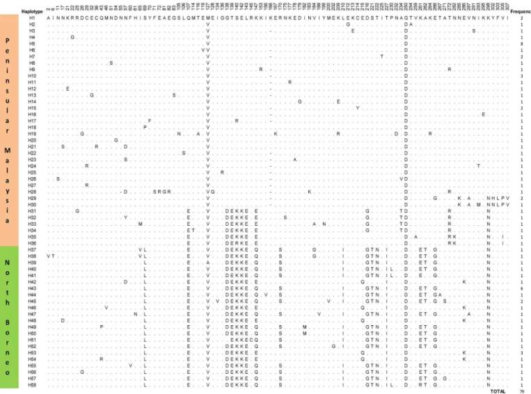
Borneo and Peninsular Malaysia [11]. In the present study, multiple alignment analysis identi-fied 58 different PkγRII haplotypes (Fig 1). Thirty-six were from Peninsular Malaysia
(H1-H36) and 22 from North Borneo (H37-H58). Haplotype H5 (frequency = 8/49) was the most abundant among the Peninsular Malaysia PkγRII haplotypes, while H43 (frequency = 6/ 30) was the most abundant in North Borneo. However, these frequencies were not significantly high to suggest that H5 and H43 were the most adapted haplotypes in the respective regions.
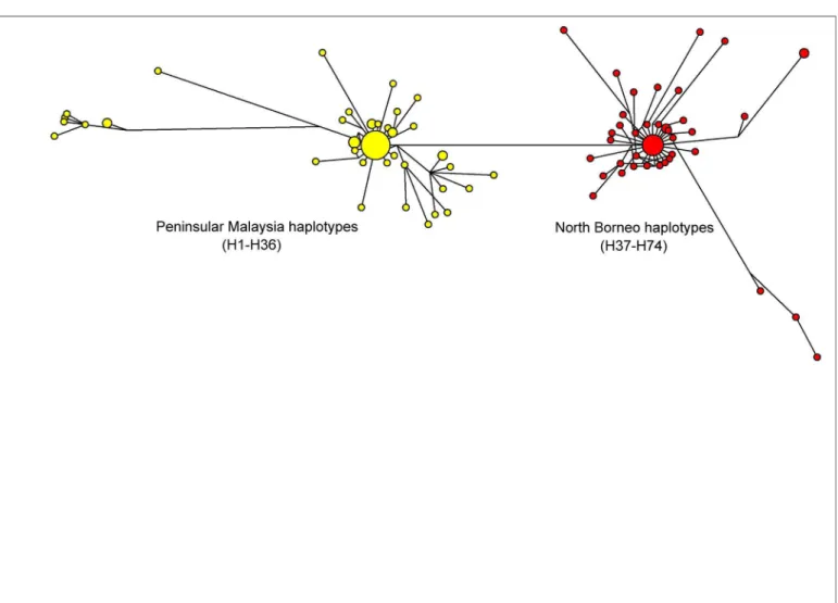
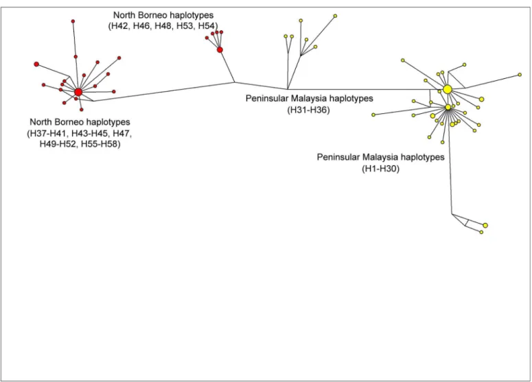
The previous studies on PkDBPαRII used Neighbour Joining to establish haplotype group-ings and relationship. The present study, however, used the Median-Joining method [17] in order to obtain a more precise depiction of haplotype relationship or network. Similar to the Neighbour Joining approach reported previously [11], the Median-Joining method generated a PkDBPαRII haplotype network consisting of distinct Peninsular Malaysia and North Borneo groups (Fig 2). For PkγRII haplotypes, a more complex network was obtained (Fig 3). Like PkDBPαRII, geographical clustering of Peninsular Malaysia and North Borneo haplotypes was evident. However, haplotypes from each region were separated into two subgroups. The large sub-groups were comparatively more complex, with high number of haplotypes. Interestingly,
Fig 1. Amino acid sequence polymorphism in the PkγRII haplotypes from Peninsular Malaysia and North Borneo.Polymorphic amino acid residues are listed for each haplotype. Residues identical to those of haplotype H1 are marked by dots. Total number of sequences for each haplotype is listed in the panel on the right.
the short link connecting the small subgroups (Peninsular Malaysia’s H31-H36, and North Borneo’s H42, H46, H48, H53, H54) suggests close relationship among the haplotypes. This was attributed to the sharing of some common amino acid residues in these haplotypes, namely, E107, D138, E139, K140, K142, E143 and E147. In contrast to PkDBPαRII and PkγRII, the highly diverseP.knowlesicircumsporozoite Th2R/Th3R epitope region displays no geo-graphical clustering of haplotypes, and many shared haplotypes from Peninsular Malaysia and Malaysian Borneo were observed in the Median Joining Network [20].
The Wright’s FSTfixation index measures population differentiation due to genetic structure
[16]. The index is also a measure of gene flow between populations. Populations with FST
val-ues of more than>0.25 are considered to be highly differentiated. A high Wright’s FSTindex
(0.61) was reported between the PkDBPαII of Peninsular Malaysia and North Borneo, indicat-ing significant genetic difference between the haplotypes groups [11]. The FSTindex obtained
in this study was equally high (0.62), thus showing high genetic difference between the PkγRII of Peninsular Malaysia and North Borneo. The amino acid substitutions in the PkγRII which most likely contribute to this genetic difference were at positions 69, 107, 138, 139, 140, 142, 143, 170, 216, 221, 222, 227, 261, 262 and 266 (Fig 1). Similarly, a recent study based on
Fig 2. Median Joining network of PkDBPαRII haplotypes.The network shows geographical clustering of PkDBPαRII haplotypes from Peninsular
Malaysia (yellow) and North Borneo (red). Amino acid sequences used for the construction of this network were from a previous study [9].
microsatellite DNA data FSTfound high level of genetic differentiation betweenP.knowlesi
human isolates from Peninsular Malaysia and Borneo [21]. The observation of high FSTvalues
betweenP.knowlesipopulations from Peninsular Malaysia and Malaysian Borneo is likely due to the separation of these two land masses since the last ice age 65,000 years ago. TheP. know-lesi, its macaque hosts and mosquito vectors in Borneo became isolated, and subsequently diverged from their respective species on mainland Asia to form distinct subpopulations. Molecular phylogeny and evolutionary studies have revealed unique history of macaque group formation in Borneo Island [22,23]. TheAnophelesmosquitoes have formed species which are uniquely found in the island, suchAn.latensandAn.balabacensis, both of which are vectors of
P.knowlesi[24,25]. Therefore, the geographical divergence ofP.knowlesiis most likely the
result of host immune selection and adaptation in the macaque and vector hosts respectively. The population substructure may also explain the Tajima’s D statistics observed in this study. The absence of a significant Tajima’s D<0 in the total sample (-1.538,P>0.10) could
be linked to the high level of subdivision between the Peninsular and Borneo Malaysia popula-tion as revealed by the distribupopula-tion of the haplotype network (Fig 3) and FSTindex. However,
since Tajima’s D tends to be>0 in the presence of subdivision, it may be the case that this Fig 3. Median Joining network of PkγRII haplotypes.The network shows geographical clustering of PkγRII haplotypes from Peninsular Malaysia
(yellow) and North Borneo (red). Note that haplotypes from each region are divided into two subgroups.
tendency is cancelling out the signal of Tajima’s D negativity observed in the Malaysia Borneo population (-1.275,P>0.10).
Although human knowlesi malaria is generally mild, there has been an increase in the num-ber of severe infections accompanied by high parasitaemias [26]. There is evidence to suggest possible human-to-human transmission [27]. Furthermore,P.knowlesihas been observed to expand its preferred host cell niche by invading older red blood cells [28]. All this may suggest increased parasite adaptation to humans. Rapid genetic changes in the invasion ligands includ-ing PkγRII may play a role in this increased adaptation in humans.
Conclusions
The region II of PkDBPαand Pkγplays an important role in the invasion ofP.knowlesiinto host erythrocytes. PkDBPαRII binds to the Duffy antigen of human andM.fascicularis eryth-rocytes, but PkγRII binds to a hitherto unidentified receptor onM.mulattaerythrocytes. Despite this difference, PkγRII was found to be almost similar to PkDBPαRII with regards to genetic diversity level, natural selection, geographical haplotype clustering and genetic differen-tiation index.
Supporting Information
S1 Fig. Full amino acid sequence alignment ofPlasmodium knowlesigamma protein region
II (PkγRII).Amino acid residues identical to those ofP.knowlesistrain H (GenBank Acces-sion No. M90695) are indicated by dots. Dash indicates amino acid deletion (highlighted blue at positions 55 and 166). The twelve conserved C residues are highlighted in yellow. The crucial amino acid residues in PkDBPαRII required for interaction with DARC are at Y94, N95, K96, R103, L168 and I175. In PkγRII, these positions have changed (highlighted orange at S95, E96, K103, K168, N175) except at Y94 (highlighted green).
(XLSX)
Acknowledgments
We express our thanks to the clinicians and nurses of the Medical Wards, University of Malaya Medical Centre, as well as the staff of Diagnostic Laboratory (Para:SEAD), Department of Par-asitology, University of Malaya. We also thank the participating hospitals in Sabah and Sara-wak for providing patient blood samples.
Author Contributions
Conceived and designed the experiments: MYF YLL. Performed the experiments: SAAR RY. Analyzed the data: MYF SAAR RY YLL. Contributed reagents/materials/analysis tools: MYF YLL. Wrote the paper: MYF YLL.
References
1. Fong YL, Cadigan FC, Coatney GR. A presumptive case of naturally occurringPlasmodium knowlesi
malaria in man in Malaysia. Trans R Soc Trop Med Hyg. 1971; 65:839–840. PMID:5003320
2. Singh B, Kim Sung L, Matusop A, Radhakrishnan A, Shamsul SS, Cox-Singh J, et al. A large focus of naturally acquiredPlasmodium knowlesiinfections in human beings. Lancet. 2004; 363:1017–1024. PMID:15051281
4. Miller LH, Mason SJ, Dvorak JA, McGinniss MH, Rothman IK. Erythrocyte receptors for (Plasmodium knowlesi) malaria: Duffy blood group determinants. Science. 1975; 189(4202):561–563. PMID: 1145213
5. Adams JH, Sim BK, Dolan SA, Fang X, Kaslow DC, Miller LH. A family of erythrocyte binding proteins of malaria parasites. Proc Natl Acad Sci USA. 1992; 89:7085–7089. PMID:1496004
6. Moon RW, Hall J, Rangkuti F, Ho YS, Almond N, Mitchell GH, et al. Adaptation of the genetically tracta-ble malaria pathogenPlasmodium knowlesito continuous culture in human erythrocytes. Proc Natl
Acad Sci USA. 2013; 110:531–536. doi:10.1073/pnas.1216457110PMID:23267069
7. Chitnis CE, Miller LH. Identification of the erythrocyte binding domains ofPlasmodium vivaxand Plas-modium knowlesiproteins involved in erythrocyte invasion. J Exp Med. 1994; 180:497–506. PMID:
8046329
8. Ranjan A, Chitnis CE. Mapping regions containing binding residues within functional domains of Plas-modium vivaxandPlasmodium knowlesierythrocyte-binding proteins. Proc Natl Acad Sci USA. 1999;
96:14067–14072. PMID:10570199
9. Dankwa S, Lim C, Bei AK, Jiang RHY, Abshire JR, Patel SD, et al. Ancient human sialic acid variant restricts an emerging zoonotic malaria parasite. Nat Commun. 2016; 7:11187 doi:10.1038/ ncomms11187PMID:27041489
10. Fong MY, Lau YL, Chang PY, Anthony CN. Genetic diversity, haplotypes and allele groups of Duffy binding protein (PkDBPαII) ofPlasmodium knowlesiclinical isolates from Peninsular Malaysia. Parasit
Vectors. 2014; 7:161. doi:10.1186/1756-3305-7-161PMID:24693997
11. Fong MY, Rashdi SA, Yusof R, Lau YL. Distinct genetic difference between the Duffy binding protein (PkDBPαII) ofPlasmodium knowlesiclinical isolates from North Borneo and Peninsular Malaysia.
Malar J. 2015; 14:91. doi:10.1186/s12936-015-0610-xPMID:25890095
12. Clustal Omega, a multiple sequence alignment program. Available:http://www.ebi.ac.uk/Tools/msa/ clustalo.
13. Librado P, Rozas J. DnaSP v5: a software for comprehensive analysis of DNA polymorphism data. Bio-informatics. 2009; 25:1451–1452. doi:10.1093/bioinformatics/btp187PMID:19346325
14. Tamura K, Stecher G, Peterson D, Filipski A, Kumar S. MEGA6: Molecular Evolutionary Genetics Anal-ysis version 6.0. Mol Biol Evol. 2013; 30:2725–2729. doi:10.1093/molbev/mst197PMID:24132122
15. Tajima F. Statistical method for testing the neutral mutation hypothesis by DNA polymorphism. Genet-ics. 1989; 123:585–595. PMID:2513255
16. Wright S. The genetical structure of populations. Ann Eugenics. 1951; 15:323–354.
17. Bandelt HJ, Forster P, Rohl A. Median-joining networks for inferring intraspecific phylogenies. Mol Biol Evol. 1999; 16:37–48. PMID:10331250
18. NETWORK v4.6.1.3, a programme for haplotype analysis. Available:http://www.fluxus-engineering. com.
19. Singh SK, Hora R, Belrhali H, Chitnis CE, Sharma A. Structural basis for Duffy recognition by the malaria parasite Duffy-binding-like domain. Nature. 2006; 439: 741–744. PMID:16372020
20. Fong MY, Ahmed MA, Wong SS, Lau YL, Sitam F. Genetic diversity and natural selection of the Plas-modium knowlesicircumsporozoite protein nonrepeat regions. PLoS One. 2015; 10:e0137734. doi: 10.1371/journal.pone.0137734PMID:26379157
21. Divis PCS, Singh B, Anderios F, Hisam S, Matusop A, Kocken CH, et al. Admixture in humans of two divergentPlasmodium knowlesipopulations associated with different macaque host species. PLoS Pathog. 2015; 11:e1004888. doi:10.1371/journal.ppat.1004888PMID:26020959
22. Ziegler T, Abegg C, Meijaard E, Perwitasari-Farajallah D, Walter L, Hodges JK, et al. Molecular phylog-eny and evolutionary history of Southeast Asian macaques forming theM.silenusgroup. Mol Phylo-genet Evol. 2007; 42:807–816. PMID:17208017
23. Abdul-Latiff MAB, Ruslin F, Vun VF, Mohd-Hashim A, Rovie-Ryan JJ, Abdul-Patah P, et al. Phyloge-netic relationships of Malaysia’s long-tailed macaques,Macaca fascicularis, based on cytochrome b sequences. ZooKeys. 2014; 407:121–140. doi:10.3897/zookeys.407.6982PMID:24899832
24. Vythilingam I, Tan CH, Asmad M, Chan ST, Lee KS, Singh B. Natural transmission ofPlasmodium knowlesito humans by Anopheles latens in Sarawak, Malaysia.
25. Wong ML, Chua TH, Leong CS, Khaw LT, Fornace K, Wan-Sulaiman WY, et al. Seasonal and spatial dynamics of the primary vector ofPlasmodium knowlesiwithin a major transmission focus in Sabah, Malaysia. PLoS Negl Trop Dis. 2015; 9:e0004135. doi:10.1371/journal.pntd.0004135PMID: 26448052
fromPlasmodium knowlesiandPlasmodium vivaxbut no mortality with early referral and artesunate
therapy. Clin Infect Dis. 2013; 56:383–397. doi:10.1093/cid/cis902PMID:23087389
27. Fornace KM, Nuin NA, Betson M, Grigg MJ, William T, Anstey NM, et al. Asymptomatic and submicro-scopic carriage ofPlasmodium knowlesimalaria in household and community members of clinical
cases in Sabah, Malaysia. J Infect Dis. 2015; 213:784–787. doi:10.1093/infdis/jiv475PMID: 26433222