Arq Neuropsiquiatr 2008;66(2-A):274-275
274
CliniCal-radiologiCal aspeCts of
primary extraCranial meningioma
of the ethmoid sinus in a Child
José Roberto Lopes Ferraz-Filho
1, Valdeci Hélio Floriano
2,
Leonardo Franco Felipe
2, José Alves Rocha-Filho
3aspeCtos ClÍniCo-radiolÓgiCos de meningioma primÁrio do seio etmoidal em CrianÇa
Image Department, Medical School, São José do Rio Preto SP, Brazil (FAMERP): 1MD, Image Department, FAMERP; 2MD, Fellow at the Image Department,
FAMERP; 3MD, Fellow at the Cardiac Image, INCOR , Hospital das Clínicas, Faculdade de Medicina da Universidade de São Paulo, Brazil (HCFMUSP).
Received 18 September 2007, received in inal form 7 December 2007. Accepted 8 February 2008.
Dr. José Roberto Lopes Ferraz-Filho - Avenida Brigadeiro Faria Lima 5544 - 15090-000 São José do Rio Preto SP - Brasil. E-mail: [email protected]
Meningiomas are tumors of benign histological nature which represent 13 to 26% of all primary intracranial neo-plasia1. Approximately 20% of intracranial meningiomas
present extracranial dissemination at sites such as the or-bit, middle ear, nasal cavity, nasopharynx and paranasal si-nuses2.Primary extracranial meningiomas are
histological-ly identical to intracranial meningiomas. They usualhistological-ly oc-cur in 40 to 60-year-old patients and are rare in the pedi-atric age group. Primary extracranial meningiomas repre-sent 1 to 2% of all meningiomas1-5.
We to report the case of a 13-year-old girl with a pri-mary extracranial meningioma of the ethmoid sinus, in-cluding the main indings of the imaging examination and a brief review of the literature.
Case
A 13-year-old girl was referred to our service with a 30-day history of constant and strong frontal headache, nasal obstruc-tion, and progressive proptosis of the left eye without signs of phlogiston or alterations in visual acuity.
At rhinoscopy, a lesion of the soft tissues was observed occupying the left superior meatus of the nasal cavity with a smooth pink surface.
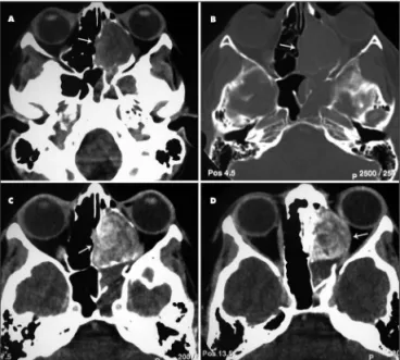
In light of these indings, a CT of the skull and face sinus-es was performed showing an extensive hypodense non-calci-ied lesion measuring 2.0x2.0 cm located in the left ethmoid sinus. The lesion presented heterogeneous iodinated contrast enhancement and demonstrated remodeling of the adjacent bone structures (Fig 1).
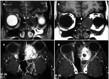
MRI of the facial sinuses was requested to evaluate the in-volvement of the soft structures as well as the invasion of the adjacent structures and resection planes. The lesion in the left ethmoid sinus was hypointense in T1 and T2-weighted sequenc-es, with heterogeneous paramagnetic contrast enhancement, determining a mass effect on the adjacent structures, lateral di-vergence of the medial rectus muscle of the left orbit, but
with-Fig 1. CT: (A,B) axial image showing lesion in the left ethmoid sinus with CT hypodense aspect, without calciication and with remodel-ing of the adjacent bone structures. (C,D) axial image showremodel-ing iodin-ated contrast heterogeneous enhancement of the lesion.
out invasion (Fig 2). Additionally, maxillary and sphenoidal sinus disease was observed.
The patient was submitted to excision of the lesion by exter-nal and endoscopic ethmoidectomy with a complete cure of the exophthalmus in the postoperative period. Diagnosis of atypical meningioma was attained by histopathological examination.
disCussion
Arq Neuropsiquiatr 2008;66(2-A)
275
Ethmoid sinus meningioma Ferraz-Filho et al.
According to published data, the irst description of pri-mary extracranial meningioma of the paranasal sinuses was in 1931, after which 40 cases have been described. And only three cases involved the ethmoid sinuses with only one case in the pediatric population1,3,4-7.
The symptoms are speciic for paranasal sinuses or re-lated to the involvement of adjacent structures. The most common complaints are sinusitis, nasal mass and epistax-is2.Nasal obstruction, headaches and proptosis are also
frequent complaints as were observed in the current case. Although meningioma is a tumor generally found at an intracranial location derived from meningocytes found in the meninges, in approximately 20% of cases it presents an extracranial extension due to continuity2,3. The nasal
cavity, as well as the paranasal sinuses, does not have this type of tissue, hence the rarity of the involvement of these regions. Some theories have been proposed to explain the primary origin of meningiomas at these loca-tions. The main ones are related to: a) the presence of arachnoid cells in nerve sheaths or in vessels where they emerge from the central nervous system, b) the migra-tion of tissue from the meninges to extracranial regions during embryogenesis, c) traumatic events or intracranial hypertension that displaces arachnoid cells and inally, d) undifferentiated mesenchymal cells1-3.
Meningiomas in children tend to present atypical char-acteristics in imaging examination, including cysts, hemor-rhage, aggressiveness and uncommon locations4.
Computed tomography better demonstrates the pres-ence of intratumoral calciication, the hyperdense aspect
of the lesion and homogeneous iodinated contrast en-hancement. The MRI supplies more precise information related to the extension and tumoral invasion of adja-cent structures, characterized by a increased T1 and T2-weighted signal intensity and with the same pattern of the contrast enhancement of CT8,9. However, in the case
described herein, the lesion presented a heterogeneous contrast enhancement, hypodense aspect at CT and with-out intratumoral calciication, which may be related to the atypical aspects of meningiomas in children.
The differential diagnosis of extracranial meningiomas located in the nasal sinus region, should include muco-cele, olfactory neuroblastoma, carcinoma, hemangioma, sarcoma and angioibroma2,3.
Treatment of primary extracranial meningioma of pa-ranasal sinuses is based on total surgical resection of the lesion through endoscopy or open surgery. When it is not possible to perform total surgical resection of the lesion as a result of the complex anatomy of both nasal cavity and paranasal sinuses, adjuvant therapeutic options is re-strict to radiation therapy and chemotherapy1-3,10,11.
In general, the prognosis is good. In cases of tumor recurrence, the tumor usually arise in the same anatomic site as the primary tumor and probably represent the re-sidual disease rather than recurrent tumor2.
In the present case, the patient was submitted to a to-tal surgical exeresis of the lesion through an external eth-moidectomy and endonasal combined approach. There was no evidence of tumor recurrence during the two years of post-surgical follow-up.
referenCes
1. Kainuma K, Takumi Y, Uehara T, Usami S. Meningioma of the parana-sal sinus: a case report. Auris Nasus Larynx 2007;34:397-400. 2. Thompson LDR, Gyure KA. Extracranial sinonasal tract meningiomas.
Am J Surg Pathol 2000;24:640-650.
3. Gökduman CA, Iplikcioglu AC, Kuzdere M, Bek S, Cosar M. Primary meningioma of the paranasal sinus. J Clin Neurosc 2005;12:832-834. 4. Rondinelli PIP, Viana CR, Osório CAM, Sredni ST. Extenso meningio-Extenso
meningio-ma atípico na infância. Ar� Neuropsi�uiatr 2003;61:695-698.Ar� Neuropsi�uiatr 2003;61:695-698. 5. García-Purriños FJ, Cervilla AR, Lemberg P, Moya JC. Meningioma ma-Meningioma
ma-ligno nasal. Acta Otorrinolaringol Esp 2005;56:373-375.
6. Petrulionis M, Valeviciene N, Paulauskiene I, Bruzaite J. Primary extra-cranial meningioma of the sinonasal tract. Acta Radiol 2005;46:415-418. 7. Zhou X, Li J, Peng T. Report of 4 cases ectopic meningioma of maxillary
sinus in children and the review of relative literatures. Lin Chuang Er Bi Yan Hou Ke Za Zhi 2005;19:490-491.
8. Hanada M, Kitajima K. Primary ectopic meningioma in the right eth-moid sinus: a case report. Auris Nasus Larynx 1997;24:321-324. 9. Whittle IR, Smith C, Navoo P, Collie D. Meningiomas. Lancet 2004;363:
1535-1543.
10. Swain RE, Kingdom TT, Delgaudio JM, Muller S, Grist W J. Meningio-mas of the paranasal sinuses. Am J Rhinology 2001;15:27-30. 11. Goyal LK, Suh JH, Mohan DS, Prayson RA, Lee J, Barnett GH. Local
control and overall survival in atypical meningioma: a retrospective study. Internat J Radiation Oncol Biol Physics 2000;46:57-61.