33
O
RIGINALA
RTICLESubmitted on: 08/17/2009 Accepted on: 12/01/2009
Corresponding author: Luis Alberto Batista Peres Rua São Paulo, 769 apt. 901- Centro Cascavel – PR – Brasil CEP: 85801-020 Fax: (45) 3327 3413 E-mail: [email protected] We declare no confl ict of interest.
Authors
Luis Alberto Batista Peres1
José Roberto Leonel Ferreira2
Ana Paula Kazue Beppu3
Everaldo Roberto de Araújo Junior3 Gustavo Vicenzi3 Ricardo Yukiharu Tsuge Yamamoto3
1Discipline of
Nephrol-ogy of the Medical School of the Universi-dade Estadual do Oeste do Paraná (UNIOESTE), Cascavel, Paraná, Brazil 2Discipline of Imaging
Diagnosis of the Medical School of the UNIOESTE, Cascavel, Paraná, Brazil 3Medical School of the
UNIOESTE, Cascavel, Paraná, Brazil
Anatomical alterations in patients with nephrolithiasis
A
BSTRACTIntroduction: Nephrolithiasis is a multi-factorial disease related to genetic disor-ders and environmental factors. Kidney stones are more common in adults and are associated with several metabolic and an-atomical disorders. The major anan-atomical abnormalities, such as obstruction of the ureteropelvic junction, horseshoe kidney, complete or incomplete duplicated ureter, bifid pelvis, and medullary sponge kidney, are known to be responsible for stone for-mation. The objective of this study is to evaluate anatomical alterations in patients with nephrolithiasis in our region. Meth-ods: Retrospective study on 1,378 patients with evidence of recent formation of kid-ney stones. Laboratory investigation and chemical analysis were performed when stones were available. Renal imaging techniques comprised at least renal ultra-sound and excretory urography. Results:
Of the 1,378 patients with nephrolithiasis cared for, only 367 (26.5%) (mean age, 36.8 ± 4.3 years) underwent anatomical investigation, of whom 198 (54.5%) were females. At least one anatomical altera-tion was found in 132 (36%) patients, the most common being renal cyst, complete-ly or incompletecomplete-ly duplicated ureter, and obstruction of the ureteropelvic junction.
Conclusions: Anatomical alterations were found in 36% of the investigated patients. Renal cyst, ureteral duplication, and ob-struction of the ureteropelvic junction were the most frequently found anatomi-cal alterations in the group.
Keywords: nephrolithiasis, anatomical al-terations.
[J Bras Nefrol 2010;32(1):33-36]©Elsevier Editora Ltda.
I
NTRODUCTIONNephrolithiasis is one of the most com-mon diseases of the urinary tract, with an incidence ranging from 5% to 15% of the population for both sexes, and prevalence of 2% to 3% in the general population.1 Several metabolic disor-ders, such as hypercalciuria, hypocitra-turia, and hyperuricosuria, are associa-ted with renal lithiasis.2
The following congenital or acqui-red anatomical alterations cause uri-nary stasis, predisposing to stone for-mation in the urinary tract: stenosis of the ureteropelvic junction; horseshoe kidney; vesical diverticula; medullary sponge kidney; caliceal cysts.3
This study aimed at describing ana-tomical alterations in a cohort of pa-tients admitted for metabolic investiga-tion of urinary lithiasis in the western region of the State of Paraná, Brazil.
M
ATERIAL ANDM
ETHODS34 J Bras Nefrol 2010;33(1):33-36
Anatomical alterations and nephrolithiasis
alterations, such as obstruction of the ureteropel-vic junction, horseshoe kidney, complete or in-complete ureteral duplication, medullary sponge kidney, and pelvic kidney, occur in up to 40% of the patients with nephrolithiasis.7
The imaging exams available for investigating anatomical alterations of the urinary tract are as follows: renal ultrasound; excretory urography; computed tomography; voiding urethrocystogra-phy; radioisotopic cystograurethrocystogra-phy; renal scintigraurethrocystogra-phy; and magnetic resonance imaging.8 The ultrasound is the first method to be applied due to its low cost and lack of risks. Excretory urography assesses, in addition to bilateral function, the anatomy of the urinary tract, and can diagnose nephrolithiasis, obstructions, duplication and position anomalies; the use of iodine contrast medium, however, may be a risk for allergic reactions and nephrotoxicity.9 Both exams were used routinely in this study to as-sess the anatomical alterations of the urinary tract in patients with nephrolithiasis.
Computed tomography assesses the anatomy of the urinary tract even in patients with renal dysfunction, and the possibility of tridimensional reconstruction provides further information. It may assess nephrolithiasis without using contrast medium and, unlike excretory urography, is not affected by the superposition of bone or intesti-ne.10 Voiding urethrocystography assesses the ana-tomy of the urinary bladder and urethra, and the presence of vesicoureteral reflux.11 Radioisotopic cystography is used in the follow-up of vesicou-reteral reflux. Renal scintigraphy provides infor-mation about the renal function and anatomy. Magnetic resonance imaging provides potential advantages in children and is a good method for assessing multiple congenital anomalies.12 Those exams have not been routinely performed in our study.
The prevalence of kidney stones in patients with urinary tract malformations and renal cys-tic diseases is greater than in the general popu-lation, suggesting a causal association. Urinary stasis is the pathogenic explanation for nephro-lithiasis associated with anatomical alterations, because of a delay in the elimination of crystals and an increase in the risk of urinary tract infec-tion. Metabolic investigation should be performed for the specific diagnosis and clinical treatment of Data were stored in a Microsoft Excel database
and were analyzed by use of descriptive statistics as follows: arithmetic mean; standard deviation; minimum and maximum values; and gross and percentage frequency. The anatomical alterations found are shown in Table 1.
R
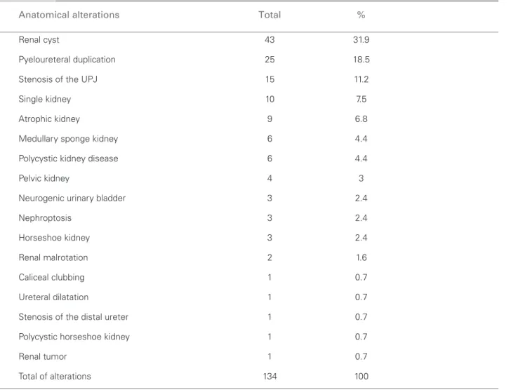
ESULTSIn the Nephrology Outpatient Clinic, 1378 pa-tients with nephrolithiasis were cared for, 738 (53.4%) being of the female sex. Their ages ran-ged from 1 to 85 years (mean age, 37.5 ± 19.1 years). Complete investigation regarding the pos-sibility of anatomical alterations was performed in 367 patients [198 (54.5%) females], whose age ranged from 1 to 77 years (mean age, 36.8 ± 4.3 years). Anatomical alteration was diagnosed in 132 (36%) patients, 72 (54.5%) being of the fe-male sex, with ages ranging from 11 to 77 years (mean age, 41 ± 13.4 years). The total of anato-mical alterations found was 134, the most com-mon being as follows: renal cysts; pyeloureteral duplication; stenosis of the ureteropelvic junction; single kidney; atrophic kidney; medullary sponge kidney; and polycystic renal disease. Two patients had two alterations as follows: one had stenosis of the ureteropelvic junction and renal cyst, and the other had pyeloureteral duplication and renal cyst. Family history of nephrolithiasis was posi-tive in 50% of the patients. Hypercalciuria was the most commonly associated metabolic disorder, diagnosed in 50% of all patients investigated for anatomical alterations. Regarding the chemical analysis of the stones, one was composed of pure uric acid, four were of calcium oxalate, and one, struvite. Table 1 shows the frequencies of the ana-tomical alterations found.
D
ISCUSSIONThis study assessed 1378 patients with nephroli-thiasis, 367 of whom completed the investigation for anatomical alterations, among which the most common were renal cysts, pyeloureteral duplica-tion, and stenosis of the pyeloureteral junction.
35
J Bras Nefrol 2010;33(1):33-36
Anatomical alterations and nephrolithiasis
the metabolic disorders, reducing the recurrence of urinary stones, even in individuals with anato-mical alterations.13
The limitations of the present study comprise its retrospective character and performing only two imaging exams in the patients. However, most anatomical alterations reported in the literatu-re may be assessed by use of both methods. Such imaging exams were performed for diagnosing and managing nephrolithiasis. The risk of exposure to radiation and radiological contrast medium should be considered.
In conclusion, renal cysts, pyeloureteral dupli-cation, and stenosis of the ureteropelvic junction were the most frequently found anatomical altera-tions in patients with nephrolithiasis in our study.
R
EFERENCES1. Hiatt RA, Dales LG, Friedman GD, Hunkeler EM. Frequency of urolithiasis in a prepaid medical ca-re program. Am J Epidemiol 1982; 115:255-65. 2. Heilberg IP, Schor N. Renal stone disease: Causes,
Evaluation and Medical Treatment. Arq Bras Endocrinol Metab 2006; 50:823-31.
3. Mouriquand PD, Whitten M, Pracros JP. Pathophysiology, diagnosis and management of pre-natal upper tract dilatation. Prenat Diagn 2001; 21: 2177-80.
4. Peres LAB. Investigação Metabólica de 578 Pacientes com Litíase Urinária no Oeste do Paraná. J Bras Nefrol 2005; 27:196-200.
5. Miliner DS, Muphy ME. Urolithiasis in pediatric patients. Mayo Clin Proc 1993; 68:241-8.
6. Stapleton FB, McKay CP, Noe HN. Urolithiasis in children: the role of hypercalciuria. Pediatr Ann 1987; 16:980.
7. Novick AC, Campbell SC. Renal tumors. In:
Table 1 ANATOMICALALTERATIONSFOUNDINPATIENTSWITHLITHIASISOFTHEURINARYTRACT
Anatomical alterations Total %
Renal cyst 43 31.9
Pyeloureteral duplication 25 18.5
Stenosis of the UPJ 15 11.2
Single kidney 10 7.5
Atrophic kidney 9 6.8
Medullary sponge kidney 6 4.4
Polycystic kidney disease 6 4.4
Pelvic kidney 4 3
Neurogenic urinary bladder 3 2.4
Nephroptosis 3 2.4
Horseshoe kidney 3 2.4
Renal malrotation 2 1.6
Caliceal clubbing 1 0.7
Ureteral dilatation 1 0.7
Stenosis of the distal ureter 1 0.7
Polycystic horseshoe kidney 1 0.7
Renal tumor 1 0.7
Total of alterations 134 100
UPJ: ureteropelvic junction.
ANATOMICALALTERATIONSFOUNDINPATIENTSWITHLITHIASISOFTHEURINARYTRACT
36 J Bras Nefrol 2010;33(1):33-36
Anatomical alterations and nephrolithiasis
Campbell’s Urology, 8th ed. Edited by Walsh PC, Retik AB, Vaughan ED, Wein AJ. Philadelphia: W. B. Saunders Co; 2002. p. 2718.
8. Omoloja AA, Patel H, Ey e, Jackson E. Common renal problems in pediatric medicine. Curr Probl Pediatr Adolesc Health Care 2007; 1:153-94. 9. Dhar M, Denstedt JD. Imaging in diagnosis,
treat-ment, and follow-up of stone patients. Advances in Chronic Kidney Disease 2009; 16:39-47.
10. Kuhn JP, Berger PE. Computed tomography of the kidney in infancy and childhood. Radiol Clin North Am 1981; 19: 445-61.
11. Bisset GS, Strife JL, Dunbar JS. Urography and voi-ding cystourethrography: finvoi-dings in girls with uri-nary tract infection. AJR Am J Roentgenol 1987; 148:479-82.
12. Jones RA, Perez-Brayfield MR, Kirsch AJ, Grattan-Smith JD. Renal transit time with MR urography in children. Radiology 2004; 233:41-50.