963
DOI: 10.1590/0004-282X20130176
ARTICLE
Asymmetric optic nerve sheath diameter as
an outcome factor following cranioplasty
in patients harboring the ‘syndrome of the
trephined’
Assimetria do diâmetro da bainha do nervo óptico como fator prognóstico após
cranioplastia em pacientes portadores da ‘síndrome do trefinado’
Antonio Santos de Araujo Junior1, Pedro Alberto Arlant1, Arnaldo Salvestrini Jr1, Carlos Eduardo Altieri2,
Jasper Guimarães Santos2, Lauro Figueira Pinto2, Mirella Martins Fazzito2, Hae Won Lee3,
Luis Felipe de Souza Godoy3
1Neurocirurgião, Hospital Sírio-Libanês, Sao Paulo SP, Brazil; 2Neurologista, Hospital Sírio-Libanês, Sao Paulo SP, Brazil; 3Neurorradiologista, Hospital Sírio-Libanês, Sao Paulo SP, Brazil.
Correspondence: Antônio Santos de Araújo Júnior; Rua Peixoto Gomide 515 / cj 96 / Cerqueira César; 01409-001 São Paulo SP - Brasil; E-mail: [email protected]
Conflict of interest: There is no conflict of interest to declare.
Received 26 February 2013; Received in final form 20 June 2013; Accepted 27 June 2013.
ABSTRACT
Decompressive craniectomy (DC) is gaining an increasing role in the neurosurgical treatment of intractable intracranial hypertension, but not without complications. A rare complication is the “syndrome of the trephined” (ST). It occurs when the forces of gravity overwhelm intracranial pressures, leading the brain to become sunken. Objective: To determine the usefulness of asymmetric optic nerve sheath diameter (ONSD) as an outcome factor after cranioplasty. Method: We followed-up 5 patients submitted to DC and diagnosed with ST. All were submitted to brain MRI to calculate the ONSD. Results: Only two patients presented an asymmetric ONSD, being ONSD larger at the site of craniectomy. Surprisingly these patients had a marked neurological improvement after cranioplasty. They became independent a week after and statisti-cally earlier than others. Conclusion: It is presumed that the presence of an asymmetric ONSD in trephined patients is an independent factor of good outcome after cranioplasty.
Keywords: decompressive craniectomy, syndrome of the trephined, optic nerve.
RESUMO
A craniectomia descompressiva (CD) tem papel fundamental no tratamento da hipertensão intracraniana refratária, mas não é isenta de complicações. Uma complicação rara é a “síndrome do trefinado” (ST). Ela ocorre quando as forças gravitacionais se sobrepõem à pressão intracraniana. Objetivo: Determinar a utilidade do diâmetro da bainha do nervo óptico (DBNO) como fator prognóstico após cranioplastia. Método: Foram acompanhados 5 pacientes trefinados portadores da ST. Estes pacientes foram submetidos à ressonância magnética com medida do diâmetro da bainha do nervo óptico (DBNO). Resultados: Dois pacientes apresentaram uma assimetria do DBNO, sendo o diâ-metro maior do lado craniectomizado. Para nossa surpresa estes evoluíram melhor do que os que apresentavam o DBNO simétrico. Estes pacientes se tornaram independentes uma semana após, e estatisticamente mais cedo do que os outros. Conclusão: Há evidências de que a assimetria do DBNO sirva como fator de bom prognóstico após cranioplastia no pacientes portadores da ST.
Palavras-chave: craniectomia descompressiva, síndrome do trefinado, nervo óptico.
Decompressive craniectomy is gaining an increasing role in the neurosurgical treatment of intractable intracranial hypertension in patients with head injury, acute stroke, and severe brain edema1-3.
hough technically straightforward, the procedure is not without signiicant complications2-5. Herniation through the
cranial defect, seizures and subdural efusion are common
964 Arq Neuropsiquiatr 2013;71(12):963-966
hydrocephalus is the most common late complication2,3.
Others rare late complications are sinking skin laps, para -doxical herniation and external brain tamponade6,7.
During the late phases of recovery, patients may develop
a new cognitive, neurological, or psychological deicit termed
“syndrome of the trephined” (ST)5. his syndrome clinically may include dizziness, tinnitus, orthostatic headaches8, cen-tral sleep apnea7, dysarthria, limb rigidity, parkinsonian levo-dopa-resistant tremor, diplopia (abducens nerve palsy)9,10, reversible monoparesis11, fatigue and luctuating levels of consciousness up to coma states7-10. It may occur following a unilateral craniectomy or a bilateral bifrontal craniectomy13.
Increasing the intracranial pressure by placing the pa-tient into Trendelenburg position or increasing hydration may reverse the symptoms7,12. However up to now there is no clinical or radiological signal responsible to the diagnosis of the “syndrome of trephined”.
In some patients harboring the ST, sinking skin laps, para
doxical herniation and external brain tamponade may be seen6. Paradoxical herniation consists of brain midline shift up to the contralateral side from the craniectomy, easily visuali-zed by head CT or brain MRI, and may suggest the diagnosis.
Cerebral blood low changes, the efect of the atmospheric pressure on the brain, as well as cerebrospinal luid (CSF) hy -drodynamic changes have been postulated as the possible rea-sons for this syndrome14,15. A moderate increase in venous
out-low as well as a twofold increase in craniocaudal cerebrospinal luid systolic low velocity has already been measured after the skull closure, reairming the hydrodynamic hypothesis15.
METHOD
We followed-up 5 patients submitted to decompressive craniectomy, secondary to head injury or acute stroke, up to 6 months postoperatively, among whom was noted altered
levels of consciousness, sinking skin lap at the site of cra
niectomy, or paradoxical herniation. All patients were opera-ted from 2008 to 2012 at our hospital by the same surgical crew. All trephined patients who died or evolved without symp-toms or signs compatible to ST were excluded from this patient
case series. Epidemiological data, reason to decompression, time to decompressive craniectomy, symptoms and signs com-patible to ST, and time to cranioplasty may be seen in Table.
All patients included in this study were routinely sub-mitted to laboratory essays, radiographic exams and vi deoelectroencephalography to exclude others causes of neu -rological impairment. All were submitted to brain MRI scan, with small coronal fat-suppression cuts, in order to calcu-late the optic nerve sheath diameter, before and after cranio-plasty. Cranioplasty was routinely planned after 3 months from decompression.
Our goal was to determine the usefulness of the asymmetric optic nerve sheath diameter in craniectomy patients as a diag-nostic signal and as a good outcome factor after cranioplasty.
RESULTS
Among these 5 patients, all have developed neurologi-cal deterioration after unilateral decompressive craniectomy at a mean follow-up of 6 months, including orthostatic hea-dache, diplopia, contralateral hemiparesis, gait ataxia, exces-sive sleepiness up to periods of minimal consciousness. All patients had their symptoms reversed by placing then into Trendelenburg position. At ectoscopy all patients presented
sinking skin lap at the site of craniectomy. On radiologic exa
mination all presented paradoxical herniation.
After ruling out all other possible disorders, all patients have been presumably diagnosed with ST.
However, on MRI scanning only two patients (patients B and E) presented an asymmetric optic nerve sheath dia-meter (ONSD), being the ONSD larger at the side of
craniec-tomy (Figure 1). Surprisingly these two patients had a
marked neurological improvement after cranioplasty, with normalization of the ONSD and disappearance of the brain
shift a day after procedure (Figure 2), becoming indepen -dent a week after procedure.
he others three patients (patients A, C and D), who had
a symmetric ONSD at MRI, have also improved their con-sciousness level, but in a very slowly way, becoming indepen-dent after a few months.
Table 1. Epidemiological and clinical data from patients submitted to decompressive craniectomy (DC), symptoms and signs suggestive of “syndrome of the trephined” (ST), and time to cranioplasty.
Patient Age Gender Diagnosis Time to DC ST symptoms ST signs Time to cranioplasty
A 23 y M TBI 24 h Headache, ALC and diplopia Paradoxical herniation 3 m
B 67 y M RCSH 1 m ALC, hemiparesis Sinking skin flap,
paradoxical herniation
3 m
C 53 y F AMCHS 12 h ALC, ataxia Paradoxical herniation 4 m
D 19 y M TBI 5 d ALC Sinking skin flap,
paradoxical herniation
3.5 m
E 58 y M AMCHS 16 h ALC Paradoxical herniation 3 m
965
Antonio Santos de Araujo Junior et al. Syndrome of the trephined
he measurement of the ONSD is an important indirect
method to suppose or diagnose increased levels of
intracra-nial pressure (ICP). he ultrasonographic visualization of the
optic nerve is feasible, costless, and world-wide applied to diagnose intracranial hypertension16,17.
By its time, the use of MRI to detect raised ICP is a more recent method. It seems to bring more precision to the measurement of the ONSD. ONSD is strongly related to ICP, a
inding that relects distension of the nerve sheath during in
-creases in CSF pressure. On T2weighted sequences, CSF ex -hibits a high signal (white), whilst fat and gray matter appear as light grey18. he perioptic CSF is surrounded by orbital
fat. Contrast between CSF and orbital fat can be improved
with fat suppression, increasing the image resolution for the ONSD measurement18,19.
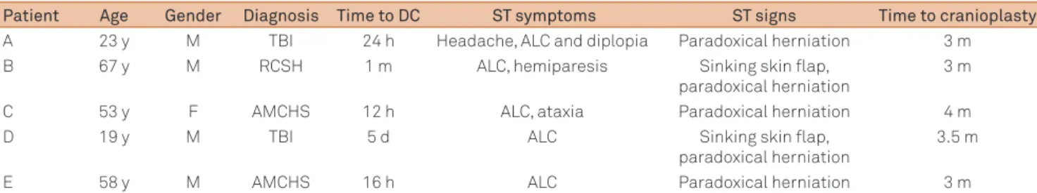
Figure 1. (A-B) Axial T1-weighted MRI, FLAIR sequence, showing sinking skin flap at the left side of craniectomy, paradoxical herniation to the right side, and subfalcine herniation; (C) Axial T1 MRI, cerebral sulci effacement at the left side; (D-E) Axial T2-weighted MRI, asymmetry of the optic nerve sheath diameter (ONSD), being larger at the left side (6.8 mm) vs right side (5.8 mm); (F) Coronal fat-suppressed T2-weighted MRI, asymmetric ONSD.
A B C
D E F
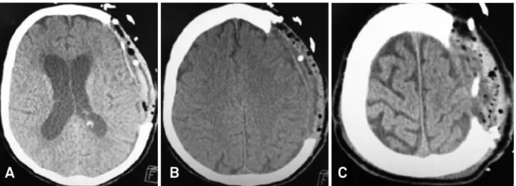
Figure 2. Axial brain CT one day after methyl methacrylate cranioplasty of the trephined patient from Figure 1. (A-B) Disappearance of the brain shift and subfalcine herniation; (C) Recovery from the cerebral sulci effacement.
A B C
Patients harboring the ST with symmetric ONSD needed statistically more time to become independent after cranio-plasty than patients with asymmetric ONSD (mean time of 38 days vs 8 days, t-test, p<0,05).
DISCUSSION
It is well known the usefulness of the decompressive cra-niectomy in the treatment of refractory intracranial
pres-sure, but not without signiicant complications. One of the
966 Arq Neuropsiquiatr 2013;71(12):963-966 References
1. Gadde J, Dross P, Spina M. Syndrome of the trephined (sinking skin flap syndrome) with and without paradoxical herniation: a series of case reports and review. Del Med J 2012;84:213-218.
2. Honeybul S, Ho KM. Long-term complications of decompressive
craniectomy for head injury. J Neurotrauma 2011;28:929-935.
3. Yang XF, Wen L, Shen F, et al. Surgical complications secondary to decompressive craniectomy in patients with a head injury: a series of 108 consecutive cases. Acta Neurochir (Wien) 2008;150:1241-1248.
4. Honeybul S. Complications of decompressive craniectomy for head injury. J Clin Neurosci 2010;17:430-435.
5. Stiver SI. Complications of decompressive craniectomy for traumatic brain injury. Neurosurg Focus 2009;26:7.
6. Akins PT, Guppy KH. Sinking skin flaps, paradoxical herniation, and external brain tamponade: a review of decompressive craniectomy management. Neurocrit Care 2008;9:269-276.
7. Choi JJ, Cirivello MJ, Neal CJ, Armonda RA. Paradoxical herniation in wartime penetrating brain injury with concomitant skull-base trauma. J Craniofac Surg 2011;22:2163-2167.
8. Mokri B. Orthostatic headaches in the syndrome of the trephined: resolution following cranioplasty. Headache 2010;50:1206-1211.
9. Bijlenga P, Zumofen D, Yilmaz H, Creisson E, de Tribolet N. Orthostatic mesodiencephalic dysfunction after decompressive craniectomy. J Neurol Neurosurg Psychiatry 2007;78:430-433.
10. Gottlob I, Simonsz-Tòth B, Heilbronner R. Midbrain syndrome with eye movement disorder: dramatic improvement after cranioplasty. Strabismus 2002;10:271-277.
11. Stiver SI, Wintermark M, Manley GT. Reversible monoparesis following decompressive hemicraniectomy for traumatic brain injury. J Neurosurg 2008;109:245-254.
12. Chalouhi N, Teufack S, Fernando Gonzalez L, Rosenwasser RH, Jabbour PM. An extreme case of the syndrome of the trephined requiring the use of a novel titanium plate. Neurologist 2012;18:423-425.
13. Janzen C, Kruger K, Honeybul S. Syndrome of the trephined following bifrontal decompressive craniectomy: implications for rehabilitation. Brain Inj 2012;26:101-105.
14. Fodstad H, Love JA, Ekstedt J, Fridén H, Liliequist B. Effect of cranioplasty on cerebrospinal fluid hydrodynamics in patients with the syndrome of the trephined. Acta Neurochir (Wien) 1984;70:21-30.
15. Dujovny M, Fernandez P, Alperin N, Betz W, Misra M, Mafee M. Post-cranioplasty cerebrospinal fluid hydrodynamic changes: magnetic resonance imaging quantitative analysis. Neurol Res 1997;19:311-316.
16. Dudea SM. Ultrasonography of the eye and orbit. Med Ultrason 2011;13:171-174.
17. Foster T, Tayal VC, Saunders T, Norton J. Emergency ultrasound optic nerve sheath measurement to detect increased intracranial pressure in head injury patients: preliminary study of interobserver variability in normal human subjects. Acad Emerg Med 2003;10:487-488.
18. Welling LC, Figueiredo EG, Machado FS, Andrade AF, Guirado VM, Teixeira MJ. Noninvasive evaluation of intracranial hypertension? Is there a gold standart? Arq Bras Neurocir 2012;31:224-230.
19. Geeraerts T, Newcombe VF, Coles JP, et al. Use of T2-weighted magnetic resonance imaging of the optic nerve sheath to detect raised intracranial pressure. Crit Care 2008;12:114.
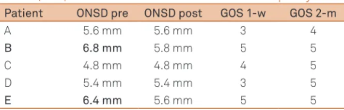
Table 2. Optic nerve sheath diameter (ONSD) at the side of cranioplasty from patients harboring the “syndrome of the trephined”, pre and post cranioplasty, and Glasgow Outcome Scale (GOS) one week and two months after cranioplasty.
Patient ONSD pre ONSD post GOS 1-w GOS 2-m
A 5.6 mm 5.6 mm 3 4
B 6.8 mm 5.8 mm 5 5
C 4.8 mm 4.8 mm 4 5
D 5.4 mm 5.4 mm 3 5
E 6.4 mm 5.6 mm 5 5
w: week; m: months.
he most useful clinical message derived from current
data may be summarized as followed: an ONSD less than 5.3 mm is unlikely to be associated with raised ICP; an ONSD above 5.82 mm indicates that the probability of raised ICP is 90%18,19.
In our series of trephined patients, those that presented ONSD asymmetry (patients B and E) had at the side of cra-niectomy an ONSD greater than 6mm (strongly suggesting raised ICP), and had their diameter decreased to normal va-lues one day after cranioplasty, see Table 2. Surprisingly those
two patients evolved better than patients without ONSD asymmetry, becoming independent (Glasgow Outcome Scale 5) just one week after cranioplasty (Table 2).
hat ONSD asymmetry may be explained by local chan ges of the CSF hydrodynamics, resulting in distension of the nerve sheath ipsilateral to the craniectomy side. his disten
-sion by its turn may relect that the intracranial pressures inally have been overwhelmed by the forces of atmospheric
pressure and gravity. Correction of this distension just after
cranioplasty may indicate the recovery from the CSF hydro -dynamics and normalization of intracranial pressures from a cranium converted back to a ‘closed box’ system.
It is presumed that the ONSD asymmetry may be used as a prognostic factor in patients with ST following the cranio-plasty, but the small number of patients enrolled in this study and the rarity of this disease may compromise further con-clusions. Larger prospective studies are needed and should dissolve major doubts.