I mmunophenotype of hematopoietic
stem cells from placental/umbilical cord
blood after culture
1Laboratório de Hematologia, Faculdade de Farmácia, 2Departamento de Genética, Universidade Federal do Rio Grande do Sul, Porto Alegre, RS, Brasil
3Faculdade de Farmácia, Pontifícia Universidade Católica do Rio Grande do Sul, Porto Alegre, RS, Brasil
4Stem Cell Biology, 5Immunogenetics, New York Blood Center, New York, NY, USA P. Pranke1,3,4, J. Hendrikx4,
G. Debnath4, G. Alespeiti4, P. Rubinstein5, N. Nardi2 and J. Visser4
Abstract
Identification and enumeration of human hematopoietic stem cells remain problematic, since in vitro and in vivo stem cell assays have different outcomes. We determined if the altered expression of adhe-sion molecules during stem cell expanadhe-sion could be a reason for the discrepancy. CD34+CD38- and CD34+CD38+ cells from umbilical
cord blood were analyzed before and after culture with thrombopoietin (TPO), FLT-3 ligand (FL) and kit ligand (KL; or stem cell factor) in different combinations: TPO + FL + KL, TPO + FL and TPO, at concentrations of 50 ng/mL each. Cells were immunophenotyped by four-color fluorescence using antibodies against CD11c, CD31, CD49e, CD61, CD62L, CD117, and HLA-DR. Low-density cord blood con-tained 1.4 ± 0.9% CD34+ cells, 2.6 ± 2.1% of which were
CD38-negative. CD34+ cells were isolated using immuno-magnetic beads
and cultured for up to 7 days. The TPO + FL + KL combination presented the best condition for maintenance of stem cells. The total cell number increased 4.3 ± 1.8-fold, but the number of viable CD34+
cells decreased by 46 ± 25%. On the other hand, the fraction of CD34+CD38- cells became 52.0 ± 29% of all CD34+ cells. The
absolute number of CD34+CD38- cells was expanded on average 15 ±
12-fold when CD34+ cells were cultured with TPO + FL + KL for 7
days. The expression of CD62L, HLA-DR and CD117 was modulated after culture, particularly with TPO + FL + KL, explaining differences between the adhesion and engraftment of primary and cultured candi-date stem cells. We conclude that culture of CD34+ cells with TPO +
FL + KL results in a significant increase in the number of candidate stem cells with the CD34+CD38- phenotype.
Correspondence P. Pranke
Laboratório de Hematologia Faculdade de Farmácia, UFRGS Av. Ipiranga, 2752
90160-000 Porto Alegre, RS Brasil
Fax: +55-51-3316-5437 E-mail: ppranke@adufrgs.ufrgs.br
Research supported by New York Blood Center, New York, USA, and CAPES.
Received January 21, 2005 Accepted August 15, 2005
Key words
•Hematopoietic stem cells
•CD34+CD38- cells •Human umbilical cord
blood
•Ex vivo expansion •Adhesion molecules
Introduction
A small population of primitive hemato-poietic stem cells (HSCs) is present in the bone marrow. These cells are defined by their ability to self-renew as well as to differ-entiate into committed progenitors of the
different myeloid and lymphoid compart-ments generating all of the blood cell lin-eages (1). The complexity of this system is enormous, since as many as 1010
erythro-cytes and 108-1010 white blood cells are
human umbilical cord blood (HUCB) has been clinically investigated as an alternative source of HSCs for allogeneic transplanta-tion of patients lacking a human leukocyte antigen-matched marrow donor. However, the number of HSCs in HUCB samples is limited. Identification of conditions that sup-port the self-renewal and expansion of hu-man HSCs remains a major goal of experi-mental and clinical hematology. Expansion of human stem cells in ex vivo culture will likely have important applications in trans-plantation, stem cell marking, and gene thera-py (2). The CD34+ protein is a surface
glyco-protein expressed on HSCs and progenitor cells in early developmental stages in HUCB and bone marrow, as well as on endothelial cells. The CD34+CD38- immunophenotype
defines a primitive subpopulation of pro-genitor cells in fetal liver and fetal or adult bone marrow (3-5). About 1% of bone mar-row cells express CD34, and generally less than 1% of these cells are CD38-negative. Hence, the frequency of the CD34+CD38
-population is about 1 in 10,000, or even lower. Phenotypic analyses of several cell surface markers reveal that even this rare population is heterogeneous (6). Ex vivo
culture is a crucial component of several clinical applications of stem/progenitor cells. A single stem cell has been proposed to be capable of more than 50 cell divisions or doublings in vivo and as such has the
capac-ity to generate up to 1015 cells, or sufficient
cells for up to 60 years. The proliferation and differentiation of cells is controlled by a group of hematopoietic growth factors. Rep-lication of this enormous cell amplification with hematopoietic growth factors in vitro
would allow the generation of large numbers of cells that could be used for a variety of clinical applications (7).
Several culture systems have been devel-oped to expand HSCs (5). Piacibelloet al. (1) reported the differential ability of FLT-3 ligand (FL), thrombopoietin (TPO), kit ligand (KL), and interleukin-3 (IL-3), alone or combined,
to support different stages of hematopoiesis in long-term stroma-free suspension cultures of HUCB CD34+ cells. Several studies have
de-scribed the effects of TPO alone in culture, which can stimulate early proliferation, sur-vival or differentiation of progenitor cells in cord blood or bone marrow (8).
The proliferation and differentiation of HSCs are controlled not only by soluble growth factors, but also by adhesion to stro-mal cells and matrix molecules. The expres-sion of adheexpres-sion molecules has attracted special attention, as their expression on HSCs and on endothelial and stromal cells plays a pivotal role in the process (9). These mol-ecules permit the interaction with various regulatory elements present in the microen-vironment, which includes stromal cells, extracellular matrix molecules and soluble regulatory factors such as cytokines and growth/differentiation factors (10).
Adhesion molecules include integrins, selectins and molecules from the immuno-globulin superfamily.
The objective of the present study was to investigate the behavior of umbilical cord blood CD34+CD38+ and CD34+CD38- cells
cultured with different combinations of growth factors, with respect to their viabil-ity, immunophenotype and self-renewal and differentiation capacities. Adhesion mol-ecules representing the integrins (CD11c or integrin α-chain, CD49e or α-5 chain and CD61 or ß-3 chain), selectins (CD62L or LECAM-1) and the immunoglobulin super-family (CD31 or PECAM-1) were analyzed. The expression of HLA-DR and CD117 (c-kit or stem cell factor receptor), which repre-sent differentiation markers for CD34+ cells,
was also investigated.
Material and Methods
Human umbilical cord blood cells
samples obtained after deliveries (≥29 weeks) were collected in sterile bags containing ci-trate-phosphate-dextrose. Samples were ob-tained at the Umbilical Cord Blood Bank of the New York Blood Center (New York, NY, USA). Blood was collected according to an Institutional Review Board-approved protocol. Units that are not used in the Pla-cental Blood Program are destined to re-search. Since collection is performed on de-livered placentas, the blood is considered discarded tissue. Blood not used for clinical transplants is not identified and is used with-out informed consent.
Isolation of CD34+ cells
Low-density mononuclear cells (MNCs) were isolated using density gradient cen-trifugation on Ficoll-Paque 1.077 g/cm2
(Amersham Pharmacia, Piscataway, NJ, USA), modified by the addition of 1 M phosphate buffer, pH 7.6, to the Dulbecco’s phosphate-buffered saline (GibcoBRL, Gai-thersburg, MD, USA) used to dilute the blood. This modification improved the isolation of the mononuclear fraction, since the harvested cell population contained 50% less reticulo-cytes and less than 50 to 60% erythroreticulo-cytes. CD34+ cells were harvested from the MNCs
using automated magnetic cell sorting (MACS) High Gradient Magnetic Separa-tion Columns for positive selecSepara-tion (Miltenyi Biotec, Bergisch Gladbach, North Rhine-Westphalia, Germany). The magnetically labeled cells were enriched by passing them twice through positive selection columns.
Antibodies
For the analysis of CD34+ cells,
mono-clonal antibodies (PharMingen/Becton Dick-inson, San Jose, CA, USA) specific for the following human antigens were used: CD34/ FITC (clone 34374X lot MO46959), CD38/ APC (clone HIT2), CD11c/PE (clone B-IY6), CD31/PE (clone WM59), CD49e/PE
(clone IIA1), CD61/PE (clone VI-PL2), CD62L/PE (clone Dreg 56), CD117/PE (clone YB5.B8), and HLA-DR/PE (clone G46-6), as well as isotype control antibodies (clones MOPC-21): mouse IgG1,k/FITC, IgG1,k/PE, and IgG1,k/APC.
Flow cytometry analyses
Processing for four-color fluorescence flow cytometry was done within 36 h of collection using at least 10,000 CD34+ cells,
before culture and after 4 and 7 days of culture. Cells were incubated with anti-CD34/ FITC and anti-CD38/APC antibodies com-bined with PE-conjugated antibodies specif-ic for CD11c, CD31, CD49e, CD61, CD62L, CD117, or HLA-DR. All incubations were done for 30 min at 4ºC, and cells were washed with phosphate-bufered saline. 7-Aminoac-tinomycin D (Molecular Probes, Inc., Eu-gene, OR, USA) at a final concentration of 1 µg/mL was used to identify dead cells. Flow cytometry was performed on a FACScalibur instrument (Becton Dickinson) equipped with an argon-ion laser tuned at 488 nm. The CELLQuest software (Becton Dickinson) was used for data analysis. Between 5,000 and 50,000 events were collected for each analysis. The gating strategy used can be summarized as follows. First, viable cells were gated, followed by a gating of the cell cluster in forward and side scatter, and using the FITC channel of CD34+ cells. Among
the CD34+ cells, negative and
CD38-positive cells were gated and the frequency of cells positive for the third antibody was analyzed.
plotted per cluster. In addition, data and cell suspension are shown as third and fourth dimensions, identified as symbol shape and symbol color.
Ex-vivo expansion cultures
MACS-isolated CD34+ cells were
cul-tured in 24-well plates (Multiwell™ Tissue
Culture Plate, Becton Dickinson) in 2 mL Iscove’s modified Dulbecco’s medium with L-glutamine and 25 mM HEPES buffer (GibcoBRL), supplemented with hydro-cortisone (10-5 M, Sigma, St. Louis, MO,
USA), 2-mercaptoethanol (5.5 x 10⁻⁵ M,
GibcoBRL), penicillin G (100 units/mL, GibcoBRL), streptomycin (0.1 mg/mL,
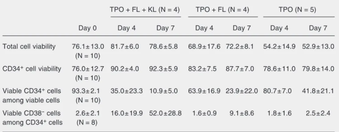
Figure 1. Effect of different com-binations of growth factors on cell proliferation after 4 and 7 days in culture. Sixteen different units of cord blood were used, and samples 2, 3, 4 and 11 were split into two different cultures (a and b), using 2 combinations of growth factors. TPO = thrombo-poietin; FL = FLT-3 ligand; KL = kit ligand.
Table 1. Volume of the umbilical cord blood samples and frequency of mononuclear and CD34+ cells obtained after the isolation on Ficoll-Paque and automated magnetic cell sorting columns, respectively.
Mean ± SD Range
Blood volume (mL) 45.2 ± 5.6 32-57
MNC (x 107) 10.4 ± 5.7 2.5-23.0
MNC (/mm3 blood) 2287 ± 1132 532-4510
CD34+ cells (x 106) 1.3 ± 0.8 0.3-4.0
CD34+ cells (/mm3 blood) 28.0 ± 16.7 6.4-85.0
% CD34+ cells among MNCs 1.4 ± 0.9 0.4-4.9
Data are reported as mean ± SD for N = 27. MNC = mononuclear cells.
GibcoBRL) and 1% bovine serum albumin (Sigma). Cell concentrations varied between 2.5 and 5.5 x 10⁵/well. Human growth
fac-tors used were: TPO (Kirin Brewery, Kirin Brewery, Chuo-ku, Tokyo, Japan), FL (Amgen Inc., Thousand Oaks, CA, USA) and KL (Amgen Inc.), also called steel factor or stem cell factor, at concentrations of 50 ng/mL each. The combination of hemato-poietic growth factors used were: TPO + FL + KL; TPO + FL and TPO. The cultures were maintained at 37ºC and 5% CO2 in a
humidi-fied atmosphere and analyzed on days 4 and 7.
Results
CD34+ cells were isolated from a total of
27 cord blood samples, and the number of MNCs as well as CD34+ cells was analyzed
in a Neubauer chamber. As shown in Table 1, the concentration of CD34+ cells was 28
cells/mm3 of cord blood (yield after Ficoll
and MACS procedure). The purity of this cell fraction was 93.2 ± 3.6% (87-99%; N = 10). Since 22.2 ± 11.3% (12.5-50.6%) of the CD34+ cells were dead, the average
fre-quency of viable CD34+ among total cells
was 71.0 ± 13.1% (39.4-86.5%).
combinations of growth factors (Figure 1). With TPO + FL + KL, the average increase in cell number in 9 samples studied was 3.55 ± 1.6-fold after 7 days of culture. Only 1 sample (3.a) showed a 0.56-fold decrease in cell number on day 4, but by day 7 the number of the cells in this sample had in-creased. The observation of individual cul-tures (Figure 1) showed some heterogeneity among samples, particularly when TPO + FL was used. With this combination of growth factors, the total number of cells changed very little until day 4. From day 4 until day 7 of culture, however, in one sample the cell number decreased 0.65-fold, in 2 samples it showed no major variation, and in the remaining 2 a considerable increase (2.57 ± 0.5-fold) in total cell number was ob-served. Considering the total culture period, in 2 samples the cell number decreased 0.59 ± 0.11-fold and in 3 of them it increased 1.92 ± 0.56-fold. In all 6 samples cultured with TPO, the total cell number decreased during the first 4 days. After that, in 4 of them there was an increase, not enough, however, to reach the original number. Taken as a whole, cell numbers decreased 0.55 ± 0.26-fold with TPO only.
Further analyses were done in 10 of the samples. Three of them were separated into two cultures submitted to different
treat-ments. The analysis of cell viability during the period of culture (Table 2) showed that, for cultures in the presence of TPO + FL + KL, the absolute number of viable cells in-creased 4.27 ± 1.82-fold in the 4 samples studied. In one sample (3.a) the number of viable cells decreased 0.5-fold from day 0 to day 4, but increased 3.4-fold from the 4th to the 7th day. In cultures with TPO + FL, the number of viable cells increased 1.94 ± 0.56-fold in 3 samples, and decreased 0.76-0.56-fold in one sample. In the 5 samples cultured with TPO, the number of viable cells decreased 0.35 ± 0.28-fold.
The viability of CD34+ cells was increased
after culture with TPO + FL + KL and with TPO + FL, and maintained in the presence of TPO, but the frequency of viable CD34+
cells among total viable cells decreased (Table 2). From day 0 to day 7 of culture with TPO + FL + KL, the number of viable CD34+ cells decreased 0.46 ± 0.25-fold in
the 4 samples studied, whereas with TPO + FL and TPO alone this decrease was 0.30 ± 0.12- and 0.13 ± 0.08-fold, respectively.
Of particular interest are the results re-garding the frequency of CD34+CD38- cells
during the culture period. As shown in Table 2, the number of viable CD34+CD38- cells
increased in some of the culture conditions. With TPO + FL + KL, this increase was
Table 2. Total and CD34+ cell viability and frequency of CD34+ and CD34+CD38- cells on day 0 and after culture with three different combinations of growth factors.
TPO + FL + KL (N = 4) TPO + FL (N = 4) TPO (N = 5)
Day 0 Day 4 Day 7 Day 4 Day 7 Day 4 Day 7
Total cell viability 76.1± 13.0 81.7±6.0 78.6 ±5.8 68.9± 17.6 72.2±8.1 54.2± 14.9 52.9±13.0 (N = 10)
CD34+ cell viability 76.0± 12.7 90.2±4.0 92.3 ±5.9 83.2± 7.5 87.7±7.0 78.6± 11.0 79.8±14.0 (N = 10)
Viable CD34+ cells 93.3± 2.1 35.0±23.3 10.9 ±5.0 63.9± 16.9 23.9±22.0 80.7± 7.0 41.8±21.1 among viable cells (N = 10)
Viable CD38- cells 2.6± 2.1 16.0±19.9 52.0 ±28.8 1.6± 0.9 9.1±8.6 1.8± 1.6 2.5±2.4 among CD34+ cells (N = 8)
14.59 ± 11.81-fold in 3 samples. In one sample, cell numbers were not determined on day 0, but from day 4 to day 7 the number of viable CD34+CD38- cells increased
4.27-fold. In one sample (3.a), the number of CD34+CD38- cells decreased 0.23-fold from
day 0 to day 4, but increased 23-fold until day 7. For cultures with TPO + FL, these cells increased 2.79 ± 2.29-fold in the 3 samples studied from day 0 to day 7 and in one sample for which the analysis was not done on day 0, they increased 1.22-fold from day 4 to day 7. In the presence of TPO, however, the number of viable CD34+CD38
-cells decreased 0.26 ± 0.31-fold in 5 samples. The expansion and proliferation of CD34+CD38- cells were significantly
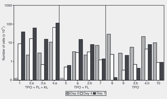
in-creased in the presence of TPO + FL + KL as compared to the other two combinations of growth factors. An interesting correlation can be seen between this effect and total increase in cell numbers, which is directly proportional to the increase or decrease in CD34+CD38- cells (Figures 1 and 2). When
TPO + FL + KL were used, in 4 samples, total cell numbers as well as CD34+CD38
-cells were gradually increased during cul-ture. In Figure 1, it can be seen that total cell
numbers increased from the first to the fourth day in 8 of 9 samples cultured in the pres-ence of TPO + FL + KL. Only one sample (3.a) showed a 0.6-fold decrease in cell num-ber until the fourth day in culture, but this number increased around 3.4-fold from day 4 to day 7. The same pattern was observed for CD34+CD38- cells (Figure 2), as well as
in cultures where TPO + FL or TPO alone were used as growth factors. On the other hand, no correlation was observed between the initial number of CD34+CD38- cells and
total cell growth or with the expansion of the CD34+CD38- cells. Similarly, no correlation
was detected between the initial number of viable CD34+ cells and cell growth or
num-ber of CD34+CD38- cells (results not shown).
The immunophenotypic profile of freshly isolated CD34+CD38+ and CD34+CD38- cells
was also investigated. In 7 of 8 samples analyzed, the number of CD34+CD38+ cells
positive for CD11c was low (less than 20%) and the fluorescence was dim or, alterna-tively, all cells were negative. Only one sample showed around 45% of positive cells with dim fluorescence. The CD34+CD38
-population could be analyzed in only 5 samples due to low event numbers. The
samples presented the same predominant pattern as observed for CD34+CD38+ cells.
All 8 samples showed 100% of the cells positive for CD31, with a bright fluores-cence, and about 70-100% of the cells with regular fluorescence for CD49e. For CD61, however, among CD34+CD38+ cells either
the number of positive cells was low (less than 15%), with regular fluorescence, or 100% of the cells were negative. In one of the samples, the positive population con-tained a small (around 3%) subpopulation of bright cells. Because the number of the events was low in CD34+CD38- cells, we could
analyze only 4 of 8 samples.
Cells were heterogeneous for CD62L expression. About 43 ± 17 and 27 ± 17% of CD34+CD38+ and CD34+CD38- cells,
re-spectively, were positive with a regular mean fluorescence intensity. In one sample, a sec-ond population of around 20%, among CD34+CD38+ cells, presented bright
fluo-rescence for CD62L. In 2 samples the num-ber of events among CD34+CD38- cells was
too low to be analyzed.
The pattern for HLA-DR was very het-erogeneous among samples. This marker was present in about 54 ± 28 and 34 ± 31% of CD34+CD38+ and CD34+CD38- cells,
re-spectively, with a regular mean fluorescence intensity. In one of the samples, a small population of the bright cells was also seen among the CD34+CD38+ population, whereas
in another two populations, very few posi-tive cells with dim and bright cells were observed.
The fluorescence pattern observed for CD117 was complex. Among CD34+CD38+
cells, two clusters could be observed, one with a high frequency of cells (80 ± 10%, range: 59-91%) with regular fluorescence and another composed of few cells (6 ± 5%) with bright or very bright fluorescence. Among the CD34+CD38- cells, 56 ± 24%
presented regular mean fluorescence inten-sity, with no bright cells observed.
The immunophenotypic profile of
um-bilical cord blood CD34+ cells was analyzed
after 4 and 7 days of culture with TPO + FL + KL, TPO + FL and TPO. In some of the samples, particularly among CD34+CD38
-cells, the analysis was not possible due to very small numbers of events. Culture with TPO + FL or TPO alone was also a factor which decreased the cell number to a level below meaningful analysis in some cases.
The patterns for CD11c, CD31, CD49e, and CD61 between CD34+CD38+ and
CD34+CD38- cells were not modified after
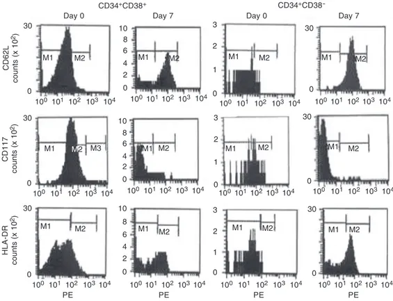
the culture in all combinations of growth factors (data not shown). For CD62L, how-ever, the number of positive cells and the fluorescence intensity increased from day 0 to day 4 and again from day 4 to day 7 in all culture conditions (Figure 3). In only one sample, cultured with TPO alone, did the number of positive cells increase from day 0 to day 4 but it decreased a little from day 4 to day 7 (data not shown).
In cultures with TPO + FL + KL, the number of cells positive for HLA-DR and the intensity of fluorescence increased from the day 0 to day 4, but decreased a little from day 4 to day 7 between CD34+CD38+ and
CD34+CD38- cells (Figure 3). In cultures
with TPO + FL, the reactivity pattern did not change among CD34+CD38+ cells, except
for one sample in which the number of posi-tive cells increased from day 0 to day 4 but remained unaltered until the end of the cul-ture. Similar patterns were observed in CD34+CD38- cells, but the number of events
was very low. The results were heteroge-neous in cells cultured with TPO alone (data not shown). Among CD34+CD38+ cells, the
number of positive cells increased signifi-cantly in 3 samples, whereas in the remain-ing 2 no changes were observed. Two of the 4 samples analyzed for CD34+CD38- cells
remained the same and in 2 the number of HLA-positive cells increased.
comparison with day 0. The presence of TPO + FL + KL, however, induced a de-crease in CD117-positive cells (Figure 3). Among CD34+CD38- cells, the results were
more heterogeneous. After culture with TPO + FL + KL, a cluster of few cells with dim or regular fluorescence could be observed, but no bright cells. The number of positive cells increased after culture with TPO + FL but no bright cells were seen. When TPO alone was used, two clusters were observed: a high number of cells with mean fluorescence around 100 and another one with few cells of mean fluorescence around 1000.
In some samples, after culture with TPO + FL (samples 5 and 6) or TPO alone (samples 9 and 3.b) the number of CD34+CD38- cells
was too low to analyze for CD117 reactivity. On day 0, the average frequency of CD117-negative cells was 44.2 ± 26.3% (N = 5). After culture with TPO + FL + KL, 84.5 ± 11.8% of the cells were negative for CD117 on day 4 (N = 3) and 96.0 ± 2.7% (N = 4) on day 7. After culture with TPO + FL, 11.3 ±
5.1 and 7.5 ± 9.2% of the CD34+CD38- cells
were CD117-negative on day 4 (N = 3) and day 7 (N = 2) respectively, whereas culture with TPO resulted in 7.3 ± 2.1 and 7.5 ± 6.4% CD117-negative cells on days 4 (N = 3) and 7 (N = 2), respectively.
Discussion
Lack of CD38, HLA-DR and lineage-committed antigens, as well as the co-ex-pression of Thy-1 (CDw90) and c-kit recep-tor (CD117), have been shown to identify the candidate hemopoietic stem cells (12). However, a better knowledge and standard-ization of the phenotype of umbilical cord blood CD34+ cells is critical since HUCB
volume is limited (13). The present study aimed to contribute to the characterization of CD34+ cells from umbilical cord blood,
analyzing their phenotype and behavior be-fore and after culture with different combi-nations of growth factors.
The frequency of CD34+ cells among
Figure 3. Frequency of cells positive for CD62L/PE, CD117/ PE and HLA-DR/PE among CD34+CD38+ and CD34+CD38 -cells on day 0 and after 7 days of culture with TPO + FL + KL. TPO = thrombopoietin; FL = FLT-3 ligand; KL = kit ligand. PE = fluo-rochrome phycoerythrin. M1 = isotype control; M2 = cells with regular fluorescence; M3 = cells with bright fluorescence.
CD62L
counts (x 10
2)
30
0
100 101 102 103 104 10
0
100101102 103 104 8
6 4 2
3
0 2
1
100 101 102 103 104 100101 102103104
100 101 102 103 104 100101102103 104
100 101102103 104 100 101 102 103 104
100 101 102 103104 100101 102103104 100 101 102 103 104 100101102103104 30
0
0
30 10
0 8 6 4 2
3
2
1
0
30
0
30
0 3
2
1
0 10
0 8
6 4 2
0 30
CD117
counts (x 10
2)
HLA-DR
counts (x 10
2)
PE PE PE PE
M1 M2 M1 M2 M1 M2 M1 M2
M1 M2
M1 M2
M1 M2 M3
M1 M2 M1
M2 M1 M2 M1 M2
CD34+CD38+
Day 0 Day 7 Day 0 Day 7
CD34+CD38
HUCB MNCs in our study was 1.4% (0.4-4.9%), in agreement with other studies. This frequency is similar to that in harvested pel-vic bone marrow (1.0 ± 0.3 vs 0.8 ± 0.4%)
(14). About 1% of bone marrow cells ex-press CD34, and generally less than 1% of these cells are CD38-negative (6,11,15). In other studies, Campagnoli et al. (16) showed that the concentration of CD34+ cells in whole
blood samples in term fetal blood was 0.4 ± 0.03% of total CD45+ cells, and Hao et al. (3)
showed that the frequency of CD34+ cells
among total MNCs in cord blood was 0.36 ± 0.33% with a large variation among samples (range 0.02 to 1.43%; N = 30).
In the present study, we found a large variation in the frequency of CD34+ cells
among HUCB MNCs, from 0.4 to 4.9%. Although correlation between total nucle-ated cell and CD34+ cells in HUCB has been reported, within groups of samples with simi-lar total nucleated cell counts a high degree of variation (at times exceeding 10-fold) in CD34+ cells is observed. CD34 counts in
HUCB can be as low as 0.1% of total nucle-ated cell as reported by Yap et al. (17), and D’Arena et al. (18) observed 0.01-1.71% CD34+ cells among HUCB cells. Different
explanations have been given for the varia-bility found in the frequency of CD34+ cells
in HUCB. There is evidence that, although the CD34 population frequency is a reliable indicator of the progenitor potential of HUCB, it is nevertheless heterogeneous in nature. On the other hand, these heteroge-neous results can reflect differences in the sensitivity of the methods employed by the different groups. CD34+ HSCs have also
been shown to vary with gestational age, mode of delivery and positioning of the de-livered neonate after delivery. Yap et al. (17) found that CD34+ cells accounted for 5.1 ±
1.0% of CD45+ cells in first trimester blood,
significantly more than in term cord blood (0.4 ± 0.03%).
Controversial results have been published regarding the frequency of CD38- cells
among cord blood CD34+ cells. We found
that 2.6 ± 2.1% (range 0.55-5.57) of the CD34+ cells were CD38-negative on day 0,
which agrees with reports showing that most CD34+ cells present the CD38 antigen in
HUCB (4) and in mobilized peripheral blood cells (19). Campagnoli et al. (16) reported that the percentage of CD34+ cells which are
CD38- was 3.9 ± 0.9% (N = 5), whereas
CD34+CD38- cells have been reported to
comprise 0.05 ± 0.08% of the MNCs present in cord blood (3). Timeus et al. (9) observed that the number of CD34+CD38- cells was
significantly higher in cord blood than in bone marrow (16 ± 8.8 and 4.7 ± 3% of total CD34+ cells, respectively). However, the
number of CD38- cells among HUCB CD34+
cells was reported as 11% (18), or 34.9 ± 3.4% (20).
Studies comparing the three different compartments have shown that the propor-tion of CD34+CD38- cells is greater in HUCB
as compared to peripheral blood (9,21,22). This might explain the successful clinical use of HUCB even when a small number of cells is used, making the presence of these antigens candidate predictive parameters for clinical use of HUCB samples (13).
The frequency of viable CD34+ cells
af-ter isolation was lower than that found, for instance, in samples processed in cord blood centers. It is possible that the extensive ma-nipulation involved in the immunomagnetic procedure, not performed in cord blood cen-ters, decreases cell viability. It is known that different factors involved in the processing of hematopoietic cells, such as a 48-h delay in their analysis or freezing and thawing have a negative impact on their biology (23). Although cell numbers were higher after culture, particularly in the presence of TPO + FL + KL, and cell viability was increased or did not show a difference, the number of CD34+ cells showed a marked decrease.
we used three different combination of he-matopoietic growth factors. TPO is a pri-mary regulator of megakaryocyte and plate-let production and might also play an impor-tant role in early hematopoiesis (24). It is an important cytokine in the early proliferation of human primitive as well as committed hematopoietic progenitors, and in the ex vivo
manipulation of human hematopoietic pro-genitors (9). TPO has also been observed to suppress apoptosis of CD34+CD38- cells in
culture, showing a potential role in main-taining quiescent primitive human progeni-tor cells viable (25). Liu et al. (26) used a combination of growth factors with and with-out TPO and showed a significant expansion of CD34+ cells from HUCB and neonatal
blood to early and committed progenitors, in the presence of this factor. This potential role of TPO in the early hematopoietic dif-ferentiation was explored in the present study, in which TPO was used in all combinations of growth factors.
FL, a class III tyrosine kinase receptor expressed on primitive human and murine hematopoietic progenitor cells, is able to induce proliferation of CD34+CD38- cells
that are non-responsive to other early acting cytokines and to improve the maintenance of progenitors in vitro (5). The expansion of
nonadherent cells from umbilical cord blood, for instance, has been shown to be greater with TPO + FL + KL than TPO + FL, and greater in this combination than with TPO alone. Similarly, the expansion of CD34+
CD38- was greater with TPO + FL than with
TPO alone, and the percentage of CD34+
cells was greater with TPO + FL than with TPO + FL + KL (1). Our data agree with these results, since the absolute number of CD34+CD38- cells increased considerably
when TPO, FL and KL were used as growth factors. The number of those cells increased in a few samples when we used TPO and FL and decreased when we used just TPO. It has already been shown that, although TPO alone can stimulate limited clonal growth, it
synergizes with the KL, FL, or IL-3 to po-tently enhance clonogenic growth. Ramsfjell et al. (24) showed that whereas KL and FL in combination stimulate the clonal growth of only 3% of CD34+CD38- cells, 40% of those
cells are recruited by TPO + FL + KL, dem-onstrating that TPO promotes the growth of a large fraction of CD34+CD38- progenitor
cells.
An interesting correlation can be made between the number of CD34+CD38- cells
and total increase in cell numbers, which was directly proportional to the increase or decrease in CD34+CD38- cells. When TPO
+ FL + KL were used total cell numbers as well as CD34+CD38- cells presented a gradual
increase during culture. These results indi-cate that self-renewal and differentiation of CD34+CD38- cells were significantly
in-creased in the presence of TPO + FL + KL as compared to the other two combinations of growth factors.
Finally, it is interesting to observe that, although an increase in total cell counts and in CD34+CD38- cell number was induced by
the growth factors, particularly in the TPO + FL + KL combination, this increase was smaller than reported in other studies (1). In those studies, as well as in several other ones, however, cells were cultured with fetal calf serum or pooled human serum, whereas we employed serum-free media. We believe that serum-free medium allows a better con-trol of the role that individual cytokines and their combination have on cell growth and differentiation. Furthermore, many of these studies analyze the behavior of the cells in long-term culture, whereas in the present study cells were cultured for only one week. Our aim was to expand CD34+CD38- cells
within a short period of time to use the expanded population for transplants.
We investigated the expression of sev-eral cell adhesion molecules and other pro-teins among CD34+CD38+ and CD34+CD38
or TPO. Adhesion molecules play a role in the migration of hematopoietic progenitor cells and in the regulation of hematopoiesis. Cell adhesion molecules are highly expressed in both HUCB and bone marrow CD34+
CD38- cells. It has been shown that
mol-ecules such as CD11a and CD62L are more expressed in HUCB than in the bone marrow CD34+CD38- subset, suggesting a possible
advantage in homing and engraftment of more undifferentiated HUCB as opposed to bone marrow HSCs (27).
The expression of CD11c on HUCB CD34+ cells in fresh samples was rare, as
already reported for bone marrow (28) and HUCB (6) CD34+ cells. This adhesion
mol-ecule has a role in the linkage to receptors on stimulated endothelium (Nancy Hogg, www.ncbi.nlm.nih.gov/prow). PECAM-1 expression was observed on all CD34+ cells,
with high fluorescence, in all samples. Other reports have also shown high expression of CD31 on bone marrow (28) and cord blood (6) CD34+ cells. This molecule is involved
in the adhesion between cells such as endo-thelial cells and leukocytes (Muller WA, www.ncbi.nlm.nih.gov/prow), as well as in the interaction between hematopoietic cells and extracellular matrix components in bone marrow. CD11c and CD31 were homoge-neously expressed, presenting the same pat-tern among CD34+ CD38+ or CD34+CD38
-before and after culture.
We found a large number of CD34+
CD38+ and CD34+CD38- cells positive for
CD49e in all samples both before and after culture. This molecule corresponds to the α
-chain of the VLA-5 integrin, and is strongly involved in the binding of bone marrow progenitor cells to extracellular matrix com-ponents (29). It is interesting, however, that different studies report conflicting results. Asosingh et al. (28) showed that all CD34+
cells in normal bone marrow expressed CD49e, while cord blood and mobilized CD34+ cells had a lower expression of this
molecule than bone marrow CD34+ cells.
On the other hand, Timeus et al. (9) showed greater expression of this molecule on CD34+
of cord blood than bone marrow. In other studies, cord blood CD34+ cells have been
reported to express VLA-5 in a pattern re-markably similar to that of bone marrow CD34+ cells. Denning-Kendall et al. (30)
showed that the expression of VLA-5 on CD34+ and CD34+CD38- cells increased
af-ter 7 days of culture with KL, FL, TPO, and G-CSF.
CD61 has been observed in small levels on HUCB CD34+ cells: less than 20% (13) or
20.2 ± 16.1% (4). In our study, the expres-sion of this antigen on HUCB CD34+ cells
was also rare in CD34+CD38+ or CD34+
CD38- cells before and after culture.
L-se-lectin is involved in the homing of CD34+
cells after peripheral blood MNC transplan-tation. The majority of the CD34+ cells had
CD62L on the membrane surface. HUCB and mobilized blood CD34+ have been shown
to present a higher expression of CD62L than bone marrow CD34+ cells (28). CD62L
was also more frequently expressed in the HUCB than in the bone marrow CD34+CD38
-subset, suggesting a possible advantage in homing and engraftment (9,27). In the pres-ent study, CD62L expression was heteroge-neous, and the CD34+CD38+ population
pre-sented a slightly higher frequency of posi-tive cells. Surbek et al. (31) showed that CD62L on CD34+ stem and progenitor cells
in umbilical cord blood change during gesta-tion. This could explain the great variability in the frequency of CD62L-positive cells observed in different samples, since in the present study the gestational age presented a range from 29 weeks to term.
An interesting effect was observed for the expression of CD62L after culture. The number of CD62L-positive CD34+CD38
after 7 days of culture with KL, FL, TPO, and G-CSF.
Timeus et al. (9) have shown that a short exposure to cytokines increases L-selectin expression in the more differentiated he-matopoietic progenitors, CD34+CD38+ cells,
which could improve their homing in a trans-plant setting. After transtrans-plantation of HSCs, adhesion molecules play a major role in the multistep process of engraftment in which L-selectin is suggested to be of relevance. Gigant et al.(32) showed a higher frequency of CD62L-positive cells in peripheral blood than in bone marrow or cord blood, and Young et al. (33) reported increased expres-sion of CD62L expresexpres-sion on mobilized pe-ripheral blood CD34+ cells cultured with
TPO + FL + KL. The present study showed a significant increase of L-selectin-positive cells, suggesting improved homing ability in HUCB cultured with growth factors in all combinations.
There is evidence that cord blood, bone marrow and peripheral blood-derived HSCs are highly heterogeneous for a number of antigens useful for HSC enumeration by flow cytometry (6). HLA-DR is expressed in the majority of HUCB (4,13) and peripheral blood (13,19) CD34+ cells. While De Bruyn
et al. (21) showed that the co-expression of CD34 with HLA-DR was not significantly different in HUCB and bone marrow (86.3 ± 2.7 and 92.7 ± 5.1%, respectively), Cho et al. (22) showed that CD34+HLA-DR+ cell
fre-quencies did not differ significantly between those two compartments and MNCs. Very heterogeneous results were found for HLA-DR in the present study which, due to the small number of cells in some of the experi-mental conditions, made their interpretation difficult. The great heterogeneity of positive cells in fresh samples, as well as small differ-ences after culture, could be explained by different gestational ages. Fetal liver cells, for instance, have been shown to present lower proportions of CD34+HLA-DR+ than
HUCB, showing that the composition of
fetal leukocytes changes during develop-ment and with gestational age (34). The frequency of HLA-DR-positive cells was a little higher among CD34+CD38+ than
CD34+CD38- cells. This result supports the
conclusion that these molecules are more expressed in more differentiated cells.
The expression of c-kit (CD117) on CD34+CD38+ cells separated the cells into
two populations in all samples, with more than 60% of the cells showing regular fluo-rescence, and a small population of bright or very bright cells. In this way, three fractions were described: negative cells, cells with regular fluorescence, and cells with high fluorescence. Among CD34+CD38- cells,
however, the frequency of c-kit-positive cells was slightly lower and bright cells were not observed. Although the number of samples in the present study was relatively small, the results show that, after culture with TPO + FL + KL, the number of CD34+CD38-CD117
-cells increased. This number decreased when we used TPO + FL or TPO alone. Culture of CD34+ cells with TPO + FL + KL thus
signifi-cantly increases the number of candidate stem cells with the CD34+CD38- (c-kit-)
pheno-type. On the other hand, the down-regula-tion of c-kit may be due to the presence of KL in the growth factor combination, since this factor was essential to expand CD34+
CD38- cells.
The most primitive human hematopoi-etic progenitor cells have demonstrated co-expression of c-kit, FLT-3 and Thy-1, being negative for HLA-DR, CD38 and lineage markers (12,24). CD117 expression has thus been reported to characterize true stem cells (35). The c-kit receptor, a member of the Ig superfamily of adhesion molecules, is in-volved in the interactions of CD34+ cells
with other cells and stroma in bone marrow, mobilized peripheral blood and HUCB. The c-kit receptor was detected on the majority of CD34+ HSCs, particularly on HUCB in
the expression of the c-kit receptor on mobi-lized peripheral blood CD34+ cells was
ap-proximately 20% of that on bone marrow- or HUCB-derived CD34+ cells which express
high levels of c-kit receptor.
Studies about the expression of c-kit on mobilized peripheral blood CD34+ cells
showed three fractions, namely CD34+
c-kithigh, CD34+c-kitlow and CD34+c-kit- cells
(19). Different levels of CD117 antigen were also shown in HUCB. While Gunji et al. (36) demonstrated that myeloid progenitors are enriched in CD34+c-kithigh cells and
eryth-roid progenitors are more enriched in CD34+
c-kitlow cells, Sakabe et al. (19) showed that
erythroid progenitors are highly enriched in mobilized peripheral blood CD34+c-kithigh
cells, and that CFU-GM is enriched in mobi-lized peripheral blood CD34+c-kit- cells.
Primitive progenitors with self-renewal po-tential may be present in the mobilized pe-ripheral blood CD34+c-kit- or CD34+c-kit low
cell population. Laver et al. (37) reported that the HUCB-derived CD34+c-kitlow cell
population contains the majority of quies-cent progenitors and blast cell colony form-ing cells. Thus, the CD34+CD38- or CD34+
c-kit- or low immunophenotype defines
primi-tive progenitor cells in fetal liver, fetal bone marrow, adult bone marrow, mobilized pe-ripheral blood, and HUCB (19). The expres-sion of c-kit may therefore be useful in iden-tifying HUCB progenitors with long-term engraftment capability (37).
HUCB has recently been explored as an alternative HSC source for allogeneic trans-plantation in both adults and pediatric pa-tients with hematological malignancies and marrow failure syndromes. HUCB transplan-tation is particularly important in patients who lack HLA-matched marrow donors, permitting the use of HLA-mismatched grafts at 1-2 loci (or antigens) without higher risk for severe graft-versus-host disease relative to bone marrow transplantation from a full-matched unrelated donor (38). Since the first case reported in 1988, more than 3700
pa-tients have received HUCB transplants for a variety of malignant and non-malignant dis-eases. Due to the relatively low number of stem cells in HUCB, limited by the blood volume which can be collected, the vast majority of recipients (2/3) were children with an average weight of 20 kg (www.netcord.org). The ex vivo expansion of HSCs thus represents an attractive ap-proach to overcoming the current limitations regarding adult HUCB transplantation.
CD34 selection and ex vivo expansion of HUCB prior to transplantation are feasible. Serum-free media, in some cases with com-plementation of growth factors such as TPO, KL and FL (39,40), have been shown to allow the expansion and transplantation of HSCs. The ex vivo expansion of HUCB he-matopoietic stem and progenitor cells has been shown, for instance, to increase cell dose and reduce the severity and duration of neutropenia and thrombocytopenia after transplantation. Additional accrual, however, will be required to assess the clinical effi-cacy of expanded HUCB progenitors. Some studies have shown that the ex vivo expan-sion of cord blood CD34+ cells results in the
generation of increased mature cells and progenitors that are capable of more rapid engraftment in fetal sheep compared to unexpanded HUCB CD34+ cells (39).
Enu-meration of CD34+CD38- cells is correlated
with the number of committed progenitors and the capacity of generating CD34+ cells.
This is an important parameter if expansion protocols are to be used in clinical transplan-tation, since CD34+CD38- cells are a good
predictive marker of cloning ability and ex-pansion potential of CD34+ cord blood cells
(40).
In conclusion, in this study the results indicating that culture of HUCB CD34+ cells
with the combination TPO + FL + KL in-duced an increase in total cell counts as well as in CD34+CD38- cell number suggest that
use of allogeneic cord blood products as a source of cellular support for patients re-ceiving high-dose chemotherapy has been limited primarily by the low numbers of cells in a HUCB unit. The results of the present study and others, however, show that the true stem cell can be expanded in vitro. Furthermore, our data show that the
discrepancy between current in vitro and in vivo read-out systems to assess candidate
stem cells may be affected by changes in adhesion molecules. Further studies should determine what culture conditions and cell populations are needed for a range of clini-cal applications, improving the use of cord blood for transplantation in adults.
References
1. Piacibello W, Sanavio F, Garetto L et al. (1997). Extensive amplifi-cation and self-renewal of human primitive hematopoietic stem cells from cord blood. Blood, 89: 2644-2653.
2. Lakshmipathy U & Verfaillie C (2005). Stem cell plasticity. Blood Reviews, 19: 29-38.
3. Hao QL, Shah AJ, Thiemann FT et al. (1995). A functional compari-son of CD34+CD38- cells in cord blood and bone marrow. Blood, 86: 3745-3753.
4. Malangone W, Belvedere O, Astori G et al. (2001). Increased con-tent of CD34+CD38- hematopoietic stem cells in the last collected umbilical cord blood. Transplantation Proceedings, 33: 1766-1768. 5. Shah AJ, Smogorzewska EM, Hannum C et al. (1996). Flt3 ligand
induces proliferation of quiescent human bone marrow CD34+CD38 -cells and maintains progenitor -cells in vitro. Blood, 87: 3563-3570. 6. Pranke P, Failace RR, Allebrandt WF et al. (2001). Hematologic and
immunophenotypic characterization of human umbilical cord blood.
Acta Haematologica, 105: 71-76.
7. McNiece IM & Briddell R (2001). Ex vivo expansion of hematopoietic progenitor cells and mature cells. Experimental Hematology, 29: 3-11.
8. Yoshida M, Tsuji K, Ebihara Y et al. (1997). Thrombopoietin alone stimulates the early proliferation and survival of human erythroid, myeloid and multipotential progenitors in serum-free culture. British Journal of Haematology, 98: 254-264.
9. Timeus F, Crescenzio N, Basso G et al. (1998). Cell adhesion molecule expression in cord blood CD34+ cells. Stem Cells, 16: 120-126.
10. Nardi NB & Costa ZZA (1999). The hematopoietic stroma. Brazilian Journal of Medical and Biological Research, 32: 601-609. 11. Pranke P, Hendrikx J, Debnath G et al. (2001). Culture of CD34+
cells from placental/umbilical cord blood. Blood, 98 (Suppl 1) 658a: 2758 (Abstract).
12. D’Arena G, Musto P, Cascavilla N et al. (1998). Thy-1 (CDw90) and c-kit receptor (CD117) expression on CD34+ hematopoietic pro-genitor cells: a five dimensional flow cytometric study. Haematolo-gica, 83: 587-592.
13. Belvedere O, Feruglio C, Malangone W et al. (1999). Phenotypic characterization of immunomagnetically purified umbilical cord blood CD34+ cells. Blood Cells, Molecules, and Diseases, 25: 141-146. 14. Kinniburgh D & Russel NH (1993). Comparative study of
CD34-positive cells and subpopulations in human umbilical cord blood and bone marrow. Bone Marrow Transplantation, 12: 489-494. 15. Kipps TJ (2001). The cluster of differentiation antigens. In: Williams
JW (Editor), Hematology. 6th edn. McGraw-Hill, New York. 16. Campagnoli C, Fisk N, Overton T et al. (2000). Circulating
hemato-poietic progenitor cells in first trimester fetal blood. Blood, 95:
1967-1972.
17. Yap C, Loh MT, Heng KK et al. (2000). Variability in CD34+ cell counts in umbilical cord blood: implications for cord blood trans-plants. Gynecologic and Obstetric Investigation, 50: 258-259. 18. D’Arena G, Musto P, Cascavilla N et al. (1996). Human umbilical
cord blood: immunophenotypic heterogeneity of CD34+ hematopoi-etic progenitor cells. Haematologica, 81: 404-409.
19. Sakabe H, Ohmizono Y, Tanimukai S et al. (1997). Functional differences between subpopulations of mobilized peripheral blood-derived CD34+ cells expressing different levels of HLA-DR, CD33, CD38 and c-kit antigens. Stem Cells, 15: 73-81.
20. Almici C, Carlo-Stella C, Wagner JE et al. (1997). Biologic and phenotypic analysis of early hematopoietic progenitor cells in um-bilical cord blood. Leukemia, 11: 2143-2149.
21. De Bruyn C, Delforge A, Bron D et al. (1995). Comparison of the coexpression of CD38, CD33 and HLA-DR antigens on CD34+ purified cells from human cord blood and bone marrow. Stem Cells, 13: 281-288.
22. Cho SH, Chung IJ, Lee JJ et al. (1999). Comparison of CD34+ subsets and clonogenicity in human bone marrow, granulocyte colony-stimulating factor-mobilized peripheral blood, and cord blood.
Journal of Korean Medical Science, 14: 520-525.
23. Van Haute I, Lootens N, De Smet S et al. (2004). Viable CD34+ stem cell content of a cord blood graft: which measurement performed before transplantation is most representative? Transfusion, 44: 547-554.
24. Ramsfjell V, Borge OJ, Cui L et al. (1997). Thrombopoietin directly and potently stimulates multilineage growth and progenitor cell ex-pansion from primitive (CD34+CD38-) human bone marrow progeni-tor cells. Journal of Immunology, 158: 5169-5177.
25. Borge OJ, Ramsfjell V, Cui L et al. (1997). Ability of early acting cytokines to directly promote survival and suppress apoptosis of human primitive CD34+CD38- bone marrow cells with multilineage potential at the single-cell level: key role of thrombopoietin. Blood, 90: 2282-2292.
26. Liu J, Li K, Yuen PM et al. (1999). Ex vivo expansion of enriched CD34+ cells from neonatal blood in the presence of thrombopoietin, a comparison with cord blood and bone marrow. Bone Marrow Transplantation, 24: 247-252.
27. Timeus F, Crescenzio N, Marranca D et al. (1998). Cell adhesion molecules in cord blood hematopoietic progenitors. Bone Marrow Transplantation, 22 (Suppl 1): S61-S62.
29. Coulombel L, Auffray I, Gaugler MH et al. (1997). Expression and function of integrins on hematopoietic progenitor cells. Acta Haema-tologica, 97: 13-21.
30. Denning-Kendall P, Singha S, Bradley B et al. (2003). Cytokine expansion culture of cord blood CD34+ cells induces marked and sustained changes in adhesion receptor and CXCR4 expressions.
Stem Cells, 21: 61-70.
31. Surbek DV, Steinmann C, Burk M et al. (2000). Developmental changes in adhesion molecule expressions in umbilical cord blood CD34 hematopoietic progenitor and stem cells. American Journal of Obstetrics and Gynecology, 183: 1152-1157.
32. Gigant C, Latger-Cannard V, Bensoussan D et al. (2003). CD34+ cells homing: quantitative expression of adhesion molecules and adhesion of CD34+ cells to endothelial cells exposed to shear stress. Biorheology, 40: 189-195.
33. Young JC, Lin K, Travis M et al. (2001). Investigation into an engraft-ment defect induced by culturing primitive hematopoietic cells with cytokines. Cytotherapy, 3: 307-320.
34. Kilpatrick DC, Atkinson AP, Palmer JB et al. (1998). Developmental variation in stem-cell markers from human fetal liver and umbilical cord blood leukocytes. Transfusion Medicine, 8: 103-109.
35. Papayannopoulou T, Brice M, Broudy VC et al. (1991). Isolation of c-kit receptor-expressing cells from bone marrow, peripheral blood, and fetal liver: functional properties and composite antigenic profile.
Blood, 78: 1403-1412.
36. Gunji Y, Nakamura M, Osawa H et al. (1993). Human primitive hematopoietic progenitor cells are more enriched in KITlow cells than KIThigh cells. Blood, 82: 3283-3289.
37. Laver JH, Abboud MR, Kawashima I et al. (1995). Characterization of c-kit expression by primitive hematopoietic progenitors in umbili-cal cord blood. Experimental Hematology, 23: 1515-1590. 38. Laughlin MJ, Eapen M, Rubinstein P et al. (2005). Outcomes after
transplantation of cord blood or bone marrow from unrelated donors in adults with leukemia. Obstetrical and Gynecological Survey, 60: 295-296.
39. McNiece IK, Almeida-Porada G, Shpall EJ et al. (2002). Ex vivo
expanded cord blood cells provide rapid engraftment in fetal sheep but lack long-term engrafting potential. Experimental Hematology, 30: 612-616.