Role of nitric oxide of the median
preoptic nucleus (MnPO) in the
alterations of salivary flow, arterial
pressure and heart rate induced by
injection of pilocarpine into the
MnPO and intraperitoneally
Departamentos de 1Odontologia UNITAU, and 2Fisiologia e Patologia,
Faculdade de Odontologia, Universidade Estadual Paulista, Araraquara, SP, Brasil Departamentos de 3Anestesiologia, Hospital 9 de Julho, and 4Cirurgia,
Faculdade de Medicina, Universidade de São Paulo, São Paulo, SP, Brasil
5Departamento de Fisiologia, Faculdade de Medicina de Ribeirão Preto,
Universidade de São Paulo, Ribeirão Preto, SP, Brasil Wilson A. Saad1,2,
I.F.M.S. Guarda3,
L.A.A. Camargo2,
T.A.F.B. Santos1,
R.S. Guarda3,
Willian A. Saad4,
S. Simões1 and
J. Antunes Rodrigues5
Abstract
We investigated the effect of L-NAME, a nitric oxide (NO) inhibitor and sodium nitroprusside (SNP), an NO-donating agent, on pilo-carpine-induced alterations in salivary flow, mean arterial blood pres-sure (MAP) and heart rate (HR) in rats. Male Holtzman rats (250-300 g) were implanted with a stainless steel cannula directly into the median preoptic nucleus (MnPO). Pilocarpine (10, 20, 40, 80, 160 µg) in-jected into the MnPO induced an increase in salivary secretion (P<0.01). Pilocarpine (1, 2, 4, 8, 16 mg/kg) ip also increased salivary secretion (P<0.01). Injection of L-NAME (40 µg) into the MnPO prior to pilocarpine (10, 20, 40, 80, 160 µg) injected into the MnPO or ip (1, 2, 4, 8, 16 mg/kg) increased salivary secretion (P<0.01). SNP (30 µg) injected into the MnPO or ip prior to pilocarpine attenuated salivary secretion (P<0.01). Pilocarpine (40 µg) injection into the MnPO increased MAP and decreased HR (P<0.01). Pilocarpine (4 mg/kg body weight) ip produced a decrease in MAP and an increase in HR (P<0.01). Injection of L-NAME (40 µg) into the MnPO prior to pilocarpine potentiated the increase in MAP and reduced HR (P<0.01). SNP (30 µg) injected into the MnPO prior to pilocarpine attenuated (100%) the effect of pilocarpine on MAP, with no effect on HR. Administration of L-NAME (40 µg) into the MnPO potentiated the effect of pilocarpine injected ip. SNP (30 µg) injected into the MnPO attenuated the effect of ip pilocarpineon MAP and HR. The present study suggests that in the rat MnPO 1) NO is important for the effects of pilocarpine on salivary flow, and 2) pilocarpine interferes with blood pressure and HR (side effects of pilocarpine), that is attenuated by NO.
Correspondence
Wilson A. Saad
Departamento de Fisiologia e Patologia
Faculdade de Odontologia, UNESP Rua Humaitá, 1680
14801-903 Araraquara, SP Brasil
Fax: +55-16-201-6488 E-mail: [email protected]
Research supported by CNPq (No. 520408/96.9), FAPESP (No. 99/ 065822) and FUNDUNESP (No. 01/06756-3).
Received December 6, 2001 Accepted February 20, 2003
Key words
•Pilocarpine •Nitric oxide
•Central nervous system •Salivation
Introduction
The overall quality of life is affected by the symptoms of a permanently dry mouth, the inability to enjoy foods, and the trouble and expense of frequent dental treatment. Most systemic treatments have not been tested adequately or have not proved to be effective. Some procedures should be fol-lowed to relieve these symptoms, to control oral disease and to improve salivary func-tion. With a systematic approach and aggres-sive control, most patients with a dry mouth can achieve oral comfort and adequate oral function (1).
Pilocarpine, a muscarinic cholinergic ago-nist, induces vasodilatation and copious sali-vation when administered systemically by activating the parasympathetic system (2-4). Pilocarpine stimulates labial salivary gland flow in patients with Sjögren’s syndrome (5). The damage produced by irradiation in the salivary glands is reduced by prior ad-ministration of pilocarpine (6). Pilocarpine has a sympathomimetic (probably ß-recep-tor mediated) stimulaß-recep-tory effect, which is also implicated in cardiovascular regulation and salivary secretion. The involvement of hypothalamic areas in the control of salivary secretion in rats has been shown by several studies (7-10). The anteroventral third ven-tricle (AV3V) region is important for the effect of pilocarpine on salivary secretion in rats (11). Morphological, morphometric and stereological changes of submandibular glands were observed after lesion of the AV3V and ventromedial nucleus of hypo-thalamus (12).
Nitric oxide (NO) plays an excitatory role in the regulation of parasympathetic nerves inducing salivary secretion in the
sub-mandibular gland of rats (13). L-NG
-nitro-arginine methyl ester (L-NAME), an NO synthase inhibitor, and L-arginine increase the salivation induced by pilocarpine (14,15). Pilocarpine has been used extensively as the best sialogogue rather than other
cholinomi-metic agents but produces cardiovascular alterations as side effects. NO plays an impor-tant role in the hydromineral and cardiovascu-lar regulation induced by angiotensin (16,17). Different data indicate that the median preop-tic nucleus (MnPO) is indeed the target of afferents from osmosensitive and barosensi-tive systems involved in fluid homeostasis and cardiovascular regulation (18-20).
The aim of the present study was to deter-mine whether NO present in the central ner-vous system (CNS) interferes with the sial-ogogue and cardiovascular changes induced by pilocarpine injected into the MnPO or
intraperitoneally (ip).
Material and Methods
Animals
Male Holtzman rats (250-300 g) were housed in individual metabolic cages, with free access to food pellets and tap water.
Brain surgery
The rats were anesthetized with 2,2,2-tribromoethanol (20 mg/100 kg body weight)
ip and restrained in a stereotaxic apparatus
Intracerebral injection
Pilocarpine, L-NAME, sodium nitroprus-side (SNP), and 0.15 M NaCl (as control) were injected into the MnPO with a Hamil-ton microsyringe (5 µl) connected by a PE-10 polyethylene tubing (25 cm) to a needle (0.3 mm, OD) which was introduced into the brain through the cannula previously fixed to the animals’ head. The volume of injec-tion was always 1 µl injected over a period of 30 to 60 s.
Salivary secretion
Salivary flow was stimulated with pilo-carpine (10, 20, 40, 80 and 160 µg) in a volume of 1.0 µl injected into the MnPO and
with pilocarpine injected ip (1, 2, 4, 8 and 16
mg/kg). The animals were anesthetized with
urethane, 1.25 g/kg body weight,injected ip.
Saliva was collected with preweighed small cotton wool balls inserted into the animals’ mouth, according to the technique used by Schallert et al. (8) which permitted us to collect the saliva. Saliva was collected with four cotton balls weighing approximately 20 mg each, two of which were placed on either side of the oral cavity, with the other two placed under the tongue. The amount of saliva secreted was measured 5 min before the injection of pilocarpine (baseline saliva secretion) and 5 min after the injection of
pilocarpine into the MnPO or ip (stimulated
salivary secretion). L-NAME and SNP were injected into the MnPO 5 min before
pilo-carpine injection into the MnPO or ip or
were injected alone, without pilocarpine.
Mean arterial blood pressure and heart rate recordings
Mean arterial blood pressure (MAP) and heart rate (HR) were recorded after injection
of pilocarpine into the MnPO or ip and after
injection of L-NAME or SNP into the MnPO prior to injection of pilocarpine into the
MnPO or ip. Polyethylene tubing (PE-10)
connected to PE-50 tubing was inserted into the abdominal aorta through the femoral ar-tery under 2,2,2-tribromoethanol anesthesia (20 mg/100 kg body weight) on the day before the recordings. The polyethylene tube was connected to a Statham (P23 Db) pres-sure transducer (Statham-Gould, Valley View, OH, USA), coupled to a multichannel recorder (DATAq Instruments, Inc., Akron, OH, USA) and connected to a computer system. HR was obtained from arterial pres-sure pulses using a biotachometer. The val-ues are reported as MAP increase or de-crease and changes in HR, as absolute values.
Histology
At the end of each experiment, the ani-mal was anesthetized with ether and injected with 1 µl fast green dye via an intracranial cannula, followed by perfusion with saline and buffered formalin. The brains were re-moved, fixed in 10% formalin, frozen at -20ºC and sections of 20 to 30 µm were prepared. The presence or absence of dye in the regions near the MnPO was determined visually. Only the data from animals in which the presence of the dye was restricted to the MnPO were used. Figure 1 illustrates the histology at the site of injection into the MnPO.
The MnPO injection sites for the salivary flow, MAP and HR responses terminated in the dorsal portion of the MnPO and in por-tions of the anterior commissure. Data from animals whose injection sites were anterior
or anterior and dorsal to the MnPO in the medial septal area were excluded from anal-ysis. It has been demonstrated that the neu-ronal elements mediating pressor activity are confined to a small region along the midline centered near the anterior commis-sure (22). The animals whose injection sites were outside the MnPO did not show any changes in the salivary or cardiovascular parameters.
Statistical analysis
Data are reported as means ± SEM.
ANOVA and the Dunnett t-test were used to
determine statistical significance. Compari-sons were considered to be statistically sig-nificant when P<0.05.
Experimental procedures
The study of salivary flow and of MAP and HR measurement was initiated 5 days after brain surgery. Each animal was submit-ted to 3 or 4 experimental sessions at 3-day
intervals. The parameters for MnPO were obtained from different experimental ses-sions and from several groups of animals after the injection of the following drugs into
the MnPO or ip in satiated animals: i) saline,
0.15 M NaCl injected into the MnPO (con-trol); ii) pilocarpine, 10, 20, 40, 80 and 160 µg injected into the MnPO; iii) pilocarpine, 1, 2, 4, 8 and 16 mg/kg body weight injected
ip; iv) L-NAME, 40 µg injected into the
MnPO alone or before pilocarpine into the
MnPO or ip; v) SNP, 30 µg injected into the
MnPO alone or before pilocarpine into the
MnPO or ip.
Salivary secretion, MAP and HR were measured in all experiments.
Results
Effect of L-NAME and SNP on salivary secretion induced by injection of pilocarpine into the MnPO
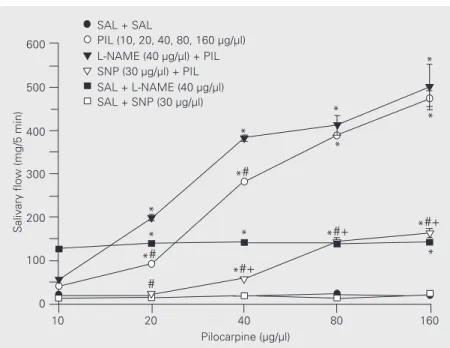
Salivary secretion after injection of iso-tonic saline (0.15 M NaCl) into the MnPO was 19.6 ± 1.6 mg/5 min. The injection of pilocarpine (10, 20, 40, 80, 160 µg) into the MnPO increased salivary secretion to 41 ± 3, 92 ± 4, 282 ± 5, 381 ± 4 and 474 ± 18 mg/ 5 min, respectively. ANOVA showed that there were significant differences among all concentrations (F(4,32) = 57.14, P<0.01). The basal values of salivary flow after injec-tion of L-NAME (40 µg) after 5 periods of
observations were 126 ± 4, 139 ± 6, 141 ± 7,
139 ± 6 and 142 ± 7 mg/5 min. L-NAME (40 µg/µl) injected into the MnPO before pilo-carpine potentiated the sialogogue effect of pilocarpine with values of 54 ± 5, 199 ± 7, 382 ± 6, 412 ± 23 and 501 ± 52 mg/5 min (F(4,32) = 29.13, P<0.01). The basal values of salivary secretion after SNP (30 µg) injec-tion into the MnPO after 5 periods of obser-vations were 12 ± 1, 14 ± 2, 18 ± 6, 13 ± 4 and 20 ± 7 mg/5 min. SNP (30 µg/µl) injected into the MnPO before pilocarpine attenuated the sialogogue effect of pilocarpine with values of Figure 2. Effect of L-NAME and SNP injected into the median preoptic nucleus (MnPO) on
salivary flow induced by injection of pilocarpine into the MnPO. Data are reported as means ± SEM. *P<0.001 compared to SAL + SAL; #P<0.001 compared to L-NAME + pilocarpine; +P<0.001 compared to pilocarpine (Dunnet t-test; N = 40 for each group). SAL, saline; PIL, pilocarpine; L-NAME, L-NG-nitro-arginine methyl ester; SNP, sodium nitroprusside.
Salivary flow (mg/5 min)
600
500
400
300
200
100
0
10 20 40 80 160
Pilocarpine (µg/µl) SAL + SAL
PIL (10, 20, 40, 80, 160 µg/µl) L-NAME (40 µg/µl) + PIL SNP (30 µg/µl) + PIL SAL + L-NAME (40 µg/µl) SAL + SNP (30 µg/µl)
*
*
*#
# *#+
*#+ *#+
*
*
*
* *#
*
*
14 ± 1, 23 ± 2, 58 ± 3, 143 ± 9 and 163 ± 8 mg/ 5 min. ANOVA showed significant differ-ences among the effects of various concentra-tions (F (4,32) = 74.48, P<0.01) (Figure 2).
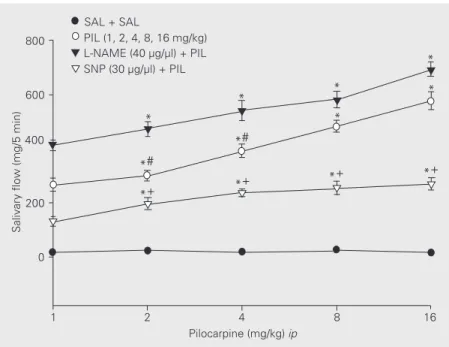
Effect of L-NAME and SNP on salivary secretion induced by ip injection of pilocarpine
The ip administration of pilocarpine (1,
2, 4, 8, and 16 mg/kg body weight) increased salivary secretion (268 ± 24, 300 ± 20, 392 ± 24, 483 ± 22 and 578 ± 33 mg/5 min, respec-tively; F(4,30) = 12.34, P<0.01). After L-NAME injection (40 µg) into the MnPO, the salivary secretion induced by various
con-centrations of pilocarpine injected ip was
potentiated, with values of 413 ± 20, 473 ± 27, 541 ± 37, 582 ± 49 and 694 ± 28 mg/5 min, respectively (F(4,32) = 12.22, P<0.01). SNP (30 µg/µl) injected into the MnPO be-fore various concentrations of pilocarpine
injected ip attenuated the pilocarpine effect
on salivary secretion to values of 132 ± 18, 197 ± 23, 237 ± 14, 255 ± 25 and 272 ± 22 mg/5 min, respectively (F(4,32) = 1.99, P<0.01) (Figure 3).
Effect of L-NAME and SNP on changes of MAP and HR induced by injection of pilocarpine into the MnPO
Pilocarpine injection into the MnPO
in-duced an increase in MAP of ∆29 ± 6 mmHg
and a decrease in HR to 320 ± 4 bpm, that differed significantly (P<0.05) from control
values (∆+4 ± 1 mmHg and 365 ± 7 bpm).
After administration of L-NAME the basal
values were ∆-15 ± 3 mmHg and 305 ± 5
bpm. L-NAME (40 µg) injected into the MnPO before pilocarpine (40 µg in a volume of 1.0 µl) injected into the same region po-tentiated the pressor effect of pilocarpine
(∆+39 ± 7 mmHg with a decrease in HR to
301 ± 8 bpm, P<0.05). The basal values after
SNP (30 µg) at a volume of 1.0 µl were ∆+9
± 2 mmHg and 370 ± 8 bpm. SNP injected
Change in MAP (mmHg)
40 35 30
10 25 20 15
5
-5 -10 0
SAL L-NAME SNP SAL + PIL
L-NAME + PIL
SNP + PIL
7
*#+
7
7
*
8
9 #8
12345 12345 12345
Figure 3. Effect of L-NAME and SNP injected into the median preoptic nucleus on salivary flow induced by intraperitoneal (ip) administration of pilocarpine (PIL). Data are reported as means ± SEM. *P<0.001 compared to SAL + SAL; #P<0.001 compared to L-NAME + PIL; +P<0.001 compared to PIL (Dunnet t-test; N = 38 for each group))))). For abbreviations, see legend to Figure 2.
Figure 4. Effect of L-NAME and SNP injected into the median preoptic nucleus (MnPO) on mean arterial pressure (MAP) during injection of pilocarpine (PIL) into the MnPO. The number of animals is given at the top of each column. Data are reported as means ± SEM. *P<0.001 compared to SAL (control); #P<0.001 compared to SAL + L-NAME; +P<0.001 compared to SAL + PIL (Dunnett t-test). For abbreviations, see legend to Figure 2.
Salivary flow (mg/5 min)
800
600
400
200
0
1 2 4 8 16
Pilocarpine (mg/kg) ip SAL + SAL
PIL (1, 2, 4, 8, 16 mg/kg) L-NAME (40 µg/µl) + PIL SNP (30 µg/µl) + PIL
*+ *+
*+ *#
*+
*#
*
* *
*
*
*
into the MnPO before pilocarpine induced a decrease of MAP (-7 ± 2 mmHg, P<0.05) without any changes in HR (369 ± 7 bpm), as shown in Figures 4 and 5.
*+
Figure 5. Effect of L-NAME and SNP injected into the median preoptic nucleus (MnPO) on heart rate during injection of pilocarpine (PIL) into the MnPO. The num-ber of animals is given at the top of each column. Data are reported as means ± SEM. *P<0.001 compared to SAL (control); +P<0.001 compared to SAL + PIL (Dunnett t-test). For abbreviations, see legend to Fig-ure 2.
Figure 7. Effect of L-NAME and SNP injected into the median preoptic nucleus (MnPO) on heart rate during intraperitoneal (ip) administration of pilocarpine (PIL). The number of animals is given at the top of each column. Data are reported as means ± SEM. *P<0.001 compared to SAL + SAL (control); #P<0.05 compared to SAL + L-NAME; +P<0.001 compared to SAL + PIL (Dunnett t-test). For abbreviations, see legend to Fig-ure 2. 480 400 320 240 160 80 0 1234 1234 1234 1234 1234 1234 1234 1234 1234 1234 1234 1234 1234 1234 1234 1234 1234 1234 1234 1234 1234 1234 + 7 *+ 7 * 7 + 8 * 8 9
SAL L-NAME SNP SAL + PIL L-NAME + PIL SNP + PIL
Heart rate (bpm)
Heart rate (bpm)
500 400 300 200 100 0 SAL (MnPO) + SAL
(ip)
L-NAME (MnPO)
+ SAL
(ip)
SNP (MnPO)
+ SAL
(ip)
SAL (MnPO)
+ PIL
(ip)
L-NAME (MnPO)
+ PIL
(ip)
SNP (MnPO)
+ PIL
(ip) 8 8 +# 8 *# 8 *+ 8 *+ 7 1234 1234 1234 1234 1234 1234 1234 1234 1234 1234 1234 1234 1234 1234 1234 1234 1234 1234 1234 1234 1234 1234
Figure 6. Effect of L-NAME and SNP injected into the median preoptic nucleus (MnPO) on mean arterial pres-sure (MAP) during intraperitoneal (ip) administration of pilocarpine (PIL). The number of animals is given at the top of each column. Data are reported as means ± SEM. *P<0.001 compared to SAL (control); #P<0.001 compared to SAL + L-NAME; +P<0.001 compared to SAL + PIL (Dunnett t-test). For abbreviations, see leg-end to Figure 2.
Change in MAP (mmHg)
40 30 20 10 0 -10 -20 -30 1234 1234 1234 SAL (MnPO) + SAL
(ip)
L-NAME (MnPO)
+ SAL
(ip) SNP (MnPO)
+ SAL
(ip)
L-NAME (MnPO)
+ PIL
(ip) SNP (MnPO)
+ PIL
(ip) SAL
(MnPO) + PIL
Effect of L-NAME and SNP injected into the MnPO on changes in MAP and HR induced byinjection of pilocarpine
Pilocarpine injected ip induced a decrease
in MAP but an increase in HR, with values of
∆-21 ± 3 mmHg and421 ± 7 bpm. L-NAME
injected into the MnPO before pilocarpine
injected ip reversed the effect of pilocarpine,
with values of ∆+21 ± 3 mmHg and 311 ± 6
bpm (P<0.05). SNP injected into the MnPO
before pilocarpine injected ip blocked the
effect of pilocarpine, with values of ∆+5 ± 1
mmHg and 349 ± 8 bpm (Figures 6 and 7).
Discussion
The present results show that the
injec-tion of pilocarpine into the MnPO and ip
affects salivary flow in a concentration-de-pendent manner. Pilocarpine at a concentra-tion of 40 µg produced a medium salivary flow, whereas the flow rate increased in a linear fashion up to 160 µg. It has been demonstrated that pilocarpine, when injected
intracerebroventricularly (icv), produced
sali-vary secretion at a significantly higher level than control (11,16). It has also been re-ported that electrolytic lesion of the AV3V produced a decrease in salivary flow
in-duced by pilocarpine injected icv (11). These
results indicate that other areas of the CNS, such as the areas surrounding the third ven-tricle, are important for pilocarpine-induced salivary secretion. The areas surrounding the third ventricle are important for the regu-lation of the water-salt and cardiovascular balance as well as for the control of salivary composition and salivary flow. This is in agreement with results showing that the MnPO is important for the central regulation of salivary gland function (23).
Difficulty in swallowing (dysphagia) is a common upper gastrointestinal disorder caused by salivary malfunction (24). The present results show the participation of the CNS in attenuating these disorders,
demon-strating that a controlled-release form of pilo-carpine may overcome the therapeutic weak-nesses of current pilocarpine preparations by prolonging salivary secretion and reduc-ing undesirable side effects. Pilocarpine
in-jected ip or into the MnPO produced changes
in salivary flow and in cardiovascular pa-rameters, such as an increase in MAP and a
decrease in HR. When injected ip it caused a
decrease in MAP and an increase in HR. These results are important, since several drugs used by patients with cardiovascular diseases may alter salivary secretion and interfere with the effects of pilocarpine when it is used as a drug. When injected centrally, pilocarpine produced alterations in salivary secretion, MAP and HR. An oral dose of pilocarpine increased salivary flow rates in patients with xerostomia (dry mouth). Pilo-carpine induced submandibular and parotid salivary flow, which remained constant over a period of time (25).
The recognition of the role of NO in cell-to-cell communication has changed the con-cept of traditional neurotransmission. N-methyl-D-aspartate receptors mediate the dip-sogenic response of c-Fos expression
in-duced by icv infusion of angiotensin II (26).
re-gion of the periaqueductal gray also decreased MAP, whereas L-NAME increased it (20). SNP injected into the MnPO before carpine reduced the sialogogue effect of pilo-carpine, with a decrease in MAP and an increase in HR. We infer that pilocarpine may also act in areas of the CNS such as the MnPO that regulates salivary flow and car-diovascular alterations. A regulatory mech-anism may also exist between salivary flow and the cardiovascular parameters.
Pilo-carpine injected ip produced a copious
sali-vary flow that was potentiated by the previ-ous central injection of L-NAME into the MnPO. Therefore we postulate that the sys-temic effects of pilocarpine on salivary flow and cardiovascular regulation are influenced by central NO release, in agreement with the
resultsreported by others (16,17). Pilocarpine
injected ip decreased the MAP and increased
the HR, with an opposite effect when in-jected into the MnPO, where it acted by increasing salivary flow and blood pressure, with a decrease in HR. The release of NO in
the CNS continuously reduces the salivary secretion, playing an important role in the central and peripheral effects of pilocarpine. The cardiovascular changes produced by pilo-carpine were mediated by the release or inhi-bition of NO. These results, taken together with those reported by others (22,23,32), suggest that the MnPO is involved in body fluid regulation not only by controlling vaso-pressin secretion and water intake but also by modulating central sympathetic outflow which regulates body fluid balance through an effect on the kidney and on salivary gland function (23,32).
Acknowledgments
The authors thank Silas Pereira Barbosa, Reginaldo da Conceição Queiroz and Silvia Foglia for excellent technical assistance, and Silvana A.D. Malavolta for typing the manu-script. We would also like to thank Luciana R. Saad for revising the English text.
References
1. Fox PC (1997). Management of dry mouth. Dental Clinics of North America, 41: 863-875.
2. Ferguson MM (1993). Pilocarpine and other cholinergic drugs in the management of salivary gland dysfunction. Oral Surgery, Oral Medi-cine, and Oral Pathology, 75: 186-191.
3. Garret JR, Suleiman AM, Anderson LC & Proctor GB (1991). Secre-tory responses in granular ducts and acini of submandibular glands in vivo to parasympathetic nerve stimulation in rats. Cell and Tissue Research, 264: 117-126.
4. Baum BJ (1987). Neurotransmitter control secretion. Journal of Dental Research, 66: 628-632.
5. Rhodus NL (1997). Oral pilocarpine HCl stimulates labial (minor) salivary gland flow in patients with Sjögren’s syndrome. Oral Dis-eases, 3: 93-98.
6. Nagler R, Marmry Y, Fox PC, Baum BJ, Har-El R & Chevion M (1997). Irradiation-induced damage to salivary gland. Radiation Research, 147: 468-476.
7. Hainsworth FR & Epstein AN (1966). Severe impairment of heat-induced saliva-spreading in rats recovered from lateral hypothalamic lesions. Science, 153: 1255-1257.
8. Schallert T, Leach LR & Braun JJ (1978). Saliva hypersecretion during aphagia following lateral hypothalamic lesions. Physiology and Behavior, 21: 461-463.
9. Flynn FW, Evey LA & Mitchell JC (1981). Heat-induced saliva secre-tion and thermoregulasecre-tion in female rats with ventromedial hypotha-lamic lesions. Physiology and Behavior, 26: 779-782.
10. Kanosue K, Nakayama T, Tanaka H, Yanase M & Yasuda H (1990). Modes of action of local hypothalamic and skin thermal stimulation on salivary secretion in rats. Journal of Physiology, 424: 459-471. 11. Renzi A, Colombari E, Mattos Filho TR, Silveira JE, Saad WA, De
Luca Jr LA, Derobio JG & Menani JV (1993). Involvement of the central nervous system in the salivary secretion induced by pilo-carpine in rats. Journal of Dental Research, 72: 1481-1484. 12. Renzi A, Lopes RA, Sala MA, Camargo LAA, Menani JV, Saad WA &
Campos GM (1990). Morphological, morphometric and stereological study of submandibular glands in rats with lesion of the anteroven-tral region of the third ventricle (AV3V). Experimental Pathology, 38: 177-187.
13. Takai N, Uchihashi K, Higuchi K, Yoshida Y & Yamaguchi M (1999). Localization of neuronal-constitutive nitric oxide synthase and secre-tory regulation by nitric oxide in the rat submandibular and sublin-gual glands. Archives of Oral Biology, 44: 745-750.
14. Damas J (1994). Pilocarpine-induced salivary secretion, kinin system and nitric oxide in rats. Archives Internationales de Physiologie, de Biochimie et de Biophysique, 102: 103-105.
Saad WA (2002). Role of nitric oxide and beta receptors of the central nervous system on the salivary flow induced by pilocarpine injection into the lateral ventricle. Pharmacology, Biochemistry and Behavior, 72: 220-235.
16. Saad WA, Camargo LAA, Saad R, Pereira AF & Simões S (1999). Effect of injection of L-NAME on drinking response. Brazilian Journal of Medical and Biological Research, 32: 1413-1416.
17. Camargo LAA, Saad Wilson A, Simões S, Santos TAFB & Saad Willian A (2002). Interaction between paraventricular nucleus and septal area in the control of physiological responses induced by angiotensin II. Brazilian Journal of Medical and Biological Research, 35: 1017-1023.
18. Cunningham JT & Johnson AK (1989). Decreased norepinephrine in the ventral lamina terminalis region is associated with angiotensin II drinking response deficits following local 6-hydroxydopamine injec-tions. Brain Research, 480: 65-71.
19. Gardiner TW & Stricker EM (1985). Imparied drinking responses of rats with lesions of nucleus medianus: circadian dependence. Ameri-can Journal of Physiology, 248: R224-R230.
20. Lind RW & Johnson AK (1982). Subfornical organ-median preoptic connections and drinking and pressor responses to angiotensin II. Journal of Neuroscience, 2: 1043-1051.
21. Paxinos G & Watson C (1986). The Rat Brain in Stereotaxic Coordi-nates. Academic Press, New York.
22. O’Neill TP & Brody MJ (1987). Role of the median preoptic nucleus in centrally evoked pressor responses. American Journal of Physiol-ogy, 252: R1165-R1172.
23. Hübschle T, McKinley MJ & Oldfield BJ (1998). Efferent connec-tions of the lamina terminalis, the preoptic area and the insular cortex to submandibular and sublingual gland of the rat traced with pseudorabies virus. Brain Research, 806: 219-231.
24. Valdez IH & Fox PC (1991). Interactions of the salivary and
gas-trointestinal system. II. Effects of salivary gland dysfunction on the gastrointestinal tract. Digestive Diseases, 9: 210-218.
25. Fox PC & Mandel ID (1992). Sjogren’s syndrome. Oral Surgery, Oral Medicine, and Oral Pathology, 74: 315-318.
26. Zhu B & Herbert J (1997). Angiotensin II interacts with nitric oxide-cyclic GMP pathway in the central control of drinking behavior: Mapping with c-fos and NADPH-diaphorase. Neuroscience, 79: 23-29.
27. Arnais SL, Coronel MF & Boveris A (1999). Nitric oxide, superoxide, and hydrogen peroxide production in brain mitochondria after halo-peridol treatment. Nitric Oxide, 3: 235-243.
28. Bredt DS, Glatt CE, Hwang PM, Fotuhi M, Dawson TM & Snyder SH (1991). Nitric oxide protein and mRNA are localized in neuronal populations of the mammalian CNS together with NADPH diapho-rase. Neuron, 7: 615-624.
29. Ignarro LJ, Bug GM, Wood KS, Byrns RE & Chaudhuri G (1987). Endothelium-derived relaxing factor produced and released from artery and vein is nitric oxide. Proceedings of the National Academy of Sciences, USA, 84: 9265-9269.
30. Kadekaro M, Terrell ML, Harmann P & Summy-Long JY (1994). Central inhibition of nitric oxide synthase attenuates water intake but does not alter enhanced glucose utilization in the hypothalamo-neurohypophysial system of dehydrated rats. Neuroscience Letters, 173: 115-118.
31. Rees DD, Palmer RM & Moncada S (1989). Role of endothelium-derived nitric oxide in the regulation of blood pressure. Proceedings of the National Academy of Sciences, USA, 86: 3375-3378. 32. Yashuda Y, Honda K, Negoro H, Higuchi T, Goto Y & Fukuda S