Seasonal morphological variation of the vas deferens of scorpion mud turtle
(Kinosternon scorpioides)
Diego Carvalho Viana1, Leandro Almeida Rui1, Amilton Cesar dos Santos1, Maria Ange´lica Miglino1,
Antoˆnio Chaves de Assis Neto1, Lianne Polliane Fernandes Araujo2, Antonia Santos Oliveira2& Alana Lislea Sousa2,3
1Anatomy of Domestic and Wild Animals, University of Sao Paulo, CEP 05508-270, Sa˜o Paulo, SP, Brazil. 2
Clinical Veterinary, State University of Maranha˜o, Sa˜o Luı´s, MA, Brazil. 3
Corresponding author: Alana Lislea Sousa, e-mail:alana@elo.com.br
VIANA, D.C., RUI, L.A., SANTOS, A.C. dos, MIGLINO, M.A., ASSIS NETO, A.C., ARAUJO, L.P. F., OLIVEIRA, A.S., SOUSA, A.L.Seasonal morphological variation of the vas deferens of scorpion mud
turtle (Kinosternon scorpioides).
http://dx.doi.org/10.1590/1676-06032014006413
Abstract:This study aimed to characterize the morphology of the vas deferens ofKinosteron scorpioides
by macroscopic and microscopic analysis. Were used 20 adult male jurara´s collected at regular intervals during the year and divided into four experimental groups in the rainy and dry seasons, being processed for light microscopy, scanning electron microscopy and transmission electron microscopy. Morphometry for tubular and luminal diameters and epithelial height were also performed. On rainy season, vas deferens presented pseudostratified epithelium with cylindrical cells, spermatozoids and milky fluid in the lumen, with cytoplasmic organelles and lipid vesicles. On dry season, epithelium was pseudostratified with cuboid cells, with cellular debris and no spermatozoids. There was significant variation (p,0,05) for morphometry of vas deferens, with lower values of tubular and luminal diameters on rainy season, and higher epithelial height on dry season.
Keywords:Kinosternon, Reproduction, Vas deferens, Morphometry, Ultrastructural.
VIANA, D.C., RUI, L.A., SANTOS, A.C. dos, MIGLINO, M.A., ASSIS NETO, A.C., ARAUJO, L.P.F., OLIVEIRA, A.S., SOUSA, A.L.Variac¸a˜o morfolo´gica sazonal dos ductos deferentes de tartaruga
jurara´ (Kinosternon scorpioides).
http://dx.doi.org/10.1590/1676-06032014006413
Resumo:Este estudo teve como objetivo caracterizar a morfologia dos ductos deferentes deKinosteron scorpioides por meio de ana´lise macrosco´pica e microsco´pica. Foram utilizados 20 machos adultos, coletados em intervalos regulares durante o ano, os quais foram divididos em quatro grupos experimentais nas estac¸o˜es chuvosa e seca. Os ductos deferentes foram processados para ana´lise por microscopia de luz, microscopia eletroˆnica de varredura e microscopia eletroˆnica de transmissa˜o. Morfometria para diaˆmetros tubular e luminal e altura epitelial tambe´m foram realizadas. Na e´poca das chuvas, o ducto deferente apresentou epite´lio pseudoestratificado com ce´lulas cilı´ndricas, espermatozoides e lı´quido leitoso no lu´men, ale´m de com organelas citoplasma´ticas e vesı´culas lipı´dicas. Na estac¸a˜o seca, o epite´lio do ducto deferente foi do tipo pseudoestratificado com ce´lulas cuboides e debris celulares, sendo que nenhum espermatozoide foi encontrado nesta estac¸a˜o. Houve variac¸a˜o significativa (p ,0,05) para a morfometria dos ductos deferentes, com menores valores de diaˆmetros tubular e luminal na estac¸a˜o chuvosa, e maior altura do epite´lio na estac¸a˜o seca.
Palavras-chave:Kinosternon, Reproduc¸a˜o, Ducto deferente, Morfometria, Ultraestrutural.
Introduction
Brazil has 35 species of chelonians distributed in its various terrestrial and aquatic ecosystems, of which 28 species are freshwater, two are terrestrial (land turtles), and 5 are marine turtles (SBH, 2005). The family Kinosternidae is composed of semi-aquatic species of small to medium size, being distributed from Canada to South America (Erns & Barbour 1989). It is composed of 22 species subdivided in four genus: Kinosternon,
Sternotherus, Staurotypus and Claudius. In the Brazilian Amazon it is possible to found only one species of this family,
Kinosternon scorpioides, also known as scorpion mud turtle (Molina & Rocha 1996).
The scorpion mud turtle is preferably an aquatic species, and inhabits both stagnant and flowing water, being also able to develop semi-aquatic behavior (Pritchard & Trebbau 1984). It displays a shell with three evident keels, especially the median, which runs through the shell in the longitudinal
article
Biota Neotropica. 14(3): e20130064.
direction (Vanzolini et al. 1980). It also has a strong jaw and a structure similar to a nail at the end of the body, like a scorpion’s stinger, which termed scientifically this species.
Kinosternon scorpioides is well distributed in the coast of South America, including Colombia, the Guianas and Trinidad. In Brazil it is found in the states of Para´, Maranha˜o, north of Goias, Ceara´, Rio Grande do Norte and Pernambuco (Pritchard & Trebbau 1984). In Maranhao its presence is confirmed on the edge of rivers (Pereira 2007), and it is considered an important species, both economically and as a source of protein.
The male reproductive system consists on a pair of oval testes of variable size, between light yellow to golden yellow color, and being fixed by mesorchium and mesocolon; epididymis located along the dorsal part of medial surface of each testis, being very delicate, presented as very convoluted structures of whitish color; and the vas deferens, which are continuous to the epididymis and culminate in the region of the cloaca. The vas deferens in jurara´ are a pair of simple structures, with a convoluted path that extends from the epididymis (Viana 2013) to the cloaca, with the function of transporting and storing spermatozoids. The penis, in turn, is grooved, and composed by the root, body and gland, located in the ventral floor of the cloaca, in which it attaches via a retractor muscle, and is protected by the foreskin (Carvalho et al. 2010).
To our knowledge, this is the first study aimed to characterize the morphology of the vas deferens ofKinosteron scorpioides by macroscopic and microscopic analysis, contri-buting to describe the reproductive characteristics of this species and elucidate the spermatogenic cycle, strategies of sperm storage, and morphology of the reproductive tract.
Materials and methods
Were used 20 adult male Kinosternon scorpioides, from the capture ex-situ in the city of Sa˜o Bento, state of Maranha˜o, Brazil, as authorized by IBAMA for the purpose of scientific activities with number 26136-1, and approval of the Ethics and Animal Research Committee of the Course of Veterinary Medicine (EAEC/UEMA), protocol number 011/2010.
The research was developed in the Laboratory of Veterinary Anatomy and Anatomopathology of the Course of Veterinary Medicine in the Center for Agricultural Sciences of State University of Maranha˜o - UEMA, Sao Luis - MA, and Laboratories of Light and Electron Microscopy, of the School of Veterinary Medicine and Animal Science of University of Sao Paulo - USP.
The animals were collected at regular intervals during the year and divided into four experimental groups in the rainy and dry seasons, the rainy season being understood by the collections of March/2011 and June/2011, and dry season being December/2010 September/2011.
The twenty animals were anesthetized with xylazine 2% (40mg/kg/IM) and ketamine hydrochloride 1% (60mg/kg/IM) and euthanized by administration of thiopental sodium 2.5% (60mg/kg/EV) by catheterization of the cervical venous sinus.
Subsequently, was held the opening of the coelomic cavity with steel handsaw, for the disarticulation of the bone bridge that connects the carapace and the plastron, and visualization and removal of the reproductive tract and isolation of the vas deferens, the processing being specific for each microscopy.
For light microscopy, the vas deferens were fixed in buffered formaldehyde 4% for about 24 hours for paraffin embedding. They were then dehydrated in increasing alcohol concentrations (706- 1006) and diaphanized in xylol, with an interval of switching between the solutions of 1 hour. After dehydration, the fragments were embedded in paraffin, sectioned at 4mm thickness, and stained with hematoxylin-eosin (HE), Masson’s trichrome and periodic acid-Schiff - PAS, and examined under an optical microscope.
For scanning electron microscopy (SEM), the fragments were fixed in 2.5% glutaraldehyde, frozen for 72 hours and thereafter cryofracturated in liquid nitrogen, washed in 0.1 M phosphate buffer, post-fixed in osmium tetroxide 1% and dehydrated in series of alcohols (50 6– 100 6). The samples were dried in a critical point apparatus Balzers CPD 020 using liquid CO2and mounted on metal aluminum basis (stub), using
carbon paste. Subsequently, were subjected to a metallic coating ("sputting") with gold in sputter device EMITECH K550, analyzed and photographed under a scanning electron microscope LEO 435VP.
For transmission electron microscopy (TEM), the frag-ments were fixed in 2.5% glutaraldehyde, washed in 0.1 M phosphate buffer and post-fixed in osmium tetroxide 1%. Subsequently, were dehydrated in series of increasing alcohols (506- 1006), propylene oxide and resin. The resin mixture was replaced by pure resin and placed in molds. The ultrathin sections were collected on copper screens and contrasted with uranyl acetate solution at 2% and 0.5% lead citrate. The samples were analyzed in transmission electron microscopy apparatus MORGANI 268d.
Images for morphometric studies were obtained using a binocular microscope Olympus BH-41 equipped with a digital camera for the photographic record. Histomorphometric analyses were performed with the aid of the program GIMP 2 to obtain the average height of the epithelium and the luminal and tubular diameters of the vas deferens, obtained with the use of micrometric ocular adapted to the microscope. Were made ten slides with three serial sections; the tubular sections have been made around the tubules in the base of epithelium, by the level of the the basal membrane, to obtain the total tubular diameter, and adjacent to the apical edge to obtain the luminal diameter, using a 10x objective. Similarly, a 40x objective was used for the measurement of the height of epithelium from its base to the apical edge.
Results
Vas deferens are continuous to the epididymis, presenting as a small structure, resembling a sinuous tube in its final portion, following lateral to ureters, and inserting on the dorsolateral wall of the cloaca, expanding in a shape of a small bulb.
In Kinosternon scorpioides, vas deferens observed by light microscopy presents as covered by pseudostratified cylindrical epithelium with secretory cells (Figure 1). Vas deferens also has a layer of dense connective tissue with the presence of blood vessels surrounded by muscle tissue. Inside the lumen are found spermatozoids and a milky fluid on the tubular center.
with cuboid cells, absence of spermatozoa in the lumen and cellular debris.
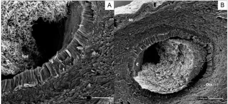
Scanning electron microscopy of vas deferens by cryofrac-ture technique showed disposition of spermatozoid on tubular epithelium and arrangement of dense connective tissue along to a muscular layer close to the blood vessels. It is believed that these elevations favor storage of sperm in the region (Figure 2). Transmission electron microscopy on rainy season showed spermatozoids, cytoplasmic organelles (mitochondria), indicat-ing high metabolic activity, and lipid vesicles responsible by nutrition of spermatozoid during storage. On dry season, ultrastructural findings were disorganized and sparse cyto-plasm, with endoplasmic reticulum, indicating protein produc-tion, and few spermatozoids (Figure 3).
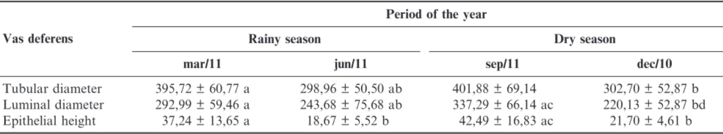
There was significant variation (p,0,05) between seasons for morphometry of vas deferens. The tubular and luminal diameters had lower values on rainy season. However, epithelial height in dry season had higher averages (Table 1).
Discussion
Although in scorpion mud turtle the vas deferens expands in a shape of a small bulb, this feature was not observed for the same species by Carvalho et al. (2010), which does not report the expansion of vas deferens before its insertion on the cloaca. Chaves (2011), however, described the same characteristic, characterizing this expansion in bulb shape on vas deferens of scorpion mud turtle.
Figure 2.Fotomicrography of scanning electron microscopy (SEM) of vas deferens of scorpion mud turtle (Kinosternon scorpioides). A: vas deferens, with pseudostratified cylindrical epithelium and spermatozoids in lumen (Spz). B: vas deferens, with evidence of tubular lumen (,), dense connective tissue (Dct) and muscular layer(Ml).
By light microscopy, in snakes (Bothrops jararaca and
Crotalus durissus), epithelial cells showed microvilli which were not observed in scorpion mud turtle, even though similar secretion was observed by the epithelium (Almeida-Sousa 2005). Similarity was described on close species, such as the crocodile (Guerrero et al. 2004), and birds, particularly in the rooster (Tingari 2001). In the snakeSeminatrix pygaeaof South Carolina, was observed arrangement of spermatozoids tangent to the epithelium, most being slightly separated from epithe-lium (Ssever 2004).
Structural morphology was also closed to that observed in snakes, presenting lots of spermatozoa in the lumen of vas deferens in rainy season, indicating copulation period, when it is used as a storage organ; and decrease of spermatozoa in the lumen on dry season, indicating a postmating stage (Rojas et al. 2013). However, in rat snakes, although vas deferens is also the main sperm storage organ, spermatozoa is present in large numbers throughout all the year, except in July (Gang et al. 2011).
Scanning electron morphology in snakes reported vas deferens as being an organ of spermatozoids storage (Almeida-Sousa 2005),. In this study of scorpion mud turtle, it is believed that vas deferens is also adapted for storage, due to its structural characteristics such as absence of cilia or stereocilia in the cells. On crocodiles (Caiman crocodilus), non-ciliated cells were also found, indicating storage function
of vas deferens in another species of reptiles (Guerrero et al. 2004).
Transmission electron microscopy also presented lipid vesicles in snakes on the rainy season, which are responsible for nutrition of spermatozoids (Rojas 2013). On dry season, the visualization of spermatozoa in the lumen demonstrates that the organ is in reproductive activity. On domestic quails, was reported that vas deferens showed little annual variability, with a significant increase in tubular caliber, intraluminal storage of spermatozoids and occurence of mitochondria, lysomes, endoplasic reticulum and variable vesicles in the cytoplasm of principal cells. These ultrastructural features of principal cells seems to be indicative of the occurence of active processes of endocytosis, and degenerative characteristics were observed at the supranuclear cytoplasm of epididimary P cells on autumn (Orsi et al. 2007). On crocodiles, endoplasmic reticulum was also abundant, indicating protein production, despite of absence of visible secretory material (Guerrero et al. 2004).
By morphometry, the decrease of tubular and luminal diameters, along with increase of epithelial heighs on rainy season, are correlated to seasonal variations in synchrony with the spermatogenic and epididymal cycles. In the same sense, was described in snakes (Cerastes vipera and Psammophis sibilans), a larger diameter and short epithelial linings during reproductive season as a result of elongation of stored spermatozoids (Sivan et al. 2012, AMER et al. 1978).
Figure 3.Fotomicrography of transmission electron microscopy (TEM) of vas deferens of scorpion mud turtle (Kinosternon scorpioides). A: vas deferens on dry season with disorganized cytoplasm, presenting endoplasmic reticulum (yellow arrow), vesicle (red circle) and spermatozoids (blue arrow). B: vas deferens on rainy season with spermatozoids (Spz), mitochondria (yellow arrow) and lipidic vesicles (red arrow).
Table 1.Mean and standard deviation of morphometry (mm) of tubular and luminal diameters and height of the vas deferens of turtle (Kinosternon scorpioides), captured in Sa˜o Bento - MA, according with the season. Sao Luis - MA –– 2012.
Vas deferens
Period of the year
Rainy season Dry season
mar/11 jun/11 sep/11 dec/10
Tubular diameter 395,72 ± 60,77 a 298,96 ± 50,50 ab 401,88 ± 69,14 302,70 ± 52,87 b Luminal diameter 292,99 ± 59,46 a 243,68 ± 75,68 ab 337,29 ± 66,14 ac 220,13 ± 52,87 bd Epithelial height 37,24 ± 13,65 a 18,67 ± 5,52 b 42,49 ± 16,83 ac 21,70 ± 4,61 b
We conclude that vas deferens is the main sperm storage organ on the scorpion mud turtle, presenting a large number of spermatozoids in reproductive season, and morphological findings that represent an adaptation to its function. However, on dry season, the storage was smaller, indicating that organ is in reproductive activity, but production of spermatozoids is reduced in comparison to the rainy season. Further studies on hormonal levels and quality of spermatozoids are suggested, in order to refine the knowledge on the reproductive biology of
Kinosternon scorpioides.
Acknowledgments
The authors would like to thank the State University of Maranha˜o (UEMA), the National Program of Academic Cooperation (Procad I-CAPES/UEMA/USP Amazon) and the Foundation for Research Support of the State of Maranha˜o (FAPEMA) for funding current research.
References
ALMEIDA-SANTOS, S.M. 2005. Modelos reprodutivos em serpentes: estocagem de esperma e placentac¸a˜o emCrotalus durissuseBothrops jararaca(Serpentes: Viperidae). Tese (Doutorado)- Programa de po´ s-graduac¸a˜o em Anatomia dos animais dome´sticos e silvestres. Faculdade de Medicina Veterina´ria e Zootecnia, Universidade de Sa˜o Paulo, Sa˜o Paulo. 204p.
AMER, F.I. & ELSHABKA, H.A. 1978. Studies on the reproductive organs of the colubrid snakes, Psammophis sibilans and spaler-osophis diadema, I. The Male Organs. Bulletin of the Faculty of Science-King Abdulaziz University, 2: 1-16.
CARVALHO, R.C., SOUSA, A., SILVA, A.L.A. & PEREIRA, J.G. 2010. Anatomia dos o´rga˜os genitais do muc¸ua˜Kinosternon scorpioides macho (Chelonia, Kinosternidae). Pes. Vet. Bras. 30: 289-294, doi: http://dx.doi.org/10.1590/S0100-736X2010000400001
CHAVES, L.P.F.A. 2011. Orga˜os genitais masculino e nı´vel se´rico de testosterona de jurara´ (kinosternon scorpioides, linnaeus, 1766) criado em cativeiro na regia˜o da baixada maranhense no estado do maranha˜o. Dissertac¸a˜o (Mestrado em Cieˆncias Animal) -Universidade Estadual do Maranha˜o, Sa˜o Luı´s, 92p.
ERNST, C.H. & BARBOUR, R.W. 1989. Turtles of the United States. Lexington: Univ. of Kentucky Press, 347p.
GANGA, L., QIAO-QIAO, L., HU-HU, Y. & QIONG, X. 2011. Histological and immunocytochemical study of deferens ducts in the Chinese rat snake (Zaocys dhumnades). Zool. Res. 32: 663-669.
GUERRERO, M.S., CALDERON, L.M., PEREZ, R.G. and PINILLA, M.P.R. 2004. Morphology of the male reproductive duct system ofCaiman crocodilus(Crocodylia, Alligatoridae). Ann. Anat. 186: 235-245, doi: http://dx.doi.org/10.1016/S0940-9602(04)80009-8
MOLINA, F.B. & ROCHA, M.B. 1996. Algumas observac¸o˜es sobre a biologia e manejo do muc¸ua˜. Aquacultura. 2: 25-26.
ORSI, A.M., DOMENICONI, F.R., SIMO˜ ES, K., STEFANINI, A.M. & BARALDI-ARTONI, S.M. 2007. Variabilidade sazonal no ducto epididima´rio de codorna dome´stica: observac¸o˜es morfo-lo´gicas. Pesq. Vet. Bras. 27: 495-500, doi:http://dx.doi.org/10.1590/ S0100-736X2007001200005
PEREIRA, L.A., SOUSA, A.L., CUTRIM, M.V.J., MOREIRA, E.G. 2007. Caracterı´sticas ecolo´gicas do habitat de Kinosternon scorpioides scorpioides Linnaeus, 1766 (Reptila, Chelonia, Kinosternidae) no Municı´pio De Sa˜ o Bento –– Baixada Maranhense (MARANHA˜ O, BRASIL). Boletim do laborato´rio de hidrobiologia, 20: 9-14.
PRITCHARD, P.C. & TREBBAU, P. 1984. The Turtles of Venezuela. Society the study of amphibians and reptiles. Ithaca, 403p. ROJAS, C.A., BARROS, V.A., ALMEIDA-SANTOS, S.M. 2013. The
Reproductive Cycle of the Male Sleep Snake Sibynomorphus mikanii (Schlegel, 1837) From Southeastern Brazil. J. Morphol. 274: 215-288, doi:http://dx.doi.org/10.1002/jmor.20099
SBH. Brazilian reptiles –– List of species. 2005. Available in: http://www. sbherpetologia.org.br. Sociedade Brasileira de Herpetologia. Acessed in 05/01/2013.
SEVER, D.M. 2004. Ultrastructure of the Reproductive System of the Black Swamp Snake (Seminatrix pygaea). IV. Occurrence of an Ampulla Ductus Deferentis. J. Morphol. 262: 714-730.
SIVAN, J., KAM, M., HADAD, S., DEGEN, A.A., ROZENBOIM, I. & ROSENSTRAUCH, A. 2012. Reproductive cycle of free-living male Saharan sand vipers,Cerastes vipera(Viperidae) in the Negev desert, Israel. Gen. Comp. Endocrinol. 179: 241-247, doi:http:// dx.doi.org/10.1016/j.ygcen.2012.08.021
TINGARI, M.D. 1971. On the structure of the epididymal region and ductus deferens of the domestic fowl (Gallus domesticus). J. Anat. 109: 423-35.
VANZOLINI, P.E.A. 1996. Contribuic¸a˜o zoolo´gica dos primeiros naturalistasviajantesdo Brasil. Revista USP, 30: 190-238. VIANA, D.C., RUI, L.A., MIGLINO, M.A., ARAUJO, L.P.F.,
OLIVEIRA, A.S., SOUSA, A.L. 2013. Morphological study of epididymides in the scorpion mud turtle in natural habitat (Kinosternon scorpioides –– Linnaeus, 1976). Biotemas, 26: 153-162.