Caffeic acid improves cell viability and
protects against DNA damage:
involvement of reactive oxygen species
and extracellular signal-regulated kinase
Y. Li
1,2*, L.J. Chen
2,3*, F. Jiang
1,2*, Y. Yang
2,3, X.X. Wang
1,2, Z. Zhang
2,3, Z. Li
1,2and L. Li
2,31Department of Nutrition and Food Hygiene, School of Public Health, Nanjing Medical University, Nanjing, China 2Department of Hygiene Analysis and Detection, School of Public Health, Nanjing Medical University, Nanjing, China 3The Key Laboratory of Modern Toxicology, Ministry of Education, School of Public Health,
Nanjing Medical University, Nanjing, China
Abstract
Hormesis is an adaptive response to a variety of oxidative stresses that renders cells resistant to harmful doses of stressing agents. Caffeic acid (CaA) is an important antioxidant that has protective effects against DNA damage caused by reactive oxygen species (ROS). However, whether CaA-induced protection is a hormetic effect remains unknown, as is the molecular mechanism that is involved. We found that a low concentration (10mM) of CaA increased human liver L-02 cell viability, attenuated hydrogen
peroxide (H2O2)-mediated decreases in cell viability, and decreased the extent of H2O2-induced DNA double-strand breaks
(DSBs). In L-02 cells exposed to H2O2, CaA treatment reduced ROS levels, which might have played a protective role. CaA also
activated the extracellular signal-regulated kinase (ERK) signal pathway in a time-dependent manner. Inhibition of ERK by its inhibitor U0126 or by its specific small interfering RNA (siRNA) blocked the CaA-induced improvement in cell viability and the protective effects against H2O2-mediated DNA damage. This study adds to the understanding of the antioxidant effects of CaA by
identifying a novel molecular mechanism of enhanced cell viability and protection against DNA damage.
Key words: Caffeic acid; Hormesis; Antioxidants; DNA double-strand breaks; Extracellular signal-regulated kinase
Introduction
Caffeic acid (3,4-dihydroxycinnamic acid, CaA), a naturally occurring hydroxycinnamic acid derivative, is an active phenolic component of propolis extract and is also found in a wide variety of plants (1). It has biological and pharmacological properties that include antiviral, antiox-idant, anti-inflammatory, anticarcinogenic, and immuno-modulatory activity (1-5). Extensive evidence from bothin vitroandin vivostudies suggests that CaA is an important antioxidant and has health benefits (2,4). CaA can inhibit lipoxygenase activity and suppress lipid peroxidation (2). Moreover, CaA alleviates alcohol-induced oxidative damage in the liver and kidney (6). It can also protect against UVB-induced DNA damage by suppressing the activation of interleukin-10 and mitogen-activated protein kinases (MAPKs) (1). However, the molecular mechanisms under-lying the CaA-induced protective effects against DNA damage remain unclear.
The MAPK pathways transduce signals that lead to diverse cellular responses such as cell growth, differ-entiation, proliferation, and apoptosis (7-9). Each of the three major MAPK pathways consists of three-tiered cascades that induce a pathway comprised of phos-phorylating proteins that mediate transduction pathways activated by a variety of extracellular signals and regulate the expression of specific genes (10,11). The extracellular signal-regulated kinase (ERK) pathway typically trans-duces growth factor signals that induce cell differentiation or proliferation, whereas cytokines and stress signals activate the c-Jun N-terminal kinase (JNK) and p38 MAPK pathways, resulting in stress responses, growth arrest, or apoptosis (9,12). A previous study indicated that CaA regulates lipopolysaccharide (LPS)-induced oxidative stress through c-Src/ERK signaling pathways in endothelial cells (4). We therefore hypothesized that ERK signaling
Correspondence: Lei Li:,[email protected]..
*These authors contributed equally to this study.
might be involved in CaA-induced protection against DNA damage.
We found that CaA activated the ERK signaling pathway by a relatively low level of reactive oxygen species (ROS), which blocked H2O2-induced DNA double-strand
breaks (DSBs), and improved the viability of human liver cells. Our study revealed a novel mechanism of CaA-induced protection against DNA damage in liver cells, which may help identify potential targets for the antioxidant and anticarcinogenic activities of CaA.
Material and Methods
Reagents
RPMI-1640 medium, fetal bovine serum (FBS), peni-cillin, and streptomycin were all purchased from Gibco Life Technologies (USA). CaA (>99% purity), H2O2, and
cata-lase were purchased from Sigma (USA). The ERK inhibitor U0126 was purchased from Cell Signaling Technology (USA). All other reagents were of analytical grade or the highest grade available.
Cells and cell culture
The human liver cell line L-02 was obtained from the Shanghai Institute of Cell Biology, Chinese Academy of Sciences (China). Cells were maintained in 5% CO2 at
376C in RPMI-1640 medium supplemented with 10% FBS, 100 U/mL penicillin, and 100mg/mL streptomycin.
Determination of cell viability
Cell viability was evaluated by WST-8 [2-(2-methoxy-4- nitrophenyl)-3-(4-nitrophenyl)-5-(2,4-disulfophenyl)-2H-tet-razolium sodium salt] hydrolysis using a cell counting kit (CCK-8, Dojindo Molecular Technologies, Inc., Japan). Briefly, cells were seeded in 96-well plates in triplicate at a concentration of 26103 per well for 24 h. The plates
were treated as indicated in the figure legends. Following treatment, 10.0mL CCK-8 solution was added to each well, and the cells were incubated for another 4 h. Absorbance at 450 nm was measured with a multiwell plate reader (Model 680, Bio-Rad, USA). Cell viability was calculated as the ratio of the absorbance of experimental and control wells, which contained only cells and medium, and is reported as a percentage.
Measurement of intracellular ROS
Intracellular ROS levels were quantified by using the DCFDA (29,79-dichloro fluorescein diacetate)-Cellular Reac-tive Oxygen Species Detection Assay Kit (Abcam, UK). DCFDA was oxidized by ROS in viable cells to 29,79-dichloro fluorescein (DCF), which is highly fluorescent at 530 nm. The cells were washed three times with phosphate-buffered saline (PBS). DCFDA, diluted to a final concentration of 10mM, was added, and the cells were incubated for 30 min at 376C in the dark. After washing three times with PBS, fluorescence was measured with a multimode microplate
reader (Tecan Trading AG, Switzerland) at excitation and emission wavelengths of 488 and 525 nm, respectively. ROS level was calculated as the absorbance ratio of experimental and control cells and expressed as a percentage.
Western blots
Cell lysates were subjected to sodium dodecyl sulfate-polyacrylamide gel electrophoresis (SDS-PAGE), and pro-teins were transferred to polyvinylidene fluoride (PVDF) membranes (Millipore, USA), which were probed with primary antibodies (1:500 dilution) overnight at 46C. The antibodies used were ERK and p-ERK (Cell Signaling Technology) and glyceraldehyde 3-phosphate dehydrogen-ase (GAPDH, Sigma). Membranes were then incubated with horseradish peroxidase-conjugated secondary antibo-dies (1:1000 dilution, Cell Signaling Technology) for 1 h at room temperature. The immune complexes were detected with enhanced chemiluminescence reagents (Cell Signaling Technology). Blots were quantified by densitometry and normalized against GAPDH to correct for differences in protein loading. For densitometric analyses, the bands on the blots were measured with the Eagle Eye II imaging system (Stratagene, USA).
RNA interference
Control, ERK1, and ERK2 small interfering RNAs (siRNA) were purchased from Santa Cruz Biotechnology (USA). Transfections were performed with the N-TERTM Nanoparticle siRNA Transfection System (Sigma). Briefly, 16106cells were seeded into each well of 6-well plates and
cultured for 48 h. Transfection was carried out for 12 h after adding a nanoparticle formation solution containing 20 nM target siRNA to each well. The cells were then maintained in conventional cultures for 24 h before conducting further experiments.
Statistical analysis
Derived values are reported as the means with 95% confidence intervals (CIs). Student’st-tests and one-way analyses of variance (ANOVAs) followed by Dunnett’s
t-tests were used to assess significant differences among groups. P values,0.05 were considered statistically signi-ficant. All tests were carried out with SPSS software (version 11.5; SPSS Inc., USA).
Results
CaA attenuated H2O2-induced inhibition of cell
viability of L-02 cells
and then declined with increasing concentrations, so 10mM was chosen for further investigation. We next exposed L-02 cells to H2O2, which induces oxidative stress and generates
DSBs, and further evaluated the antioxidant effects induced by a low concentration of CaA. As shown in Figure 1B, H2O2
decreased cell viability in a dose-dependent manner, but CaA attenuated the H2O2-mediated inhibition of cell
viability, suggesting that hormesis induced by a low concentration of CaA attenuated the decrease in L-02 cell viability induced by H2O2.
CaA decreased the H2O2-induced DSBs in L-02 cells
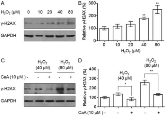
Oxidative DNA damage is the leading cause of decreased cell viability (13,14). We exposed L-02 cells to 0, 10, 20, 40, or 80mM H2O2 for 6 h and found
dose-dependent increases in the expression ofc-H2AX, which is a biomarker of DSBs (Figure 2A and B). We then evaluated the ability of CaA to protect against the DNA damage that resulted from H2O2treatment. After pretreating L-02 cells
with 0 or 10mM CaA for 24 h, they were exposed to 40 or 80mM of H2O2for 6 h. As shown in Figure 2C and D, CaA
attenuated the H2O2-induced increase inc-H2AX
expres-sion. These results indicate that a low concentration of CaA decreased H2O2-induced DSBs in L-02 cells.
CaA decreased ROS levels in L-02 cells
ROS have been implicated in a number of processes including cell proliferation, DNA damage, and apoptosis (9,11,14). At low levels, ROS modulate gene expression by acting as second messengers, but at high levels they cause oxidative injury leading to cell death (13,15). We hypothe-sized that the CaA-induced improvement of cell viability and protection against DNA damage following H2O2 treatment
were mediated by the generation of low levels of ROS. To confirm our hypothesis, L-02 cells were exposed to 10mM CaA for 0, 3, 6, 12, 24, or 48 h. As shown in Figure 3, the ROS levels induced by CaA were 112-136% compared with control cells (100%); however, in cells treated with 40mM H2O2, the relative ROS level was 236%. These results
suggest that compared with exposure to H2O2, CaA
generated relatively lower levels of ROS in L-02 cells.
CaA activated ERK signaling by ROS in L-02 cells The ERK pathway typically transduces growth factor signals that lead to cell differentiation or proliferation (7); however, the association of ERK with CaA-induced improvement of cell viability and subsequent protection against DNA damage is unclear. We exposed L-02 cells to
Figure 2.Caffeic acid (CaA) decreased H2O2-induced double-strand breads (DSBs) in L-02 cells.A,B, L-02 cells were exposed to 0, 10, 20, 40, or 80mM H2O2for 6 h.A, Western blot analysis and B, relativec-H2AX protein levels. **P,0.01 compared with control
cells.C,D, L-02 cells were pretreated with 0 or 10mM CaA for 24 h
and then exposed to 40 (lanes 2and3) or 80 (lanes 4and5)mM
H2O2for 6 h. C, Western blot analysis andD, relative c-H2AX
protein levels. *P,0.05 and **P,0.01 compared with L-02 cells treated with H2O2alone (Student’st-test). Bands were normalized to glyceraldehyde 3-phosphate dehydrogenase (GAPDH).
Figure 1. Caffeic acid (CaA) attenuated the H2O2-induced inhibition of L-02 cell viability.A, L-02 cells were treated with 0, 5, 10, 20, 40, 80, or 160mM CaA for 12, 24, or 48 h. B, After
pretreatment of L-02 cells with 0 or 10mM CaA
for 24 h, they were exposed to 0, 10, 20, 40, or 80mM H2O2for 24 h. Cell viability was measured by cell counting and comparison with control cells treated with culture medium only. *P,0.05 and **P,0.01 compared with L-02 cells treated only with H2O2(Student’st-test).
Figure 3.Caffeic acid (CaA) generated relatively lower levels of reactive oxygen species (ROS) in L-02 cells compared to cells treated only with H2O2. L-02 cells were treated with 10mM CaA for
0, 3, 6, 12, 24, or 48 h. Cells exposed to 40mM H2O2for 24 h served as positive controls. ROS levels were measured with the DCF fluorescence method. *P,0.05 compared to medium control cells; ##P
10mM CaA for 0, 3, 6, 12, or 24 h, and found that with increased time of CaA exposure, there was enhanced expression of p-ERK, a biomarker for the activation of ERK signaling (Figure 4A and B). Next, we investigated the mechanisms underlying CaA-induced activation of ERK signaling. L-02 cells were pretreated with 10 nM catalase, an H2O2scavenger, for 1 h and then exposed to 10mM
CaA for 24 h. As shown in Figure 4C-E, ROS scavenging by catalase attenuated both the CaA-induced generation of ROS and ERK activation. These results indicate that CaA generated a relatively low level of ROS in L-02 cells, which induced sustained activation of the ERK signal pathway. These results suggest that ERK played a role in the CaA-induced improvement of cell viability and protection against DNA damage that are associated with H2O2treatment.
Inhibition of ERK blocked CaA-mediated reduction of DSBs
We found evidence to support our hypothesis that ERK was involved in the CaA-mediated reduction of DSBs in H2O2-treated L-02 cells. Following pretreatment with 0 or
10mM CaA in the presence or absence of 10mM U0126 (an ERK inhibitor) for 24 h, cells were exposed to 80mM H2O2
for an additional 6 h. As shown in Figure 5A and B, CaA attenuated the H2O2-induced increase inc-H2AX expres-sion. However, inhibition of ERK by U0126 abolished this phenomenon. RNA interference confirmed the effect of ERK inhibition. In L-02 cells, knockdown of ERK1 and ERK2 by their specific siRNAs blocked the CaA-induced attenuation
of c-H2AX expression in response to H2O2 treatment
(Figure 5C and D). These results indicate that the ERK signaling pathway was involved in CaA-induced protection against DNA damage by H2O2treatment.
Inhibition of ERK blocked the CaA-induced improvement of cell viability in H2O2-treated L-02
cells
Finally, we demonstrated that ERK was involved in the CaA-induced improvement in the viability of cells treated with H2O2. L-02 cells were treated as described above for
24 h. As shown in Figure 6, CaA attenuated the H2O2
-induced decrease in cell viability; however, inhibition of ERK by U0126 or siRNA abolished this effect. These results indicate that the ERK signaling pathway was involved in the CaA-induced improvement of cell viability.
Discussion
Hydroxycinnamic acid derivatives are reported to have anticancer, anti-inflammatory, and antioxidant activities, and CaA is a well known hydroxycinnamic acid (16). Previous studies have demonstrated that daily coffee intake was associated with a reduced incidence of colon and rectal cancer. Michels et al. (17) reported that participants who regularly consumed two or more cups of decaffeinated coffee per day had a 52% lower incidence of rectal cancer than those who never consumed it. Tavani et al. (18) found that compared with coffee nondrinkers, the risk of colon Figure 4.Caffeic acid (CaA) activated extracellular signal-regulated kinase (ERK) signaling by reactive oxygen species (ROS) in L-02 cells.A,B, L-02 cells were treated with 10mM CaA for 0, 3, 6, 12, or 24 h.A, Western blots and (B) relative p-ERK protein levels.C-E,
L-02 cells were pretreated with 10 nM catalase for 1 h and then exposed to 10mM CaA for 24 h.C, ROS levels were measured with the DCF fluorescence method. The relative ROS ratios were determined by comparison with control cells.D, Western blots andE, relative p-ERK levels. *P,0.05 and **P,0.01 compared with controls;#P
,0.05 and##P
cancer was reduced in drinkers of four or more cups per day. In most coffee drinkers, the daily intake of CaA is 0.5-1 g (approximately 0.5-1 mM), and the absorption ratio of CaA is about 95%. Some of the CaA in food enters the circulation, but most passes into the colon (19), so here we used a relatively low concentration of CaA (10mM) to assess its ability to protect against DNA damage and maintain cell viability after H2O2treatment.
The present study employed L-02 cells because i) hepatocellular carcinoma, the most common liver malig-nancy, is a global health problem (20); ii) CaA protects against liver lesions and carcinogenesis in humans (21,22); andiii) the liver is thought to be the most important organ for CaA metabolism. Identification of the molecular mechanisms underlying the CaA-induced effects on cell viability and DNA damage would add to our understanding of the anti-oncogenetic effects of CaA.
Biphasic dose-response relationships have recently received considerable attention (23,24). They are character-ized by stimulation of chemical agents at low doses (hormesis) and inhibition at high doses (25). Hormesis, also known as oxidative stress adaptation, is an important mechanism by which cells and organisms respond to and cope with environmental and physiological shifts in oxidative stress levels (25). The accumulated evidence for hormesis of chemical agents derives from three different areas: cell proliferation or viability, DNA base excision repair, and telomerase activity (25-27). Here, we established that low levels of CaA act as a hormesis trigger to improve cell viability and protect against DNA damage caused by H2O2treatment.
ROS, such as superoxide anions, H2O2, and hydroxyl
radicals, are ubiquitous, highly reactive, diffusible mole-cules (16,28). It has long been recognized that ROS cause complex and irreversible damage to cellular constituents that impairs cellular homeostasis (15). Oxidative damage Figure 5. Extracellular signal-regulated kinase (ERK) inhibition blocked the caffeic acid (CaA)-induced reduction in double-strand breads (DSBs) in human L-02 cells.A,B, After pretreat-ment with 0 or 10mM CaA in the presence or
absence of U0126 (10mM) for 24 h, they were
exposed to 80mM H2O2 for 24 h. A, Western blots and B, relative c-H2AX protein levels.
**P,0.01 compared with L-02 cells treated with CaA plus H2O2. C, D, After L-02 cells were transfected with 20 nM ERK1-siRNA plus ERK2-siRNA for 12 h, they were treated with 0 or 10mM
CaA for 24 h, followed by exposure to 80mM
H2O2for an additional 24 h.C, Western blots and D, relative c-H2AX protein levels. **P,0.01
compared with L-02 cells treated by H2O2alone (one-way ANOVA followed by Dunnett’st-test).
Figure 6. Extracellular signal-regulated kinase (ERK) inhibition blocked the caffeic acid (CaA)-induced improvement of cell viability in H2O2-treated L-02 cells.A, After pretreatment with 0 or 10mM CaA in the presence or absence of U0126 (10mM) for
24 h, they were exposed to 80mM H2O2for an additional 24 h. Cell viability was measured by cell counting. Relative cell viability was determined by comparison with control cells. **P,0.01 compared with L-02 cells treated with H2O2alone.##P,0.01 compared with L-02 cells treated with CaA plus H2O2.B, After L-02 cells were transfected by 20 nM ERK1 plus ERK2 siRNA for 12 h, they were treated with 0 or 10mM CaA for 24 h, followed by exposure to
is related to the high reactivity of molecular oxygen and its intermediates, which can lead to oxidative modifications of proteins, lipids, and DNA (15). A role for oxidative damage to DNA in carcinogenesis is consistent with accumulating evidence that the rate of genome instability increases with age (4,14). Recent studies suggest that the enhanced ROS generation without cytotoxicity has a cellular protective effect (15,27). On the other hand, marked ROS formation causes oxidative stress and cellular damage (16,29). Here we found that compared with cells exposed to H2O2, a low
concentration of CaA-generated ROS may play a protective role in L-02 cells. These results are in line with previous findings that sodium arsenite acts as a hormesis trigger at low concentrations and induced enhanced ROS generation without cytotoxicity and had a cellular protective effect (15). ROS can trigger the activation of redox-sensitive signal transduction and MAPK pathways that regulate cellular mechanisms of cell survival, death, and immunity (9,30,31). MAPKs including ERK, p38 MAPK, and JNK are key components of signaling pathways that control cell differ-entiation and growth (9,30,31). There is evidence that MAPKs can be phosphorylated and activated in response to
oxidant-induced alterations of the redox state (7). After activation, each MAPK phosphorylates a distinct spectrum of substrates including key regulatory enzymes, cytoskele-tal proteins, regulators of apoptosis, nuclear receptors, and many transcription factors that bind to specific DNA sequences and induce transcriptional activation and DNA synthesis, with cellular recruitment to the S-phase (9,30,32). CaA regulates LPS-induced oxidative stress through c-Src/ ERK signaling pathways in endothelial cells (4). Notably, the ERK signal pathway is involved in the improvement of cell viability induced by a low concentration of sodium arsenite, a hormesis trigger (33). Here we found that a low concentra-tion of CaA induced sustained activaconcentra-tion of ERK signaling. Further, we confirmed that the CaA-induced activation of ERK was mediated by ROS generation. Based on these results, we hypothesized that ERK might play a role in CaA-induced improvement of cell viability and protection against DNA damage caused by H2O2treatment. To further
under-stand the role of the ERK pathway, we used U0126 and ERK1/2-siRNA to block ERK activation. Inhibition of ERK blocked the CaA-mediated reduction of DSBs and attenu-ated the CaA-induced improvement of cell viability asso-ciated with H2O2treatment.
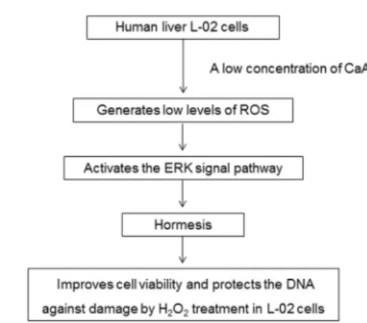
A low concentration of CaA increased the viability of human liver L-02 cells, attenuated the H2O2-associated
reduction of cell viability, and decreased the occurrence of H2O2-induced DSBs. Compared with cells exposed to
H2O2, CaA-treated cells generated lower levels of ROS that
induced ERK signaling pathway. Inhibition of ERK signaling blocked the CaA-induced improvement of cell viability and protection against DNA damage caused by H2O2treatment
(Figure 7).
Acknowledgments
This research was supported by the Natural Science Foundations of China (#81072338, #81473020, and
#81402667), a project funded by the Priority Academic Program Development of Jiangsu Higher Education Institutions (2010), and a Technology Development Fund of Nanjing Medical University (#2013NJMU021).
References
1. Kang NJ, Lee KW, Shin BJ, Jung SK, Hwang MK, Bode AM, et al. Caffeic acid, a phenolic phytochemical in coffee, directly inhibits Fyn kinase activity and UVB-induced COX-2 expression. Carcinogenesis 2009; 30: 321-330, doi: 10.1093/carcin/bgn282.
2. Karthikesan K, Pari L. Beneficial effect of caffeic Acid on alcohol-induced alterations in lipid peroxidation and antiox-idant defense in rats.Toxicol Mech Methods2007; 17: 527-534, doi: 10.1080/15376510701410476.
3. Khan AQ, Khan R, Qamar W, Lateef A, Ali F, Tahir M, et al. Caffeic acid attenuates 12-O-tetradecanoyl-phorbol-13-acetate (TPA)-induced NF-kappaB and COX-2 expression
in mouse skin: abrogation of oxidative stress, inflammatory responses and proinflammatory cytokine production.Food Chem Toxicol2012; 50: 175-183, doi: 10.1016/j.fct.2011. 10.043.
4. Kim SR, Jung YR, Kim DH, An HJ, Kim MK, Kim ND, et al. Caffeic acid regulates LPS-induced NF-kappaB activation through NIK/IKK and c-Src/ERK signaling pathways in endothelial cells.Arch Pharm Res2014; 37: 539-547, doi: 10.1007/s12272-013-0211-6.
5. Touaibia M, Jean-Francois J, Doiron J. Caffeic Acid, a versatile pharmacophore: an overview.Mini Rev Med Chem 2011; 11: 695-713, doi: 10.2174/138955711796268750. Figure 7.Caffeic acid (CaA)-induced improvement of cell viability
6. Pari L, Karthikesan K. Protective role of caffeic acid against alcohol-induced biochemical changes in rats. Fundam Clin Pharmacol 2007; 21: 355-361, doi: 10.1111/j.1472-8206. 2007.00505.x.
7. Ma Y, Jin Z, Huang J, Zhou S, Ye H, Jiang S, et al. IQGAP1 plays an important role in the cell proliferation of multiple myeloma via the MAP kinase (ERK) pathway. Oncol Rep 2013; 30: 3032-3038.
8. Li L, Eun JS, Nepal M, Ryu JH, Cho HK, Choi BY, et al. Isopsoralen induces differentiation of prechondrogenic ATDC5 cells via activation of MAP kinases and BMP-2 signaling pathways. Biomol Ther 2012; 20: 299-305, doi: 10.4062/biomolther.2012.20.3.299.
9. Sun Y, Tang S, Jin X, Zhang C, Zhao W, Xiao X. Opposite effects of JNK and p38 MAPK signaling pathways on furazolidone-stimulated S phase cell cycle arrest of human hepatoblastoma cell line.Mutat Res2013; 755: 24-29, doi: 10.1016/j.mrgentox.2013.04.015.
10. Yang Y, Li Y, Wang K, Wang Y, Yin W, Li L. P38/NF-kappaB/snail pathway is involved in caffeic acid-induced inhibition of cancer stem cells-like properties and migratory capacity in malignant human keratinocyte.PLoS One2013; 8: e58915, doi: 10.1371/journal.pone.0058915.
11. Hao C, Hao W, Wei X, Xing L, Jiang J, Shang L. The role of MAPK in the biphasic dose-response phenomenon induced by cadmium and mercury in HEK293 cells.Toxicol In Vitro 2009; 23: 660-666, doi: 10.1016/j.tiv.2009.03.005. 12. Byun MR, Kim AR, Hwang JH, Kim KM, Hwang ES, Hong
JH. FGF2 stimulates osteogenic differentiation through ERK induced TAZ expression. Bone2014; 58: 72-80, doi: 10. 1016/j.bone.2013.09.024.
13. Haberzettl P, Hill BG. Oxidized lipids activate autophagy in a JNK-dependent manner by stimulating the endoplasmic reticulum stress response.Redox Biol2013; 1: 56-64, doi: 10.1016/j.redox.2012.10.003.
14. Schumacher B. Transcription-blocking DNA damage in aging: a mechanism for hormesis. Bioessays 2009; 31: 1347-1356, doi: 10.1002/bies.200900107.
15. Yang P, He XQ, Peng L, Li AP, Wang XR, Zhou JW, et al. The role of oxidative stress in hormesis induced by sodium arsenite in human embryo lung fibroblast (HELF) cellular proliferation model. J Toxicol Environ Health A2007; 70: 976-983, doi: 10.1080/15287390701290832.
16. Chung KS, Han G, Kim BK, Kim HM, Yang JS, Ahn J, et al. A novel antitumor piperazine alkyl compound causes apoptosis by inducing RhoB expression via ROSmediated cAbl/p38 MAPK signaling. Cancer Chemother Pharmacol2013; 72: 1315-1324, doi: 10.1007/s00280-013-2310-y.
17. Michels KB, Willett WC, Fuchs CS, Giovannucci E. Coffee, tea, and caffeine consumption and incidence of colon and rectal cancer. J Natl Cancer Inst2005; 97: 282-292, doi: 10.1093/jnci/dji039.
18. Tavani A, Pregnolato A, La Vecchia C, Negri E, Talamini R, Franceschi S. Coffee and tea intake and risk of cancers of the colon and rectum: a study of 3,530 cases and 7,057 controls. Int J Cancer 1997; 73: 193-197, doi: 10.1002/(SICI)1097-0215(19971009)73:2,193::AID-IJC5.3.0.CO;2-R. 19. Olthof MR, Hollman PC, Katan MB. Chlorogenic acid
and caffeic acid are absorbed in humans.J Nutr2001; 131: 66-71.
20. Mun AR, Lee SJ, Kim GB, Kang HS, Kim JS, Kim SJ. Fluoxetine-induced apoptosis in hepatocellular carcinoma cells.Anticancer Res2013; 33: 3691-3697.
21. Liu Y, Flynn TJ, Ferguson MS, Hoagland EM, Yu LL. Effects of dietary phenolics and botanical extracts on hepatotoxicity-related endpoints in human and rat hepatoma cells and statistical models for prediction of hepatotoxicity.Food Chem Toxicol2011; 49: 1820-1827, doi: 10.1016/j.fct.2011.04.034. 22. Guerriero E, Sorice A, Capone F, Costantini S, Palladino P, D’ischia M, et al. Effects of lipoic acid, caffeic acid and a synthesized lipoyl-caffeic conjugate on human hepatoma cell lines.Molecules2011; 16: 6365-6377, doi: 10.3390/molecules 16086365.
23. Radak Z, Chung HY, Koltai E, Taylor AW, Goto S. Exercise, oxidative stress and hormesis.Ageing Res Rev2008; 7: 34-42, doi: 10.1016/j.arr.2007.04.004.
24. Wang L, Zou W, Zhong Y, An J, Zhang X, Wu M, et al. The hormesis effect of BDE-47 in HepG2 cells and the potential molecular mechanism.Toxicol Lett2012; 209: 193-201, doi: 10.1016/j.toxlet.2011.12.014.
25. Stebbing AR. Hormesis - the stimulation of growth by low levels of inhibitors.Sci Total Environ1982; 22: 213-234, doi: 10.1016/0048-9697(82)90066-3.
26. Radak Z, Chung HY, Goto S. Exercise and hormesis: oxidative stress-related adaptation for successful aging. Biogerontology 2005; 6: 71-75, doi: 10.1007/s10522-004-7386-7.
27. Kinoshita A, Wanibuchi H, Morimura K, Wei M, Shen J, Imaoka S, et al. Phenobarbital at low dose exerts hormesis in rat hepatocarcinogenesis by reducing oxidative DNA damage, altering cell proliferation, apoptosis and gene expression. Carcinogenesis 2003; 24: 1389-1399, doi: 10.1093/carcin/ bgg079.
28. Strengert M, Jennings R, Davanture S, Hayes P, Gabriel G, Knaus UG. Mucosal reactive oxygen species are required for antiviral response: role of Duox in influenza a virus infection. Antioxid Redox Signal2014; 20: 2695-2709, doi: 10.1089/ ars.2013.5353.
29. Ramyaa P, Padma VV. Ochratoxin-induced toxicity, oxida-tive stress and apoptosis ameliorated by quercetin--modula-tion by Nrf2.Food Chem Toxicol2013; 62: 205-216, doi: 10.1016/j.fct.2013.08.048.
30. Cai DT, Jin H, Xiong QX, Liu WG, Gao ZG, Gu GX, et al. ER stress and ASK1-JNK activation contribute to oridonin-induced apoptosis and growth inhibition in cultured human hepatoblastoma HuH-6 cells.Mol Cell Biochem2013; 379: 161-169, doi: 10.1007/s11010-013-1638-2.
31. Zhang H, Tan S, Wang J, Chen S, Quan J, Xian J, et al. Musashi2 modulates K562 leukemic cell proliferation and apoptosis involving the MAPK pathway.Exp Cell Res2014; 320: 119-127, doi: 10.1016/j.yexcr.2013.09.009.
32. Achiwa Y, Hasegawa K, Udagawa Y. Effect of ursolic acid on MAPK in cyclin D1 signaling and RING-type E3 ligase (SCF E3s) in two endometrial cancer cell lines.Nutr Cancer2013; 65: 1026-1033, doi: 10.1080/01635581.2013.810292. 33. He XQ, Chen R, Yang P, Li AP, Zhou JW, Liu QZ. Biphasic