Connexin domains relevant to
the chemical gating of gap
junction channels
Department of Pharmacology and Physiology, School of Medicine and Dentistry, University of Rochester, Rochester, NY 14642-8711, USA
C. Peracchia and X.G. Wang
Abstract
Most cells exchange ions and small metabolites via gap junction channels. These channels are made of two hemichannels (connexons), each formed by the radial arrangement of six connexin (Cx) proteins. Connexins span the bilayer four times (M1-M4) and have both amino-and carboxy-termini (NT, CT) at the cytoplasmic side of the mem-brane, forming two extracellular loops (E1, E2) and one inner (IL) loop. The channels are regulated by gates that close with cytosolic acidification (e.g., CO2 treatment) or increased calcium concentration, possibly via calmodulin activation. Although gap junction regulation is still unclear, connexin domains involved in gating are being de-fined. We have recently focused on the CO2 gating sensitivity of Cx32, Cx38 and various mutants and chimeras expressed in Xenopus oocytes and studied by double voltage clamp. Cx32 is weakly sensitive to CO2, whereas Cx38 is highly sensitive. A Cx32 chimera containing the second half of the inner loop (IL2) of Cx38 was as sensitive to CO2 as Cx38, indicating that this domain plays an important role. Deletion of CT by 84% did not affect CO2 sensitivity, but replacement of 5 arginines (R) with asparagines (N) at the beginning of CT (C1) greatly enhanced the CO2 sensitivity of Cx32. This suggests that whereas most of CT is irrelevant, positive charges of C1 maintain the CO2 sensitivity of Cx32 low. As a hypothesis we have proposed a model that involves charge interaction between negative residues of the beginning of IL (IL1) and positive residues of either C1 or IL2. Open and closed channels would result from IL1-C1 and IL1-IL2 interactions, respectively.
Correspondence
C. Peracchia
Department of Pharmacology and Physiology
School of Medicine and Dentistry University of Rochester 601 Elmwood Avenue Rochester, NY 14642-8711 USA
Fax: (716) 461-3259 E-mail:
Presented at the XI Annual Meeting of the Federação de Sociedades de Biologia Experimental, Caxambu, MG, Brasil, August 21-24, 1996.
Research supported by the National Institutes of Health (No. GM20113).
Received February 18, 1997 Accepted March 11, 1997
Key words
•Cell-cell interaction •Cell junctions •Gap junctions •Calcium •Calmodulin •Membrane channels
Introduction
In most tissues, cells in contact with each other exchange charged and neutral cytoso-lic molecules lower than 1 kDa in molecular mass. This exchange involves ions as well as small metabolites such as amino acids, nucle-otides, second messengers and high-energy compounds, and enables electrical and meta-bolic signals to spread widely among cell populations (reviewed in Ref. 1). This form
sei-zures (8), spreading depression (9), Chagas’ disease (10), among others.
Cell coupling is mediated by channels clustered at cell-cell contact domains known as gap junctions. Each channel is formed by the extracellular interaction of two hemichan-nels (connexons), and creates a hydrophilic pathway that spans the two apposed plasma membranes and a narrow extracellular space (gap). In turn, each connexon is an oligomer formed by the radial arrangement of six iden-tical proteins (connexins) that span the mem-brane thickness and insulate the hydrophilic pore from the lipid bilayer and the extracel-lular medium (reviewed in Ref. 11).
In recent years, structural studies have generated a portrait of gap junction architec-ture, channel framework and connexin to-pology. Studies on dye diffusion and meta-bolite exchange have defined the nature, size limit and charge characteristics of channel permeants, and the extent of metabolic co-operation among cells. Biochemistry and
molecular genetics have provided the means for identifying connexin sequences and for mapping secondary and tertiary structure. Electrophysiology, particularly double whole-cell patch clamp recording, the devel-opment of reliable channel expression sys-tems, and channel reconstitution in artificial membranes have paved the way for defining single channel attributes such as conduc-tance, gating kinetics, voltage dependence, subconductance and residual conductance states, etc., and for clarifying mechanisms of channel regulation and modulation (reviewed in Ref. 12). Nonetheless, crucial aspects of channel structure and regulation are still hy-pothetical.
In the absence of high resolution crystal-lographic information, the three-dimensional structure of connexins can only be guessed, and still unclear are the parameters that de-termine connexin-connexin interaction with-in and across junctional membranes, the structure of the channel lining, the molecular domains and the mechanisms involved in channel regulation and gating, the functional meaning of connexin diversity and differen-tial expression, the physiological conse-quences of heterotypic junction formation (junctions between cells expressing differ-ent connexins), the reason for multiple connexin expression in the same cell, etc.
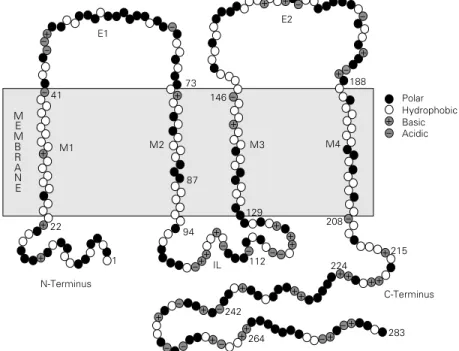
During the last decade at least 16 mem-bers of the connexin family have been cloned (reviewed in Ref. 12). Sequence analyses and studies using site-specific antibodies or selective proteolysis have defined connexin topology (13-22). Connexins span the bi-layer four times (M1-M4) and have both amino- and carboxy-termini (NT, CT) at the cytoplasmic side of the membrane, forming two extracellular loops (E1, E2) and one inner loop (IL) (Figure 1). Two connexin regions are conserved: one spans approxi-mately the first 100 residues, comprising NT (~23 residues), E1 (~35 residues), both M1 and M2 (~18 residues each), and the begin-ning of IL; the other contains M3 (18-20
Figure 1 - Model of connexin (Cx32) topology. The molecule is believed to span the bilayer four times (M1-M4) and to have both N- and C-termini (NT, CT) at the cytoplasmic side of the membrane, forming two extracellular loops (E1, E2) and one inner loop (IL). Two connexin regions are conserved: one spans approximately the first 100 residues, comprising NT, E1, M1, M2 and the beginning of IL; the other contains M3, M4, E2 and the beginning of CT. The two remaining regions, most of IL and CT, vary in sequence and length.
-Polar Hydrophobic Basic Acidic
- + - + +
+ -+
+ +
+ +
+ + +
++ + +
+ +
+ + +
+ + +
+
+
--
-283 264
242
224
215 112
94 IL 1
22
N-Terminus
C-Terminus E1
E2
+ +
+
+
-208 129
M4 M3
M2 87 M1
41 146
73 188
M E M B R A N E
+
+
-residues), M4 (~20 -residues), E2 (44-48 resi-dues), and the beginning of CT. The two remaining regions, most of IL and CT, vary in sequence and length. IL ranges from less than 30 residues (Cx31.1) to over 70 resi-dues (Cx45), and CT from 18 resiresi-dues (Cx26) to 188 residues (Cx56). M3 is believed to provide the channel lining structure, as it is the most amphiphilic of the four transmem-brane domains.
It is still unclear how connexins interact with each other within the membrane and across the gap. Recently, we have proposed a model that envisions a staggered (one-to-two) interaction between opposite connex-ins (12) (Figure 2). This model is based on the idea that each junctional membrane is unlikely to be a mirror-symmetrical image of the other, because connexins are believed to be identical rather than mirror-symmetrical images of their counterparts in a gap junc-tion. Thus, if M3 lines the channel and both E1 and E2 interact with homologous do-mains across the gap, a likely model would place E1 and E2 in a radial arrangement around the channel with their axes at ~30o
angle from each other (Figure 2). In this model, opposite connexins would not bind one-to-one but rather would be staggered with each other, such that each connexin of one membrane would interact with two con-nexins of the adjoined membrane. Indeed, a staggered (one-to-two) connexin interaction would provide a stronger junction than a matched (one-to-one) arrangement. There are two possible configurations of the stag-gered model: in one, both E1 and E2 would have the same N-to-C sequence orientation (Figure 2), centrifugal with respect to the channel, whereas in the other, only E2 would have this orientation (12).
Role of calcium and pH in gap junction channel gating
Functional gap junction channels are mostly in an open state, but can close in
response to certain changes in the ionic com-position of the cytosol. As a result of channel closure neighboring cells uncouple from each other electrically and metabolically. Although cell uncoupling is generally believed to be just a protective all-or-nothing mechanism, recent evidence for channel permeability regulation by nearly physiological changes in [Ca2+]
i (23-28) indicates that a fine
modu-lation of cell communication may play a role in normal cellular functions. For understand-ing how cell communication is modulated physiologically and how cell coupling regu-lation is linked to specific cellular activities we need to define the nature of the uncou-pling agents and the molecular basis of chan-nel gating. The latter can only be defined once we fully understand which connexin domains participate in the gating mechanism. Over the years a large body of evidence has emphasized the role of cytosolic calcium and hydrogen ions in cell coupling regula-tion. Evidence for gap junction channel sen-sitivity to internal calcium first surfaced in the mid-sixties through studies on insect gland cells (29), following an earlier observation
C
H
A
N
N
E
L
CT
NT
M2
E1
M2
NT
CT
IL
IL
Figure 2 - Model of staggered (one-to-two) interaction be-tween opposite connexins (12). Based on our present under-standing that M3 lines the chan-nel and both E1 and E2 interact with homologous domains across the gap, this model places E1 and E2 radially ar-ranged around the channel with
their axes at ~30o angle from
in cardiac myocytes (30). These findings were later confirmed in various cell systems (31-33). The role of H+, first proposed by
Turin and Warner (34,35) for amphibian embryonic cells, was later supported by Spray et al. (36) who proposed that the junctional conductance (Gj) of these cells is a simple
function of pHi. However, during the last
two decades a number of conflicting data on the role of calcium and pH have been re-ported (23,24,28,37-41), such that it is still unclear whether H+ and Ca2+ act
independ-ently from each other, and which of them regulates cell coupling under physiological circumstances.
Recently, we have tested in detail by double whole-cell patch clamp (DWCC) gap junction gating sensitivity to Ca2+ and H+ in
Novikoff hepatoma cell pairs internally buff-ered for Ca2+ with either EGTA or BAPTA,
as well as the effects on Gj of internal
solu-tions buffered to various pH values and [Ca2+]
(24). Novikoff cells express Cx43. The ef-fect of cytosolic acidification on Gj varied
depending on the Ca2+ buffer used. With
EGTA, CO2 had a large effect on Gj, whereas
with BAPTA it had virtually no effect. This observation suggested that Ca2+ mediates
the effect of low pHi on gap junctions, as
previously shown in crayfish axons (23), because the Ca2+-buffering efficiency of
EGTA is severely weakened by low pH, whereas that of BAPTA is only minimally affected. The Ca2+-EGTA affinity constant
drops by two orders of magnitude with a decrease in pH from 7 to 6, whereas that of Ca2+-BAPTA decreases only slightly with
the same pH drop.
For further testing this idea, we have monitored the single exponential decay of Gj
in cells buffered to different pCai and pHi
values. At pCai 6.9 or higher, Gj decreased
with a time constant (τ) of 28 min, whereas at pCai 6-6.3 Gj decreased with a τ of ~5 min.
A pCai of 5.5 resulted in fast uncoupling
with a τ of ~20 s. The same results were obtained at pHi 7.2 and at pHi 6.1 (24). These
data indicate that the channels of Novikoff cells are sensitive to nanomolar [Ca2+]
i and
are insensitive to pHi, at least in the range
7.2-6.1.
Recently, we have reevaluated the rela-tionship among pHi, pCai and Gj in Xenopus
oocyte pairs expressing Cx38 (28). Expo-sure to 100% CO2 for 3 min caused a rapid
drop of Gj, pHi and pCai (28). The time
course of Gj was close to that of pCai, but
contrasted sharply with that of pHi (Figure
3). This finding, also supported by the inhib-itory effect of intracellularly injected BAPTA (28), further confirmed the idea that junc-tional permeability is more closely related to [Ca2+]
i than to [H+]i. Low pHi appears to
increase [Ca2+]
i by releasing it from internal
stores, such as endoplasmic reticulum and/ or mitochondria, rather than by increasing Ca2+ entry (28,41). The lack of
correspon-dence between Gj and pHi is consistent with
data obtained in other cells (23,24,39,40). In crayfish axons (23,41), Novikoff cells (24,25) and oocytes (28), Ca2+ appears to
affect Gj at nanomolar concentrations. Over
the years, various [Ca2+]
i have been reported
to induce uncoupling. Only [Ca2+]
i as high as
40-400 µM was reported to be effective in ruptured (42) or internally perfused (43) cells, whereas low micromolar to high nanomolar concentrations were shown to induce gating in intact cells (23,32,44-49). This was re-cently confirmed in pancreatic ß-cells (26), and in Novikoff cells studied by dye cou-pling (27). A channel gating sensitivity to nearly physiological [Ca2+]
i does not conflict
with data for gap junction permeability to Ca2+ (50-52), because the gating mechanism
is relatively slow at near physiological [Ca2+] i
(24), and because the [Ca2+]
i required to
close all of the channels is in the high nM to low µM range (24), and thus above physi-ological values (70-200 nM).
Evidence for gap junction sensitivity to near physiological [Ca2+]
i (23-25,28)
phenom-Time (min)
0 4 8 12 16 20 24 28 120
100
80
60
40
20
0
Gj
and pH
i
(%, mean ± SEM)
120
100
80
60
40
20
0
Gj
and pCa
i
(%, mean ± SEM)
100% CO2
Gj (N = 16)
pCai (N = 25)
100% CO2
Gj (N = 16)
pHi (N = 18)
Time (min)
0 4 8 12 16 20 24 28
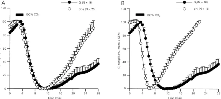
Figure 3 - Time course of changes in normalized pCai (A), pHi (B) and junctional conductance (Gj, A and B) in Xenopus oocyte pairs exposed to 100% CO2
for 3 min. pCai and pHi were measured at the oocyte periphery with fura-C18 (a membrane-associated Ca2+ indicator) and BCECF, respectively. Gj was
measured by double voltage clamp electrophysiology. Before CO2 exposure, the oocytes had a pCai of 6.66 ± 0.17 (mean ± SD; N = 25) and pHi of 7.63
± 0.115 (N = 18). With CO2, pCai dropped to 6.37 ± 0.263 (N = 25) at a maximum rate of ~23%/min (A). pCai minima were reached within 8-10 min and
pCai recovered to normal or slightly higher than normal values within ~15 min. In contrast, pHi dropped to 6.54 ± 0.113 (N = 18) at a maximum rate of
~34%/min (B). pHi minima were reached within ~4 min and pHi recovered to normal or slightly higher than normal values within ~10 min. The time
course of pHi contrasted sharply with that of Gj, which dropped at a maximum rate of ~25%/min and was lowest 8-10 min from the beginning of the
CO2 treatment (A and B), whereas the time course of Gj was very close to that of pCai during uncoupling. pCai minima preceded only slightly Gj minima,
but pCai recovered at a faster rate (A). From Ref. 28, with permission.
ena involving second messengers. Indeed, we have recently found that brief exposures to arachidonic acid uncouple Novikoff hep-atoma cells in a Ca2+-dependent manner,
whereas long exposures affect coupling in both Ca2+-dependent and Ca2+-independent
ways (25). Ca2+ participation was supported
by the exquisite sensitivity of the arachi-donic acid effect to [Ca2+]
i buffering (Figure
4). The absence of uncoupling in Ca2+-free
external solutions pointed to a role of Ca2+
entry in the uncoupling process (25).
Potential role of calmodulin in the uncoupling mechanism
In the early eighties, three independent observations suggested the existence of un-coupling intermediates. Johnston and Ramón (53) reported the inability of Ca2+ and H+ to
uncouple internally perfused crayfish axons. Peracchia et al. (54,55) suggested the par-ticipation of calmodulin (CaM) in the un-coupling mechanism, based on the ability of a CaM inhibitor (trifluoperazine) to prevent uncoupling in Xenopus embryonic cells. Hertzberg and Gilula (56) demonstrated the ability of CaM to bind to Cx32.
More recently, calmidazolium and W7, two more specific CaM blockers, inhibited uncoupling in various cells (57-61), and in-ternally perfused crayfish axons uncoupled with Ca2+ only in the presence of CaM (62).
CaM binding to Cx32 was further confirmed through gel overlay (13,63) and some evi-dence for CaM association with gap junction membranes was obtained by immunoelectron microscopy (64). In pairs of cardiac myo-cytes in which one cell was voltage clamped and Gj was measured after perforation of the
partner cell, gap junction sensitivity to Ca2+
increased from pCai 5.7 to pCai 7 upon
per-fusion with 10 µM CaM, and W7 (but not W5) prevented uncoupling (65).
To test more directly the participation of CaM in gating, we have studied CO2
-in-duced uncoupling in Xenopus oocytes in which CaM gene expression was inhibited (28). In oocytes injected with oligonucle-otides antisense to CaM mRNA, CaM mRNA was permanently degraded within 5 h, and the oocytes gradually lost junctional sensi-tivity to CO2 within 72 h. Uncoupling
com-petence recovered by ~35% following CaM injection. These data further confirm previ-ous evidence for CaM participation in cou-pling regulation (reviewed in Ref. 12).
CaM could affect coupling by directly binding to connexins or by activating CaM-dependent enzymes. Phosphorylation of Cx32 by Ca2+/CaM kinase II has been
re-ported, but only in isolated junctions (66). Phosphatases could also play a role. There is evidence that connexins can be phosphoryl-ated by various kinases (66,67) and that connexin phosphorylation decreases with Ca2+-induced uncoupling (27). Furthermore,
a difference in Cx43 phosphorylation has
been observed between communication-com-petent and -deficient cell lines (67), suggest-ing that phosphorylation may convert imper-meable hemichannels to perimper-meable cell-cell channels.
Connexin domains relevant for pH/Ca2+ gating
The molecular mechanism of CO2
-in-duced gating is still unknown, but data on the potential involvement of certain connexin domains are accumulating. The C-terminus domain has been suggested to play a role in determining the difference in CO2 gating
sensitivity between Cx43 and Cx32, because a Cx43 mutant missing over 80% of it de-creased in pH sensitivity to match that of Cx32 (40). Recently, the same group has proposed a ball-and-chain model for CO2
gating of Cx43 in which the carboxy-termi-nus (CT) end (the ball) would close the channel by binding to a receptor domain located somewhere else in Cx43. This model, similar to that proposed for K+ channels
(68,69), is based on provocative data show-ing that the reduced CO2 sensitivity of a
Cx43 mutant deleted at the CT end is
re-120 100 80 60 40 20
Gj
(%)
120 100 80 60 40 20 0
Gj
(%)
Time (min)
0 1 2 3 4 5 7 8
EGTA (mM) 2.0 1.0 0.5 0.1 0.0 AA
0 6
BAPTA (mM) 2.0 1.0 0.5 0.1 0.0
Time (min)
0 1 2 3 4 5 6 7 8 AA
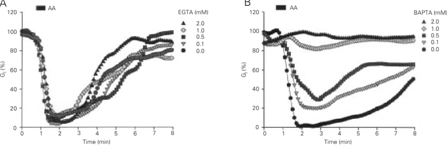
Figure 4 - Effect of arachidonic acid (AA) on electrical coupling studied in Novikoff cell pairs by double whole-cell clamp electrophysiology. The cytosol
was buffered for Ca2+ through the pipette solution with either BAPTA or EGTA. The uncoupling effect of AA (20 µM, 20 s) depends on [Ca2+]
i buffering.
EGTA at concentrations as high as 2 mM was totally ineffective in inhibiting uncoupling by AA (A). In contrast, BAPTA caused a 20% inhibition at concentrations as low as 0.1 mM and completely eliminated the uncoupling effects of AA at 1-2 mM concentrations (B). Indeed, BAPTA is known to be
a faster and more efficient intracellular Ca2+ buffer than EGTA. EGTA inhibited uncoupling by ~40% and ~80% at 5 and 10 mM concentrations,
respectively. From Ref. 25, with permission.
versed by coexpression of deleted Cx43 and the deleted CT end (70,71). However, the deleted Cx43 was not insensitive to acidifi-cation, but just less sensitive, a finding hard to explain if the postulated gating “ball” (CT end) is missing. Nonetheless, this model may only be relevant for Cx43, because a Cx32 mutant in which 84% of the CT had been deleted (D219) was as sensitive to CO2 as
wild-type Cx32 (72,73).
For defining connexin domain(s) of Cx32 participating in CO2-induced gating, we have
studied the gating sensitivities to CO2 of
Cx32, Cx38, and various chimeras and mu-tants of the above, expressed in Xenopus
oocytes (74). Cx32 is much less sensitive to CO2 than Cx38. Our data show that two
chimeras, Cx32/38I (Cx32 with an inner loop, IL, of Cx38) and Cx32/38I2 (Cx32 with the
second half of the inner loop, IL2, of Cx38),
are as sensitive to CO2 as Cx38 (74,75)
(Figure 5). This indicates that the second half of the inner loop plays an important role in pH gating sensitivity.
The mechanism by which IL2 plays a role
in CO2 gating sensitivity is still unclear. Spray
and Burt (76) have proposed that low-pH induced uncoupling follows protonation of H residues. An important role in determining the CO2 sensitivity of Cx43 has been
attrib-uted to H95 (77), a residue located at the N-terminus of IL in most connexins. Both Cx32 and Cx38 have an H residue at that location, but their neighboring residues are different and this could account for their difference in pH sensitivity. However, in view of our data on the relevance of IL2 (74), this residue may
not play a key role in determining the CO2
sensitivity of Cx32 and Cx38. More relevant to channel gating could be some of the H residues of IL2. Recently, Hermans et al.
(78) have provided preliminary evidence in-dicating that two H residues of Cx43 (H126 and H142) modulate in opposite ways the uncoupling effect of CO2. In Cx32, IL2
con-tains two H residues (H123 and H126). Al-though in preliminary experiments the
re-placement of H126 with R did not affect the CO2 sensitivity of Cx32 (75), a more
de-tailed evaluation of the potential role of these two residues is presently underway in our laboratory.
Since CT chimeras did not express func-tional channels, the potential role of CT could not be tested with chimeras, but inter-esting data were obtained with mutations of basic residues at its initial 18-residue seg-ment (C1) and with CT deletions. Although
much of the C-terminus of Cx32 seems not to play a significant role in CO2-gating
sensi-tivity, as 84% deletion of it at residue 219 (Cx32-D219) does not affect CO2 sensitivity
(72,73) (Figure 6), the C1 domain (Figure 7)
appears to have an inhibitory role. This is suggested by our recent data with mutants in which some or all of the positively charged residues (R) of C1 were replaced with neutral
(polar) residues (N or T) (73). Progressive replacement of R with N residues resulted in
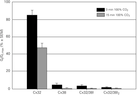
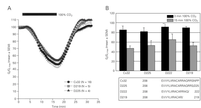
Figure 5 - Junctional sensitivity to CO2, expressed as normalized junctional conductance (Gj/
Gj max; 100% = control, pretreatment value), in oocyte pairs expressing Cx32, Cx38 or Cx32/
38 chimeras (74,75). With Cx38, a 3-min exposure to CO2 decreased Gj to nearly 0%,
whereas with Cx32, even a 15-min CO2 treatment decreased Gj by only ~55%. Two
chimeras, Cx32/38I (inner loop of Cx32 replaced by that of Cx38) and Cx32/38I2 (second half
of inner loop, IL2, of Cx32 replaced by that of Cx38), reproduced the uncoupling efficiency
of Cx38. This indicates that IL2 plays an important role in pH gating sensitivity. The
N-terminal domain does not appear to be relevant because the chimera Cx32/38N (Cx32 with NT of Cx38) behaved similarly to Cx32 (see Ref. 74).
Gj
/G
j max
(% ± SEM)
100
80
60
40
20
0
3 min 100% CO2
15 min 100% CO2
110 100 90
70 60 50 40
Gj
/G
j max
(mean ± SEM)
0 15 20 25 30 35 120
Time (min)
Cx32 D225 D222 D219 Cx32 208 EVVYLIIRACARRAQRRSNPP D225 208 EVVYLIIRACARRAQRRS/225 D222 208 EVVYLIIRACARRAQ/ 222 D219 208 EVVYLIIRACAR/ 219 100% CO2
Gj
/G
j max
(mean ± SEM)
100 80 60 40 20 0
3 min 100% CO2
15 min 100% CO2
Figure 6 - Decrease in junctional conductance (Gj) in Xenopus oocyte pairs, expressing wild-type Cx32 or Cx32 deleted of most of
the C-terminus, with exposure to 100% CO2 for either 15 min (A and B) or 3 min (B). Note that deletion of the C-terminus by over
80% (D225, D222, D219) did not affect CO2 sensitivity. With 3 min CO2, Gj dropped to 82 ± 8%, 91 ± 7% and 90 ± 3% (mean ±
SEM) with D225, D222 and D219, respectively, and with 15 min CO2, to 53.5 ± 10%, 65 ± 11% and 53 ± 7% with D225, D222 and
D219, respectively.
80
30
0 20 10
Gj
/G
j max
(mean ± SEM)
100
80
60
40
20
0
3 min 100% CO2
15 min 100% CO2
Cx32 4R/N 3R/N#1 2R/N 1R/N 3R/N#2 5R/N 5R/T D225-5R/N
Figure 7 - Summary of the effects of partial or total replacement of arginine (R) residues with asparagine
(N) or threonine (T) residues in the initial domain (C1) of
the C-terminus chain, on normalized Gj (Gj/Gj max; 100%
= control, pretreatment value), following 3-min or
15-min exposure to CO2. Note that replacement of all of
the 5 R with N or T residues greatly increased the CO2
sensitivity of Cx32, whereas partial R/N replacement
resulted in intermediate CO2 sensitivities. This
indi-cates that the R residues differ in their ability to inhibit
the CO2 sensitivity of Cx32. R215 appears to have
greater inhibitory power than R219-220. In contrast, R223-224 seems to partly counteract the inhibitory activity of both R215 and R219-220, because 2R/N and
1R/N were more sensitive to 15-min exposure to CO2
than 4R/N and 3R/N#1, respectively.
Cx32 (N = 16) D219 (N = 5) D225 (N = 4) 5 10
A
B
Cx32 215 RACARRAQRRSNPP
4R/N 215
R---NN--NN----3R/N#1 215
N---RR--NN----2R/N 215
R---NN--RR----1R/N 215
N---RR--RR----3R/N#2 215
N---NN--RR----5R/N 215
N---NN--NN----5R/T 215
a progressive increase in Cx32 sensitivity to CO2 (Figure 7). Interestingly, the 5 R
resi-dues were not all equally effective in inhibit-ing the Cx32 sensitivity to CO2. R215 has
greater inhibitory power than R219-220, whereas R223-224 seems to partly counter-act the inhibitory counter-activity of both R215 and R219-220. This is suggested by the fact that 2R/N and 1R/N were more sensitive to 15-min exposure to CO2 than 4R/N and 3R/
N#1, respectively (Figure 7).
A possible interpretation of these data is that the gating mechanism involves electro-static interactions among intracellular do-mains of Cx32. IL2 and C1 are positively
charged domains in all connexins, whereas IL1 is the only cytoplasmic domain that is
rich in negative charges. Although IL1
con-tains positive charges as well, being the most heavily charged domain of connexins, in α -helical conformation it would have positive and negative charges partitioned on opposite
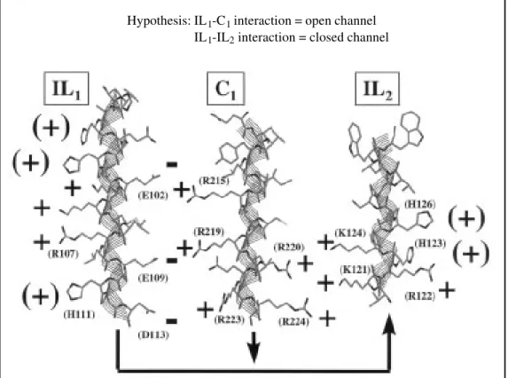
Figure 8 - Model of potential electrostatic interactions among
three cytoplasmic domains (IL1,
IL2 and C1) of Cx32, displayed in
alpha-helical conformation. In
view of the fact that 1) IL2 and
C1 are positively charged, 2) the
inhibitory action of C1 depends
on its positive charges, and 3) the only cytoplasmic domain
with negative charges is IL1 (not
considering some acidic resi-dues of the C-terminal domain that can be deleted without gat-ing consequences), we propose that open and closed channel states depend on charge
inter-actions among IL1, IL2 and C1.
In coupled conditions the
nega-tive charges of IL1 would be
un-available for interaction,
where-as with CO2 conformational
changes would expose them,
enabling IL2 and C1 to
competi-tively interact with IL1. IL1-IL2
interaction would result in
closed channel, whereas IL1-C1
interaction would maintain the channel open.
Hypothesis: IL1-C1 interaction = open channel
IL1-IL2 interaction = closed channel
sides of the helix (Figure 8); indeed, IL1, IL2
and C1 of Cx32 are likely to be α-helical and
IL may have a helix-loop-helix structure, in view of the presence of G (residues 110 and 112) and P (residue 114) residues at its mid-region.
As a working model, we are considering the possibility that in Cx32 under normal coupling conditions the negative charges of IL1 are masked or somehow unavailable for
electrostatic interaction with other domains. With CO2, conformational changes, brought
about by changes in the connexin phospho-rylation state, protonation of H residues, CaM binding, a combination of the above, or other as yet unknown factors, may expose them, allowing IL2 and C1 to competitively
interact with IL1. IL1-IL2 interaction would
result in a closed channel state, whereas IL1
-C1 interaction would maintain the channel in
de-pend on differences between IL2 and C1 in
binding affinity to IL1. In Cx32, one would
predict C1 to be a strong competitor of IL2,
whereas the opposite would be true for con-nexins more sensitive to CO2, such as Cx38,
Cx50, Cx45, etc. The gradual increase in Cx32 sensitivity to CO2 that follows the
progressive removal of positive charges in C1 would be the consequence of a gradual
decrease in the capacity of C1 and IL1 to
interact with each other. The increased CO2
sensitivity of the Cx32/38I2 chimera,
com-pared to wild-type Cx32, would indicate that IL2 of Cx38 has greater affinity for IL1 of
Cx32 than IL2 of Cx32. Therefore, IL2 of
Cx38 would compete more efficiently against the inhibitory domain (C1) for binding to IL1.
Although one should be well aware that based on the very limited amount of data this and any other potential model should not be valued more than working hypotheses, we feel that the potential participation of elec-trostatic interactions among connexin do-mains in the gating mechanism is worth care-ful study.
A puzzling question is the apparent con-tradiction between our data on Cx32 and those of Delmar’s group (40,70,71) on Cx43 regarding the relevance of CT in CO2 gating.
Whereas we found most of the CT of Cx32 to be irrelevant and its initial regions (C1) to act
as a gating inhibitor, Delmar’s group found
middle (residues 261-300) and end (residues 374-382) regions of the CT of Cx43 to be gating mediators (or activators); indeed, they have named the end of CT “the gating par-ticle” of the ball-and-chain model (71). Of course, it is quite possible that connexins are gated by different molecular mechanisms. On the other hand, we think that there might be a common denominator for Cx32 and Cx43 data. A possibility is that in Cx43 the negatively charged region of the “gating par-ticle” interacts with C1 (a basic-amphiphilic
domain, as in Cx32). By doing so, the “gat-ing particle” would eliminate the inhibitory function of C1, as it would prevent it from
interacting with IL1; its deletion would
re-duce CO2 sensitivity because C1 would then
be free to bind to IL1 and to act as inhibitor
domain by competing against IL2. Indeed,
acidic residues of the “gating particle” were found to be crucial for its function (71). The proline-glycine-rich mid-region of CT may provide the hinge that enables the “gating particle” to bend backward and bind to C1.
Based on this interpretation, the reason why coexpressing Cx32 with the “gating particle” of Cx43 increases the CO2 sensitivity of
Cx32 (70,71) would be that the “gating par-ticle” of Cx43 interacts electrostatically with C1 of Cx32 and blocks its inhibitory
func-tion. Note that in Cx32 the end of the CT chain does not contain sequences even re-motely similar to that of the “gating par-ticle”.
Does chemical gating require connexin cooperativity?
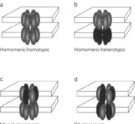
Coupling between cells expressing dif-ferent connexins has been demonstrated in many systems (1), indicating that a cell-cell channel can be homotypic (made of two connexons expressing the same connexin, Figure 9a) or heterotypic (made of two con-nexons each expressing a different connexin, Figure 9b) (1). In turn, connexons can be homomeric (made of the same connexin,
Figure 9 - Gap junction channels can be homotypic (made of two connexons expressing the same connexin) (a) or heterotypic (made of two connexons each expressing a different connexin) (b). Similarly, connexons can be homomeric (made of the same connexin) (a and b) or hetero-meric (composed of different connexins) (c and d). Therefore, cell-cell channels can be meric-homotypic (a), homo-meric-heterotypic (b), monohet-eromeric (one connexon hetero-meric and the other homohetero-meric) (c), or biheteromeric (both con-nexons heteromeric) (d).
Homomeric-homotypic Homomeric-heterotypic
Biheteromeric Monoheteromeric
a b
Figure 9a and b) or heteromeric (composed of different connexins, Figure 9c and d) (79). Therefore, cell-cell channels can be homo-meric-homotypic (Figure 9a), homomeric-heterotypic (Figure 9b), monoheteromeric (one connexon heteromeric and the other homomeric, Figure 9c), biheteromeric (both connexons heteromeric, Figure 9d), etc.
In view of this complexity, we have re-cently begun addressing questions on CO2
gating in heteromeric connexons and hetero-typic channels. We think that heteromeric connexons are an excellent tool for learning whether CO2 gating requires cooperativity
among the connexins of a connexon. Simi-larly, heterotypic channels can help deter-mining whether the two connexons of a cell-cell channel influence each other’s gating behavior. Our preliminary data indicate that connexin cooperativity within a connexon (hemichannel) may be necessary (80). If this were true, one would expect physiologically occurring heteromeric hemichannels to gate poorly, resulting in important functional con-sequences for tissue behavior.
We have tested oocyte pairs in which one oocyte expresses a 50/50 mixture of wild-type Cx32 and 5R/N mutant (mixed oocyte) and the other either wild-type Cx32 (32 oo-cyte) or 5R/N mutant (R/N oocyte), creating
mixed-32 and mixed-R/N pairs (both with monoheteromeric channels, Figure 9c). In-terestingly, these pairs were much less sensi-tive than 32-32 and R/N-R/N pairs, respec-tively (Figure 10). Since the 5R/N mutant is much more sensitive to CO2 than the
wild-type Cx32 (Figure 7), if connexins were gating independently from each other one would have expected the mixed-32 to be more sensitive than 32-32 and the mixed-R/ N to be only slightly less sensitive than R/N-R/N pairs. The presence of one or more 5R/ N in most hemichannels should have in-creased the gating sensitivity of the hetero-meric hemichannels.
Whereas connexin cooperativity within a connexon may be needed for efficient
gat-ing, cooperativity between two connexons forming a cell-cell channel may not be nec-essary. In our preliminary data, 32-R/N pairs (homomeric heterotypic, Figure 9b) were less sensitive than R/N-R/N pairs and more sensitive than 32-32 pairs (Figure 10) to a level predicted for independent hemichannel gating.
Conclusion
The chemical gating of gap junction chan-nels appears to be a complex phenomenon that involves more than one connexin do-main. Both an increase in cytosolic calcium concentration and a decrease in pHi appear
to initiate the cell-cell uncoupling process, but based on our data the effect of low pHi on
gap junction channels appears to be prima-rily mediated by an increase in cytosolic free calcium concentration. The chain of events that link the increase in calcium and/or hy-drogen ion concentration to the channel gat-ing mechanism is unclear, although indirect evidence suggests a role of calmodulin in the uncoupling process. At the molecular level, two connexin domains have been consid-ered important: the inner (cytoplasmic) loop and the carboxy-terminus chain. However, different functions have been attributed to the latter in Cx32 and in Cx43.
The absence of high resolution structural
Figure 10 - Sensitivity to CO2
presented as normalized
junc-tional conductance (Gj/Gj max;
100% = control, pretreatment value) in oocyte pairs expressing heteromeric or heterotypic chan-nels. Pairs in which one oocyte expressed a 50/50 mixture of
Cx32 and 5R/N mutant (mixed)
and the other either Cx32 (32) or
5R/N (R/N) were less sensitive
to CO2 than 32-32 and R/N-R/N
pairs, respectively. Their
sensi-tivity is consistent with the idea that in heteromeric hemichannels (mixed) gating is
impaired and suggests that gating may require connexin cooperativity. In contrast, the
sensitivity of heterotypic channels (32-R/N) was close to that theoretically predicted,
indicating that the two hemichannels of a cell-cell channel are likely to gate independently from each other.
Gj
/G
j max
(% ± SEM)
100 80 60 40 20 0
mixed-R/N mixed-32 32-R/N R/N-R/N
References
1. Bruzzone R, White TW & Paul DL (1996). Connections with connexins: The molec-ular basis of direct intercellmolec-ular signaling.
European Journal of Biochemistry, 238: 1-27.
2. Garfield RE & Hertzberg EL (1990). Cell-to-cell coupling in the myometrium:
Emil Bozlers prediction. Progress in
Clini-cal and BiologiClini-cal Research, 327: 673-682. 3. Bergoffen J, Scherer SS, Wang S, Scott MO, Bone LJ, Paul DL, Chen K, Lensch MW, Chance PF & Fischbeck KH (1993). Connexin mutations in X-linked
Char-cot-Marie-Tooth disease. Science, 262:
2039-2042.
4. Ionasescu V, Searby C & Ionasescu R (1994). Point mutations of the connexin32 (GJB1) gene in X-linked dominant
Char-cot-Marie-Tooth neuropathy. Human
Mo-lecular Genetics, 3: 355-358.
5. Fairweather N, Bell C, Cochrane S, Chelly J, Wang S, Mostacciuolo ML, Monaco AP & Haites NE (1994). Mutations in the connexin 32 gene in X-linked dominant Charcot-Marie-Tooth disease (CMTX1).
Human Molecular Genetics, 3: 29-34. 6. Spray DC (1994). CMTX1: A gap junction
genetic disease. Lancet, 343: 1111-1112.
7. Britz-Cunningham SH, Shah MM, Zuppan CW & Fletcher WH (1995). Mutations of the connexin43 gap-junction gene in pa-tients with heart malformations and
de-fects of laterality. New England Journal of
Medicine, 332: 1323-1329.
8. Perez-Velazquez JL, Valiante TA & Carlen PL (1994). Modulation of gap junctional mechanisms during calcium-free induced field burst activity: A possible role for
elec-trotonic coupling in epileptogenesis.
Jour-nal of Neuroscience, 14: 4308-4317. 9. Nedergaard M, Cooper AJL & Goldman
SA (1995). Gap junctions are required for the propagation of spreading depression.
Journal of Neurobiology, 28: 433-444.
10. Campos de Carvalho AC, Tanowitz HB, Wittner M, Dermietzel R, Roy C, Hertzberg EL & Spray DC (1992). Gap junction distribution is altered between
cardiac myocytes infected with
Trypano-soma cruzi. Circulation Research, 70: 733-742.
11. Peracchia C (1980). Structural correlates
of gap junction permeation. International
Review of Cytology, 66: 81-146. 12. Peracchia C, Lazrak A & Peracchia LL
(1994). Molecular models of channel in-teraction and gating in gap junctions. In:
Peracchia C (Editor), Handbook of
Mem-brane Channels. Molecular and Cellular Physiology. Academic Press, San Diego, 361-377.
13. Zimmer DB, Green CR, Evans WH & Gilula NB (1987). Topological analysis of the major protein in isolated intact rat liver gap junctions and gap junction-derived
single membrane structures. Journal of
Biological Chemistry, 262: 7751-7763. 14. Hertzberg EL, Disher RM, Tiller AA, Zhou
Y & Cook R (1988). Topology of the Mr
27,000 liver gap junction protein. Journal
of Biological Chemistry, 263: 19105-19111.
15. Milks LC, Kumar NM, Houghten R, Unwin N & Gilula NB (1988). Topology of the 32-kd liver gap junction protein deter-mined by site-directed antibody
localiza-tions. EMBO Journal, 7: 2967-2975.
16. Goodenough DA, Paul DL & Jesaitis L (1988). Topological distribution of two connexin32 antigenic sites in intact and split rodent hepatocyte gap junctions.
Journal of Cell Biology, 107: 1817-1824. 17. Yancey SB, John SA, Lal R, Austin BJ &
Revel J-P (1989). The 43-kD polypeptide of heart gap junctions: Immunolocaliza-tion, topology, and functional domains.
Journal of Cell Biology, 108: 2241-2254.
18. Laird DW & Revel J-P (1990). Biochemical and immunochemical analysis of the ar-rangement of connexin43 in rat heart gap
junction membranes. Journal of Cell
Sci-ence, 97: 109-117.
19. Meyer RA, Laird DW, Revel JP & Johnson RG (1992). Inhibition of gap junction and adherent junction assembly by connexin
and A-CAM antibodies. Journal of Cell
Bi-ology, 119: 179-189.
20. Yeager M & Gilula NB (1992). Membrane topology and quaternary structure of
car-diac gap junction ion channels. Journal of
Molecular Biology, 223: 929-948. 21. Zhang J-T & Nicholson BJ (1994). The
topological structure of connexin 26 and its distribution compared to connexin 32
in hepatic gap junctions. Journal of
Mem-brane Biology, 139: 15-29.
22. Leube RE (1995). The topogenic fate of the polytopic transmembrane proteins, synaptophysin and connexin, is deter-mined by their membrane-spanning
do-mains. Journal of Cell Science, 108:
883-894.
23. Peracchia C (1990). Increase in gap junc-tion resistance with acidificajunc-tion in cray-fish septate axons is closely related to changes in intracellular calcium but not
hydrogen ion concentration. Journal of
Membrane Biology, 113: 75-92. 24. Lazrak A & Peracchia C (1993). Gap
junc-tion gating sensitivity to physiological in-ternal calcium regardless of pH in Novikoff
hepatoma cells. Biophysical Journal, 65:
2002-2012.
25. Lazrak A, Peres A, Giovannardi S & Peracchia C (1994). Ca-mediated and in-dependent effects of arachidonic acid on gap junctions and Ca-independent effects
of oleic acid and halothane. Biophysical
Journal, 67: 1052-1059.
information is the major handicap for under-standing channel gating mechanisms at the molecular level. Nonetheless, important in-formation on the gating process will con-tinue to accumulate through connexin chi-meras and mutants. A thorough study of relevant connexin domains based on molec-ular genetics and biophysics, coupled to care-ful comparisons of connexin sequences and gating behaviors, should provide a good
un-derstanding of the molecular mechanism of channel gating even in the absence of a detailed portrait of the three-dimensional ar-chitecture of connexin.
Acknowledgment
26. Mears D, Sheppard Jr NF, Atwater I & Rojas E (1995). Magnitude and modula-tion of pancreatic ß-cell gap juncmodula-tion
elec-trical conductance in situ. Journal of
Mem-brane Biology, 146: 163-176.
27. Crow JM, Atkinson MM & Johnson RG (1994). Micromolar levels of intracellular calcium reduce gap junctional
permeabil-ity in lens cultures. Investigative
Ophthal-mology and Visual Science, 35: 3332-3341.
28. Peracchia C, Wang XG, Li LQ & Peracchia LL (1996). Inhibition of calmodulin expres-sion prevents low-pH-induced gap
junc-tion uncoupling in Xenopus oocytes.
Pflügers Archiv, 431: 379-387.
29. Loewenstein WR (1966). Permeability of
membrane junctions. Annals of the New
York Academy of Sciences, 137: 441-472. 30. Délèze J (1965). Calcium ions and the healing-over in heart fibers. In: Taccardi T
& Marchetti C (Editors),
Electrophysiol-ogy of the Heart. Pergamon Press, Elms-ford, New York, 147-148.
31. Délèze J & Loewenstein WR (1976). Per-meability of a cell junction during
intracel-lular injection of divalent cations. Journal
of Membrane Biology, 28: 71-86. 32. Rose B & Loewenstein WR (1976).
Per-meability of a cell junction and the local cytoplasmic free ionized calcium
concen-tration: a study with aequorin. Journal of
Membrane Biology, 28: 87-119. 33. De Mello WC (1975). Effect of
intracellu-lar injection of calcium and strontium on
cell communication in heart. Journal of
Physiology, 250: 231-245.
34. Turin L & Warner AE (1977). Carbon diox-ide reversibly abolishes ionic communica-tion between cells of early amphibian
embryo. Nature, 270: 56-57.
35. Turin L & Warner AE (1980). Intracellular
pH in early Xenopus embryos: its effect
on current flow between blastomeres.
Journal of Physiology, 300: 489-504. 36. Spray DC, Harris AL & Bennett MV (1981).
Gap junctional conductance is a simple and sensitive function of intracellular pH.
Science, 211: 712-715.
37. Ramón F & Rivera A (1987). Gap junction channel modulation: a physiological
view-point. Progress in Biophysics and
Molec-ular Biology, 48: 127-153.
38. Peracchia C (1987). Permeability and regu-lation of gap junction channels in cells and in artificial lipid bilayers. In: De Mello WC
(Editor), Cell-to-Cell Communication.
Ple-num Press, New York, 65-102.
39. Pressler ML (1989). Intracellular pH and cell-to-cell transmission in sheep Purkinje
fibers. Biophysical Journal, 55: 53-65.
40. Liu S, Taffet S, Stoner L, Delmar M, Vallano ML & Jalife J (1993). A structural basis for the unequal sensitivity of the major cardiac and liver gap junctions to intracellular acidification: The carboxyl tail
length. Biophysical Journal, 64:
1422-1433.
41. Peracchia C (1990). Effects of caffeine
and ryanodine on low pHi-induced
changes in gap junction conductance and calcium concentration in crayfish septate
axons. Journal of Membrane Biology, 117:
79-89.
42. Oliveira-Castro GM & Loewenstein WR (1971). Junctional membrane
permeabil-ity: effects of divalent cations. Journal of
Membrane Biology, 5: 51-77.
43. Spray DC, Stern JH, Harris AL & Bennett MV (1982). Gap junctional conductance: comparison of sensitivities to H and Ca
ions. Proceedings of the National
Acade-my of Sciences, USA, 79: 441-445. 44. Weingart R (1977). Action of ouabain on
intercellular coupling and conduction-ve-locity in mammalian ventricular muscle.
Journal of Physiology, 264: 341-365. 45. Dahl G & Isenberg G (1980). Decoupling
of heart muscle cells: correlation with in-creased cytoplasmic calcium activity and with changes of nexus ultrastructure.
Journal of Membrane Biology, 53: 63-75. 46. Neyton J & Trautmann A (1986). Physi-ological modulation of gap junction
per-meability. Journal of Experimental
Biol-ogy, 124: 93-114.
47. Maurer P & Weingart R (1987). Cell pairs isolated from adult guinea pig and rat
hearts: effects of [Ca2+]
i on nexal
mem-brane resistance. Pflügers Archiv, 409:
394-402.
48. Noma A & Tsuboi N (1987). Dependence of junctional conductance on proton, cal-cium and magnesium ions in cardiac
paired cells of guinea-pig. Journal of
Phys-iology, 382: 193-211.
49. Veenstra RD & DeHaan RL (1988). Car-diac gap junction channel activity in
em-bryonic chick ventricle cells. American
Journal of Physiology, 254: H170-H180. 50. Christ GJ, Moreno AP, Melman A & Spray
DC (1992). Gap junction-mediated
inter-cellular diffusion of Ca2+ in cultured
hu-man corporal smooth muscle cells.
Ameri-can Journal of Physiology, 263: C373-C383.
51. Brehm P, Lechleiter J, Smith S & Dunlap K (1989). Intercellular signaling as visual-ized by endogenous calcium-dependent
bioluminescence. Neuron, 3: 191-198.
52. Dunlap K, Takeda K & Brehm P (1987). Activation of a calcium-dependent photoprotein by chemical signaling
through gap junctions. Nature, 325: 60-62.
53. Johnston MF & Ramón F (1981). Electro-tonic coupling in internally perfused
cray-fish segmented axons. Journal of
Physiol-ogy, 317: 509-518.
54. Peracchia C, Bernardini G & Peracchia LL (1981). A calmodulin inhibitor prevents gap junction crystallization and electrical
uncoupling. Journal of Cell Biology, 91:
124 (Abstract).
55. Peracchia C, Bernardini G & Peracchia LL (1983). Is calmodulin involved in the regu-lation of gap junction permeability?
Pflügers Archiv, 399: 152-154.
56. Hertzberg EL & Gilula NB (1981). Liver gap junctions and lens fiber junctions: comparative analysis and calmodulin
in-teraction. Cold Spring Harbor Symposia
on Quantitative Biology, 46: 639-645. 57. Peracchia C (1984). Communicating
junc-tions and calmodulin: inhibition of
electri-cal uncoupling in Xenopus embryo by
calmidazolium. Journal of Membrane
Bi-ology, 81: 49-58.
58. Peracchia C (1987). Calmodulin-like pro-teins and communicating junctions. Elec-trical uncoupling of crayfish septate axons is inhibited by the calmodulin inhibitor W7 and is not affected by cyclic nucleotides.
Pflügers Archiv, 408: 379-385.
59. Wojtczak JA (1985). Electrical uncoupling induced by general anesthetics: a calci-um-independent process? In: Bennett
MVL & Spray DC (Editors), Gap Junctions.
Cold Spring Harbor Laboratory, Cold Spring Harbor, New York, 167-175. 60. Tuganowski W, Korczynska I, Wasik K &
Piatek G (1989). Effects of calmidazolium and dibutyryl cyclic AMP on the longitudi-nal interlongitudi-nal resistance in sinus node strips.
Pflügers Archiv, 414: 351-353.
61. Gandolfi SA, Duncan G, Tomlinson J & Maraini G (1990). Mammalian lens inter-fiber resistance is modulated by
cal-cium and calmodulin. Current Eye
Re-search, 9: 533-541.
62. Arellano RO, Ramón F, Rivera A & Zampighi GA (1988). Calmodulin acts as an intermediary for the effects of calcium on gap junctions from crayfish lateral
axons. Journal of Membrane Biology, 101:
119-131.
63. Van Eldik LJ, Hertzberg EL, Berdan RC & Gilula NB (1985). Interaction of calmodu-lin and other calcium-modulated proteins with mammalian and arthropod junctional
membrane proteins. Biochemical and
64. Fujimoto K, Araki N, Ogawa K-S, Kondo S, Kitaoka T & Ogawa K (1989). Ultracyto-chemistry of calmodulin binding sites in myocardial cells by staining of frozen thin sections with colloidal gold-labeled
cal-modulin. Journal of Histochemistry and
Cytochemistry, 37: 249-256.
65. Toyama J, Sugiura H, Kamiya K, Kodama I, Terasawa M & Hidaka H (1994).
Ca2+-calmodulin mediated modulation of
the electrical coupling of ventricular
myo-cytes isolated from guinea pig heart.
Jour-nal of Molecular and Cellular Cardiology,
26: 1007-1015.
66. Sáez JC, Nairn AC, Czernik AJ, Spray DC, Hertzberg EL, Greengard P & Bennett MVL (1990). Phosphorylation of connexin 32, a hepatocyte gap-junction protein, by cAMP-dependent protein kinase, protein
kinase C and Ca2+/calmodulin-dependent
protein kinase II. European Journal of
Bio-chemistry, 192: 263-273.
67. Musil LS, Cunningham BA, Edelman GM & Goodenough DA (1990). Differential phosphorylation of the gap junction pro-tein connexin43 in junctional communica-tion-competent and -deficient cell lines.
Journal of Cell Biology, 111: 2077-2088. 68. Armstrong CM (1966). Time course of
TEA+-induced anomalous rectification in
squid giant axons. Journal of General
Physiology, 50: 491-503.
69. Zagotta WN, Hoshi T & Aldrich RW (1990). Restoration of inactivation in mutants of
Shaker potassium channels by a peptide
derived from ShB. Science, 250: 568-571.
70. Morley GE, Taffet SM & Delmar M (1996). Intramolecular interactions mediate pH
regulation of connexin43 channels.
Bio-physical Journal, 70: 1294-1302. 71. Ek-Vitorin JF, Calero G, Morley GE,
Coombs W, Taffet SM & Delmar M (1996). pH regulation of connexin43:
mo-lecular analysis of the gating particle.
Bio-physical Journal, 71: 1273-1284. 72. Werner R, Levine E, Rabadan-Diehl C &
Dahl G (1991). Gating properties of connexin32 cell-cell channels and their
mutants expressed in Xenopus oocytes.
Proceedings of the Royal Society of Lon-don, Series B: Biological Sciences, 243: 5-11.
73. Wang XG & Peracchia C (1996). Molecu-lar domains relevant for chemical gating in gap junction channels made of
con-nexin32. Molecular Biology of the Cell, 7:
462 (Abstract).
74. Wang XG, Li LQ, Peracchia LL & Peracchia C (1996). Chimeric evidence for a role of the connexin cytoplasmic loop in gap
junc-tion channel gating. Pflügers Archiv, 431:
844-852.
75. Wang XG & Peracchia C (1996). Con-nexin32/38 chimeras suggest a role for the second half of the inner loop in gap
junction gating by low pH. American
Jour-nal of Physiology, 271: C1743-C1749.
76. Spray DC & Burt JM (1990). Struc-ture-activity relations of the cardiac gap
junction channel. American Journal of
Physiology, 258: C195-C205.
77. Ek JF, Delmar M, Perzova R & Taffet SM (1994). Role of histidine 95 on pH gating of the cardiac gap junction protein
con-nexin43. Circulation Research, 74:
1058-1064.
78. Hermans MMP, Kortekaas P, Jongsma HJ & Rook MB (1996). The role of histidine residues on pH sensitivity of the gap
junc-tion protein connexin 43. Molecular
Biol-ogy of the Cell, 7: 91 (Abstract). 79. Brink PR, Cronin K, Petersen-Grine E,
Beyer EC, Veenstra RD, Varadaraj K & Christ GJ (1996). Evidence for hetero-meric forms in cell systems expressing both Cx37 and Cx43: comparison with
homotypic Cx37 and Cx43. Proceedings
of the Keystone Symposium on Molecu-lar Approaches to the Function of Inter-cellular Junctions. Lake Tahoe, CA, March 1-7, 17 (Abstract).
80. Wang XG & Peracchia C (1997). Is con-nexin cooperativity necessary for
chemi-cal gating of gap junction channels?