645
Brazilian Journal of otorhinolaryngology 79 (5) SeptemBer/octoBer 2013 http://www.bjorl.org / e-mail: [email protected]
Small cell lung carcinoma metastasis to palatine tonsils
Helena Hotz Arroyo
1, Jefferson Takehara
2, Allex Itar Ogawa
3, Ronaldo Frizzarini
4, Rui Imamura
4,
Henrique Moura de Paula
51 MD, (Fellowship, Facial Plasic Surgery, Otorhinolaryngology Department, School of Medicine of the University of São Paulo, Brazil). 2 MD (Preceptor, Otorhinolaryngology Program, School of Medicine of the University of São Paulo, Brazil).
3 MD (Assistant Physician, Otorhinolaryngology-Oncology Program, São Paulo State Cancer Insitute, Brazil). 4 MD (Assistant Physician, Otorhinolaryngology Program, School of Medicine of the University of São Paulo, Brazil).
5 MD, Pathologist (Preceptor, Pathology Division, School of Medicine of the University of São Paulo, Brazil).
School of Medicine of the University of São Paulo, Brazil.
Send correspondence to: Helena Hotz Arroyo Rua Oscar Freire, nº 2040, Ap 65. São Paulo - SP. Brazil. CEP: 05409-011. E-mail: [email protected]
Paper submited to the BJORL-SGP (Publishing Management System - Brazilian Journal of Otorhinolaryngology) on June 11, 2012; and accepted on August 30, 2012. cod. 9259.
CASE REPORT
Braz J Otorhinolaryngol. 2013;79(5):645.
BJORL
Keywords: lymphatic metastasis; oropharynx; palatine tonsil; small cell lung carcinoma; tonsillar neoplasms.
.org
INTRODUCTION
Small-cell lung carcinomas (SCLC) are aggressive tumors1 which rarely metastasize to
the palatine tonsils2,3. Dissemination of disease
to the tonsils seems to occur after antegrade movements of tumor cells through the neck lymphatic system1. The reasons that led the
authors to report this case were the rarity of the occurrence in itself and the importance of carrying out thorough patient physical examination.
CASE REPORT
A 64-year-old male individual came to our service complaining of dysphagia and odynophagia evolving for six months and a lesion in his left oropharynx. He had had dysphonia for two months and mild dyspnea while performing physical effort. The subject claimed antibiotics failed to improve his condition. He had been a smoker for 50 years.
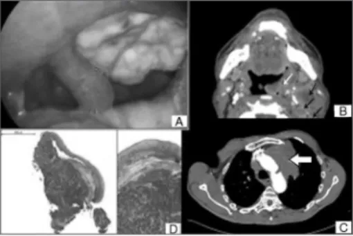
Physical examination revealed an ulcerated lesion of approximately four centimeters covered with fibrin in the left tonsillar bed (Figure 1A). Laryngoscopy showed he had left vocal fold paresis. Neck palpation showed coalescent nodes on levels II and III on the left side.
Neck CT scans revealed a tumor with heterogeneous contrast uptake infiltrating the left oropharynx and extending into the soft palate, associated with clustering nodes on levels IIA/IIB, with liquefied areas and signs of capsular leakage; other similar but smaller nodes were seen on levels III, IV, V, and VI (Figure 1B).
Chest CT scans showed lung emphysema and a tumor in the left upper lobe suggestive of lung carcinoma, in addition to en-larged mediastinal and bilateral supraclavicular lymph nodes (Figure 1C).
Pathology tests indicated the tonsillar tumor was a poorly differentiated carcinoma invading the squamous mucosa (Figure 1D). Immunohistochemistry (IHC) results were consistent with SCLC (CD45-negative, CD56-pos-itive, cytokeratin 35BH11-posCD56-pos-itive, cytokeratin 5-positive, protein p63-negative, TTF1-negative, cromogranine-negative, synaptophysin-nega-tive, ki67-positive).
Endobronchial biopsy indicated the patient had SCLC, as confirmed by IHC.
The patient died before the beginning of radiation and chemotherapy.
DISCUSSION
Small-cell lung carcinoma is a relatively common type of lung carcinoma, accounting for 15-25% of the cases1-3. It is considered to be
an aggressive tumor, as it involves a significant portion of the lung. Metastasis occurs early on and involves the liver, abdominal lymph nodes, bones, the brain, the adrenal gland, the skin, the kidneys, and the pancreas. Palatine tonsillar involvement is rare1,4.
Palatine tonsillar metastasis originating from other primary tumors account for a small portion of tonsillar tumors (0.8%), and has been more frequently associated with breast, lung, kidney, testicular, skin, and rectal tumors1,5,6.
Such pattern of disease spread may stem from antegrade movements of tumor cells through neck lymph nodes, the thoracic duct, and neck veins into the tonsils1.
A review covering 76 cases of tonsillar metastasis showed that ten of the 12 patients with lung cancer in the series had SCLC. In ten of the 12 cases there was evidence of metastasis in other tissues5.
Yaren et al.2 and Hisa et al.3 reported
one case each of tonsillar metastasis by SCLC after treatment with chemoradiotherapy.
Mastronikolis et al.6 reported cases of tonsillar
tumors with metastasis to the liver, spleen, and ribs. Seddon et al.1 reported a case of tonsillar
metastasis in a patient with SCLC and exuberant pulmonary symptoms.
Unlike other cases, our patient’s ENT complaints were more marked than the pulmonary symptoms as a result of the tonsillar metastasis, which accounted for the disease’s early manifestations.
Despite the poor prognosis usually seen for subjects with SCLC, late diagnosis may impact the chances of managing the disease. In the described case, the patient had been treat-ed with antibiotics for having signs of disease consistent with tonsillitis. Accurate diagnosis was delayed because the patient was subject to inadequate interviews, lack of follow-up, and disregard for the possibility of his disease being cancer.
CLOSING REMARKS
Given the simple nature of oropharynx examination and how often tumors in this area produce symptoms, careful oral examination must be performed even in patient with tumors in other sites. Tumors must be considered in patients with ulcerated lesions refractory to clinical management to expedite diagnosis and medical intervention.
REFERENCES
1. Seddon DJ. Tonsillar metastasis at presentation of small cell carcinoma of the lung. J R Soc Med. 1989;82(11):688. PMID: 2556580
2. Yaren A, Degirmencioglu S, Topsakal Ş, Yüksel S, Bir F, Çoban Ş. et al. Tonsillar metastasis from small cell lung cancer: Rare but occurs. Turk J Cancer. 2009;39(1):28-30.
3. Hisa Y, Yasuda N, Murakami M. Small cell carci-noma of the lung metastatic to the palatine tonsil. Otolaryngol Head Neck Surg. 1997;116(4):563-4. PMID: 9141413 DOI: http://dx.doi.org/10.1016/ S0194-5998(97)70313-9
4. Harper PG. Staging of small cell carcinoma. In: Spiro SG, ed. Clinics in oncology 4. London: WB Saunders; 1985. p.85-103.
5. Brownson RJ, Jaques WE, Lamonte SE, Zollinger WK. Hypernephroma metastatic to the palatine tonsils. Ann Otol Rhinol Laryngol. 1979;88(2 Pt 1):235-40.
6. Mastronikolis NS, Tsiropoulos GE, Chorianopou-los D, Liava AC, Stathas T, Papadas TA. Palatine tonsilar metastasis from lung adenocarcinoma. Eur Rev Med Pharmacol Sci. 2007;11(5):279-82.
DOI: 10.5935/1808-8694.20130117