UNIVERSIDADE FEDERAL DO CEARÁ
FACULDADE DE FARMÁCIA, ODONTOLOGIA E ENFERMAGEM
PROGRAMA DE PÓS-GRADUAÇÃO EM ODONTOLOGIA
MARIA DENISE RODRIGUES DE MORAES BEZERRA
ANÁLISE DA AÇÃO DE CATEQUINAS DERIVADAS DO CHÁ VERDE EM DENTINA
HUMANA EROSIVAMENTE DESMINERALIZADA: ESTUDOS IN VITRO E IN SITU
FORTALEZA
MARIA DENISE RODRIGUES DE MORAES BEZERRA
ANÁLISE DA AÇÃO DE CATEQUINAS DERIVADAS DO CHÁ VERDE EM DENTINA
HUMANA EROSIVAMENTE DESMINERALIZADA: ESTUDOS IN VITRO E IN SITU
Tese apresentada ao Programa de Pós-Graduação em Odontologia da Faculdade de Farmácia, Odontologia e Enfermagem da Universidade Federal do Ceará, como requisito parcial para obtenção do título de Doutor em Odontologia. Área de concentração: Clínica Odontológica
Orientador: Prof. Dr. Sérgio Lima Santiago
Co-orientadora: Profa. Dra. Gislaine Cristina Padovani
FORTALEZA
Dados Internacionais de Catalogação na Publicação Universidade Federal do Ceará
Biblioteca Universitária
Gerada automaticamente pelo módulo Catalog, mediante os dados fornecidos pelo(a) autor(a)
B1a BEZERRA, MARIA DENISE RODRIGUES DE MORAES BEZERRA.
ANÁLISE DA AÇÃO DE CATEQUINAS DERIVADAS DO CHÁ VERDE EM DENTINA HUMANA EROSIVAMENTE DESMINERALIZADA : ESTUDOS IN VITRO E IN SITU / MARIA DENISE RODRIGUES DE MORAES BEZERRA BEZERRA. – 2016.
95 f. : il. color.
Tese (doutorado) – Universidade Federal do Ceará, Faculdade de Farmácia, Odontologia e Enfermagem, Programa de Pós-Graduação em Odontologia, Fortaleza, 2016.
Orientação: Prof. Dr. SÉRGIO LIMA SANTIAGO.
Coorientação: Profa. Dra. GISLAINE CRISTINA PADOVANI.
1. Eletroforese. 2. Erosão dentária. 3. Inibidores teciduais de metaloproteinases. 4. Metaloproteinases da matriz. 5. Desgaste dos dentes. I. Título.
MARIA DENISE RODRIGUES DE MORAES BEZERRA
ANÁLISE DA AÇÃO DE CATEQUINAS DERIVADAS DO CHÁ VERDE EM DENTINA
HUMANA EROSIVAMENTE DESMINERALIZADA: ESTUDOS IN VITRO E IN SITU
Tese apresentada ao Programa de Pós-Graduação em Odontologia da Faculdade de Farmácia, Odontologia e Enfermagem da Universidade Federal do Ceará, como requisito parcial para obtenção do título de Doutor em Odontologia. Área de concentração: Clínica Odontológica
Aprovada em: ______________________
BANCA EXAMINADORA
______________________________________________________________________
Prof. Dr. Sérgio Lima Santiago (Orientador) Universidade Federal do Ceará (UFC)
______________________________________________________________________
Profª. Drª. Linda Wang Universidade de São Paulo (USP)
______________________________________________________________________
Profª. Drª. Lidiany Karla Azevêdo Rodrigues Gerage Universidade Federal do Ceará (UFC)
______________________________________________________________________
Profª. Drª. Vanara Florêncio Passos Universidade de Fortaleza (UNIFOR)
______________________________________________________________________
Dedico este trabalho a Deus. À minha família. Ao meu marido e meu filho.
AGRADECIMENTOS
A Deus e Maria santíssima por me conduzir por caminhos iluminados e por me acolher nos momentos mais angustiantes durante esta jornada.
Aos meus Pais, Joaquim Josualdo de Moraes e Raimunda Rodrigues de Moraes, por
serem pais tão amorosos e suficientemente bons! Sei o quanto eles se orgulham de ver sua
filha batalhando por seus sonhos. Eles sempre me incentivaram e disseram que a única
herança que eles podiam me dar era a educação. Obrigada por me ensinar valores que nenhum
curso pode ensinar.
Aos meus irmãos, Samara Rodrigues de Moraes, André Rodrigues de Moraes e
Moisés Rodrigues de Moraes. Eu já nasci irmã de dois, depois veio mais um. Vocês,
inconscientemente, me moldaram, me tornaram uma pessoa melhor do que se eu não os
tivesse. São pessoas completamente diferentes de mim, mas com características únicas que
me fazem acreditar no amor incondicional.
Ao meu marido, Tácio Pinheiro Bezerra, por ser um homem tão presente e
companheiro. Ele tem me mostrado todos os dias o quanto me apoia e incentiva para meu
crescimento pessoal e profissional. Ele foi fundamental para que eu acreditasse que superaria
os desafios do doutorado. Obrigada por ser um marido tão amável, amigo e por acreditar que
superaríamos mais um desafio juntos.
Ao meu filho, Gabriel Moraes Bezerra, por me ensinar que o mais importante da vida
é viver o presente. Ele me faz ter os sentimentos mais intensos que eu já pude sentir. Ele me
torna um ser humano com esperança de dias melhores. Acreditei que poderia ser uma mãe
suficientemente boa, embora nem sempre presente durante esse período, mas com vontade de
formar um ser humano com personalidade íntegra.
Aos meus cunhados, Cláudio Milério e Tânia Mara Pinheiro Bezerra, por vibrarem
pelas minhas conquistas.
A minha sogra, Celina Pinheiro Bezerra, por ser uma segunda mãe para mim. Pessoa
alegre e disponível que me ajuda sempre nas atividades diárias, tornando-as mais leves e
descontraídas. Ao meu sogro, Francisco das Chagas Aguiar Bezerra, por nos acolher quando
precisamos.
Aos meus amigos, Mayara Dias, Homero Carls, Camila Crispim, Jessé Fonteles, Jairo
Cavalcante, Isabel Cavalcante, Carol, Juliano, Patríca Filgueiras, Marcus Filgueiras, por
A minha querida amiga, Dra. Maria de Fátima Diógenes, que me proporcionou muita
troca de conhecimento pessoal e profissional. Ela sempre foi a minha orientadora do
consultório e da vida também.
Às minhas amigas de especialização, Renata Prado Coral, Islânia Amorim e Amorim,
Lívia Mesquita Marques, Fabianne Abrants Braga Saraiva, Giordana Albuquerque Martins,
Ana Patrícia Sousa Batista, que compartilharam comigo os desafios inerentes do período de
doutorado e das quais me privei de muitos encontros para me dedicar aos estudos.
Aos colegas de pós-graduação, Vanessa Fontenele, Jéssica Rodrigues, Cecília Atem,
Nadine Albuquerque, Marcelo Sidou, que muito me ajudaram em várias etapas laboratoriais e
que se tornaram grandes amigos.
Ao técnico de laboratório David Queiroz, pela sua ajuda e disponibilidade constantes.
À Dra. Lady Clarissa da Rocha Bezerra pela parceria durante os experimentos no
Departamento de Bioquímica. Uma pessoa incrível, de bom coração, humilde, e muito sábia.
À Profa. Dra. Ilka Maria Vasconcelos pela disponibilidade e ajuda durante as etapas
laboratoriais executadas no Laboratório de toxinas vegetais.
Às ex-alunas, voluntárias e amigas, Jéssica Josino, Hellen Suzany, Flávia Azeredo e
Marcia Feitosa pela a ajuda em etapas laboratoriais exaustivas.
Meus sinceros agradecimentos às alunas e voluntárias da etapa in situ Jéssica Josino Monteiro, Bárbara Maria Nogueira Carvalho, Emanuelle Maria Pereira Carvalho, Aline de
Oliveira Lima Nunes, Giovanna Dodt Sales, Tatiane Andrade Figueiredo Rojas, Wlhadya
Kaenny de Freitas Costa, Camila Moraes Moura Nascimento, Dayrine Silveira de Paula,
Thays Araújo Mota, Ellane de Araújo Rodrigues, Raissa Lopes Ferreira, Cynthia Soares
Martins, Ana Karina Gondim Macêdo, Ana Beatriz Rodrigues, Giúlia Myrna Peixoto
Marques, Sarenne Pacheco Barbosa Carioca, Mariana Moreira Carvalho, Luciana Nogueira,
Rúbia Grazielly Nogueira de Sousa Carvalho. Além de terem sido voluntárias, ainda me
incentivavam nos momentos atribulados.
Ao meu querido orientador, Prof. Dr. Sérgio Lima Santiago, pela credibilidade,
confiança e parceria. Pessoa que superou todas as minhas expectativas. Um homem íntegro,
humano e de bom coração. Sempre me estimulando, acalmando e me dando forças para
À minha co-orientadora, Profa. Dra. Gislaine Cristina Padovani, por me ajudar desde a
etapa de confecção do projeto de pesquisa e que me acompanhou durante o período do
pós-doutorado do Dr. Sérgio Lima Santiago. Uma pessoa muito compreensiva, amiga e prestativa.
À amiga e colega de trabalho, Profa Dra. Vanara Florêncio Passos, que realizou as
análises estatísticas e me ajudou sempre nos momentos de dúvidas e incertezas.
A todos os meus professores da graduação e pós-graduação da Universidade Federal
do Ceará pelo exemplo de competência, que contribuíram para o meu desenvolvimento
pessoal, profissional e científico.
À Universidade de Fortaleza, na figura do Prof. Dr. Fernando André Campos Viana,
Coordenador do Curso de Odontologia, pelo incentivo.
Aos meus colegas de trabalho da Universidade de Fortaleza, Polyana Maria Rocha
Novais, Marcia Rosa de Alencar Sobreira, Sergio Luis Pereira, Eduardo Diogo Gurgel Filho,
Andreia Cristina Bastos Ramos, Ana Acácia Marinho Almeida, Eliardo Silveira Santos pelo
apoio e ajuda nos momentos de ausência na disciplina de Clínica Integrada 3.
À Central Analítica - UFC/CT - INFRA/MCTI-SISNANO/Pró-equipamentos- CAPES
da Universidade Federal do Ceará, e ao Dr. José Valdenir da Silveira, pela análise da
microscopia eletrônica de varredura.
Ao Prof. Dr. Alejandro Pedro Ayala do Departamento de Física da Universidade
Federal do Ceará, pela ajuda com o preparo das amostras e uso do moinho de bolas.
À Universidade Federal do Ceará, na pessoa do Reitor Prof. Dr. Henry de Holanda
Campos, pela oportunidade de estudar gratuitamente nessa instituição e poder acompanhar o
progresso da universidade ao longo dos anos de 2002, quando ingressei na graduação, até a
presente data.
À Faculdade de Farmácia Odontologia e Enfermagem, na pessoa da Diretora Profa.
Dra. Lidiany Karla Azevedo Rodrigues Gerage. Esta querida professora e minha orientadora
do mestrado a quem eu devo muito aprendizado.
Ao programa de Pós-Graduação em Odontologia da Universidade Federal do Ceará,
nas pessoas dos Prof. Dr. Vicente de Paulo Aragão Sabóia e Prof. Dr. Fábio Wildson Gurgel
Costa, que, por meio dos recursos humanos e físicos, possibilitaram a execução de quase
Às secretárias do Programa de Pós-Graduação em Odontologia da Universidade
Federal do Ceará, Sra. Janaíne Marques Leal Barros e Sra. Lúcia Ribeiro Marques Lustosa,
pela disponibilidade e ajuda em todas as etapas do curso.
À CAPES pelo apoio financeiro com concessão de bolsa de estudos de doutorado.
Aos profissionais que cuidaram da minha saúde: Dra Lana Cristina, Dra Ionésia
Amaral, Dra. Ediara Rios, Dr. Meudo Santos, Dr. Lucas Dias, Dr. Celso Kuroki.
Aos professores Dr. Juliano Sartori Mendonça, Dra Gislaine Cristina Padovani e Dr.
Jiovanne Rabelo Neri que compuseram a banca da pré-defesa, pela valiosa contribuição no
trabalho.
Aos professores Dr. Sérgio Lima Santiago, Dra. Linda Wang, Dra. Lidiany Karla
Azevedo Rodrigues, Profa Dra. Vanara Florêncio Passos, Dra. Paula Borges Jaques pela
valiosa contribuição na banca de defesa de tese.
À bibliotecária Sra. Rosane Maria Costa pela atenção com as padronizações
bibliográficas.
À Profa. Sandra Alencar pela realização da revisão gramatical e correção do trabalho
em inglês.
A todos que de maneira direta ou indireta ajudaram na elaboração destas pesquisas.
RESUMO
O presente estudo avaliou a ação de catequinas do chá verde em dentina humana sob situação
de desafio erosivo e originou três capítulos. Nos dois primeiros, blocos de dentina humana
(n=10) coronária ou radicular (4x4x2mm) foram imersos em saliva (2h) para formação de
película adquirida. Foram submetidos de forma cíclica (3x/dia-3 dias) ao desafio erosivo pela
imersão em ácido cítrico (60s), seguido pelos tratamentos: G1-Água destilada (AD),
G2-Solução de gluconato de clorexidina 0,12% (CLX), G3-Infusão de chá verde (CV). Os
espécimens permaneciam em saliva artificial entre os desafios e sob agitação (37ºC). A
microdureza de superfície e perfilometria dos blocos foram analisadas diariamente. O estudo
in situ foi randomizado, cego, cruzado e 20 voluntários utilizaram dispositivo intra-oral,
contendo 4 blocos de dentina humana, por três fases de 5 dias cada. Foi realizada imersão
extra-oral do dispositivo em 50 mL de Coca-Cola® (4x/dia-1min), seguida por gotejamento
(1mL, 4x/dia) sobre os blocos: G1-solução de fluoreto de sódio (NaF) 0,05%,
G2-epigalocatequina-3-galato (EGCG) 0,1%, G3-CV. Foram realizadas análises de microdureza
de superfície, perfilometria e microscopia eletrônica de varredura. Para análise da inibição
enzimática, blocos de dentina humana foram congelados, moídos, submetidos à erosão por
ácido cítrico (24h) e extração de proteínas solúveis. O pool de proteínas extraído foi utilizado
como fonte de metaloproteinases (MMPs), foi tratado: G1-sem tratamento, G2-CLX 0,12%,
G3-NaF 0,05%, G4-CV, G5-EGCG 0,1% e foi submetido ao ensaio colorimétrico de inibição
enzimática. Foi realizada a eletroforese das enzimas extraídas em gel de acrilamida sob
condições não redutoras. O gel foi encubado por 3h em tampão de renaturação modificado
pelo acréscimo das soluções: G1-sem modificação, G2-CLX 0,12%, G3-NaF 0,05%, G4-CV
e G5-EGCG 0,1%, e corado em solução contendo 0,025% Comassie blue e descorado com
de Kolmogorov-Smirnov, seguido por Análise de variância e teste de Tukey (α=5%). Foi
observada redução na perda de dureza da dentina coronária in vitro com o uso de CLX e CV
em comparação com o controle. O tratamento com CV reduziu significativamente o desgaste
e a rugosidade da dentina coronária in vitro. O tratamento in vitro com CV reduziu estatisticamente o desgaste e a rugosidade da dentina radicular quando comparado ao controle
e à CLX. Não houve diferença estatisticamente significante entre os grupos em relação à
perda de dureza de superfície de dentina radicular in vitro. Quanto ao estudo in situ, os
tratamentos com EGCG e CV reduziram a perda de dureza da dentina significativamente. Por
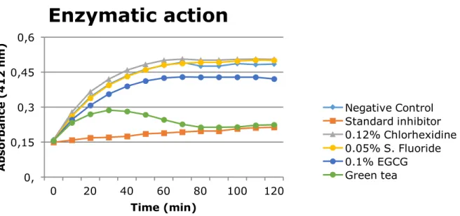
outro lado, não houve diferença estatística nos valores de desgaste e rugosidade. A zimografia
demonstrou que a CLX 0,12%, o CV e o EGCG 0,1% apresentaram ação inibitória sobre as
MMPs, enquanto que no ensaio colorimétrico o CV apresentou inibição enzimática. A
solução de CV reduz in vitro o desgaste e a rugosidade da dentina coronária e radicular
erosivamente desmineralizada. O CV e a EGCG 0,1% reduzem os danos causados pela erosão
em dentina coronária in situ e contribuem para inativação de MMPs extraídas da dentina.
Palavras chaves:
Eletroforese. Erosão dentária. Inibidores teciduais de metaloproteinases. Metaloproteinases da
ABSTRACT
The objective of this study was to evaluate and compare the action of different enzyme
inhibitors on human dentin in cyclical erosion challenging situation. To this were carried out
3 projects that yielded the following chapters: In the chapter 1 and 2, coronary or root human
(n=10) dentin blocks (4x4x2 mm) were submitted cyclically (3x/ day/3 days) to erosive
challenge by immersion in acid [dehydrated citric acid (C6H8O7), pH 3.75, 60 s] followed by
treatments depending on the group: G1-distilled water; G2- 0.12% Chlorhexidine digluconate
(CHX); G3- Green tea infusion (GT). The blocks were analyzed daily by surface profilometry
and hardness. In the chapter 3, a randomized, blinded, crossover, in situ study in which 20 volunteers used palatal intra oral device for three phases of 5 days each, containing 4 blocks
of human dentin. They were immersed in human saliva for 2 hours to acquired pellicle
formation. The erosive challenge was performed by the device immersion in 50 mL of
Coke™ (pH 2.6, 4x/ day /1 min, extraorally) followed by treatment (4x/ day) by dropping on
blocks 1 mL of the following solutions: G1- 0.05% Sodium fluoride, G2- 0.1%
Epigallocatechin-3-gallate (EGCG), G3- Green tea infusion. At each phase the volunteer used
only one substance. Quantitative analyzes were performed, such as percentage loss of surface
hardness, roughness and wear, as well as qualitative analysis by scanning electron
microscopy. Complementing the studies to analysis of enzyme inhibition, human dentin
blocks were milled, subjected to erosion by citric acid and subjected to extraction of soluble
proteins. Electrophoresis was performed and then the gel was incubated for 3 h in renaturation
buffer modified according to the groups G1: without modification, G2- 0.12% chlorhexidine
digluconate, G3- 0.05% NaF, G4- Green tea and G5- 0.1% EGCG. Gels were stained in a
solution containing 0.025% Coomassie blue and destained with acetic acid 10% solution to
enzymes. Then were subjected to colorimetric enzyme inhibition test, in which the absorbance
was measured. After extraction of dentin proteins, protein quantification by Bradford’s
method was performed, which showed 0.15 µg soluble proteins. The results were analyzed
using the Kolmogorov-Smirnov test to evaluate the normal range of results, followed by
analysis of variance (ANOVA) and Tukey test, using a significance level of 5%. Results: A
significant reduction in the dentin hardness loss in coronary dentin in vitro with the use of CHX and GT in comparison to the control (p <0.05). Treatment with GT significantly
reduced wear and roughness of dentin in vitro (p <0.05). In relation to in vitro study on root dentin, the treatment with GT reduced the wear and roughness (p <0.05). There was no
statistically significant difference between the groups regarding the loss of surface hardness of
root dentin in vitro (p> 0.05). As for the in situ study, treatment with EGCG and GT reduced the loss of dentin hardness significantly (p <0.05). On the other hand, there was no significant
difference in relation to wear and roughness values (p> 0.05). For zymography analysis,
0.12% CHX, green tea and 0.1% EGCG showed inhibitory action on the extracted
metalloproteinases dentin and, the colorimetric assay green tea has enzimatic inhibition
similar to standard inhibitor. Conclusion: Green tea solution reduces in vitro wear and roughness of coronary and root dentin erosively demineralized. The 0.1% EGCG and green
tea reduce erosion damage on coronal dentin in situ and contribute to inactivate MMPs extracted dentin.
Keywords:
Electrophoresis. Dental erosion. Tissue inhibitors of metalloproteinases. Matrix
LISTA DE ABREVIATURAS
MOD Matriz orgânica desmineralizada
MMPs Metaloproteinases da matriz
EGCG Epigalocatequina-3-galato
DW Distiled water; água destilada
CHX Chlorhexidine; Clorexidina
GT Green Tea; chá verde
Ca2+ Íon Cálcio
Zn2+ Íon Zinco
Fe2+ Íon Férro
MMP-2 Metaloproteinases da matriz-2
MMP-9
Ra
SEM
IBGE
Metaloproteinases da matriz-9
Averagen roughness; Média de
rugosidade
Scanning electron microscopy;
Microscopia eletrônica de varredura
Instituto Brasileiro de Geografia e
SUMÁRIO
1 INTRODUÇÃO GERAL 16
2 OBJETIVOS 19
3 CAPÍTULOS 20
3.1 Capítulo 1 21
3.2 Capítulo 2 37
3.3 Capítulo 3 53
4 CONCLUSÕES GERAIS 73
REFERÊNCIAS 74
APÊNDICES
ANEXOS
79
INTRODUÇÃO GERAL
A erosão dentária é uma patologia com alta prevalência em adolescentes e adultos
(36% - 61%), causada pela ação de ácidos de origem não bacteriana, os quais promovem a
dissolução dos tecidos dentais mineralizados, podendo causar dor e complicações
endodônticas (VERED et al., 2014; IMFELD, 1996; GANSS, 2014). Apresenta-se em superfícies lisas com cavidades pouco profundas, ocorrendo coronal à junção
cemento-esmalte, e na superfície oclusal observa-se aspecto de chanfrado distinto e cúspides
arredondadas (GANSS, LUSSI, 2014; (MAGALHÃES et al., 2009a).
Sabe-se que o hábito de dieta ácida, doenças gastroesofágicas, radioterapia da cabeça e
pescoço, e uso crônico de medicamentos são alguns dos fatores que elevam o risco do
indivíduo de erosão dos dentes (LIESHOUT; BOTS, 2014). A idade, composição salivar e
abrasividade da língua, bem como natureza, composição e frequência de ingestão de ácidos
são fatores que influenciam na suscetibilidade e no grau da erosão (HOOPER et al., 2015). No Brasil, segundo a pesquisa nacional de saúde, realizada em 2013 pelo Instituto Brasileiro
de Geografia e Estatística (IBGE), 23,4% da população consomem refrigerantes ou sucos
artificiais pelo menos 5 dias na semana (PNS, 2013). Esse consumo de bebidas carbonatadas é
um fator de risco para erosão dental. Estudos nacionais apontam para uma grande
variabilidade nos resultados sobre prevalência, assim como nos estudos internacionais,
apresentando uma variabilidade entre 3,4% até 58% em crianças e adolescentes (FARIAS et al., 2013). A alta frequência de ingestão de comidas e bebidas ácidas indica uma forte associação com desgaste dentário e exposição dentinária (WEI et al., 2016).
Por se tratar de uma condição multifatorial, a ocorrência de erosão dentária depende
de fatores socioeconômicos e comportamentais (hábitos dietéticos), químicos (fontes ácidas
intrínsecas e extrínsecas) e biológicos (presença de película adquirida e fluxo salivar), sendo a
interação entre esses aspectos condição fundamental para se determinar o início e a
severidade da doença (LUSSI; CARVALHO, 2014; YOUNG; TENUTA, 2011). Porém,
orientações comportamentais são difíceis de serem controladas para redução de hábidos de
dieta ácida (MAGALHÃES et al., 2009a; (COMAR et al., 2015). Portanto, diferentes abordagens podem ser utilizadas para minimizar os efeitos deletérios da doença.
A realização de uma completa anamnese, avaliando os fatores de risco individual, a
ou dentina) é crucial para o diagnóstico e o esclarecimento dessa patologia. Os fatores
químicos e biológicos, por serem passíveis de avaliação e terem grau de complexidade menor
que os outros critérios, podem ser utilizados para o direcionamento do tratamento preventivo
ou terapêutico (GANSS; LUSSI, 2014).
O desgaste causado pelas lesões de erosão pode comprometer os dentes do indivíduo
por toda a vida, além de estar relacionado à hipersensibilidade dentinária. O substrato
dentinário é exposto quando ocorre recessão gengival e desmineralização dos cristais do
esmalte, deixando as fibras colágenas desprotegidas e vulneráveis à degradação pelas enzimas
colagenolíticas (KATO, 2012). Assim, na erosão, a dentina tem sua superfície atacada, tanto
por desmineralização quanto por degradação proteolítica por meio de proteases endógenas
(metaloproteinases da matriz -MMPs e cisteino-catepsina). As MMPs são enzimas zinco e
cálcio dependentes que se encontram inativadas na saliva e dentina e, quando associadas ao
pH ácido, são ativadas e iniciam a ação proteolítica, desempenhando um papel fundamental
na progressão da cárie e erosão dentária (VISSE R, 2003); CARRILHO, 2012).
Estudos demonstram que a manutenção da matriz orgânica desmineralizada (MOD)
protege o tecido subjacente de sofrer mais erosão e perda de mineral (BUZALAF; KATO;
HANNAS, 2012). Assim, preservar a MOD após desafios erosivos dificulta a difusão dos
íons ácidos através do tecido dentinário e parece ser uma estratégia para reduzir a taxa de
desgaste da dentina (GANSS; KLIMEK; STARCK, 2004; HARA et al., 2005; KATO et al., 2012).
Substâncias inibidoras de MMPs estão sendo utilizadas como tratamento preventivo
de algumas doenças sistêmicas e bucais (DEMEULE et al., 2000; KATO et al., 2014). Dentre elas, a solução de digluconato de clorexidina, o fluoreto de sódio (KATO et al., 2009; MAGALHÃES et al., 2009b; KATO et al., 2010, KATO et al., 2012; ) e as catequinas isoladas do chá verde [Epigalocatequina-3-galato - EGCG (0,0065%)] (GENDRON et al., 1999; DEMEULE et al., 2000, VIDAL et al., 2014, de MORAES et al., 2016) foram utilizadas em estudos prévios para proteção da dentina contra erosão.
Soluções enxaguatórias de fluoreto de sódio são amplamente utilizadas em produtos
de higiene oral. Porém não apresentam substantividade, pois a camada de CaF2 é dissolvida
em baixo pH e apresentam ação parcialmente reversível na inibição de MMP-2 e -9, não
sendo eficaz na proteção da dentina contra a erosão (GANSS; SCHLUETER; KLIMEK,
utilizada em pesquisas na área da odontologia é a abordagem biomimética em que agentes
bioativos são utilizados para aumentar ou reforçar o tecido pela alteração de propriedades
biomecânicas e bioquímicas (BEDRAN-RUSSO et al., 2014).
O chá verde é proveniente da planta camélia sinensis e é um potente antioxidante e anticarcinogênico. Possui em sua composição a epigalocatequina-3-galato (EGCG), que é um
polifenol responsável por aproximadamente 59% do total das catequinas da folha do chá
verde (STEINMANN et al., 2013). Esse polifenol vem sendo avaliado como um promissor auxiliar no controle do processo de desgaste dentinário por erosão, apresentando ação
inibitória relevante sobre as MMPs (DEMEULE et al., 2000) e ação protetora do desgaste de dentina erosivamente desmineralizada (MAGALHÃES et al., 2009b; (SILVEIRA et al., 2014); (KATO et al., 2010); (DE MORAES et al., 2016). Ademais, acredita-se ser um potente
agente biomodificador, promovendo ligações cruzadas com o colágeno preservando-o,
melhorando as propriedades mecânicas e, dessa forma, mantendo a camada de matriz
orgânica que foi desmineralizada (BEDRAN-RUSSO et al., 2014). Notavelmente, a EGCG e
várias preparações de chá verde estão disponíveis como remédios de prateleiras em muitos
países e são acessíveis para a população (STEINMANN et al., 2013).
Uso de substâncias naturais, como os polifenóis provenientes de plantas, tem tido
ênfase por serem fontes renováveis, sustentáveis, de baixa toxicidade e apresentarem
bioatividade, biocompatibilidade e aplicabilidade na Odontologia. Portanto, a interação desses
compostos com o colágeno da dentina resulta em ligações de alta estabilidade, dificultando a
degradação da dentina e aumentando a proteção contra erosão (BEDRAN-RUSSO et al., 2014).
Assim, torna-se fundamental buscar estratégias como alternativas para a efetiva
proteção dos tecidos dentários mineralizados contra o desgaste causado pela erosão. Este
estudo teve como objetivo avaliar e comparar, in vitro e in situ, algumas substâncias inibidoras enzimáticas na manutenção da proteção da perda tecidual em dentina humana
desmineralizada em situação de desafio erosivo cíclico. As hipóteses do trabalho são: as
substâncias testadas serão eficazes na proteção da superfície dentinária e apresentarão ação
2. OBJETIVOS
2.1 Objetivo Geral
O objetivo deste trabalho, in vitro e in situ, foi avaliar a ação do chá verde sobre dentina humana erosivamente desmineralizada.
2.2 Objetivos Específicos
Capítulo 1: Analisar in vitro a ação da solução de clorexidina e da infusão de chá verde na proteção da perda tecidual da dentina coronária sob desafio erosivo cíclico.
Capítulo 2: Analisar in vitro a ação da solução de clorexidina e da infusão de chá verde na proteção da perda tecidual da dentina radicular sob desafio erosivo cíclico.
Capítulo 3: Analisar in situ a ação da infusão de chá verde, da solução de Epigalocatequina-3-galato e de fluoreto de sódio na proteção da perda tecidual da dentina sob
desafio erosivo cíclico, bem como verificar suas ações e da solução de clorexidina sobre as
20
3 CAPÍTULOS
REGIMENTO INTERNO
Esta tese está baseada no Artigo 46 do Regimento Interno do Programa de Pós-Graduação em
Odontologia da Universidade Federal do Ceará (Anexo A), que regulamenta o formato
alternativo para dissertações de Mestrado e teses de Doutorado e permite a inserção de artigos
científicos de autoria ou coautoria do candidato. Por se tratar de pesquisa envolvendo partes
de animais, o presente trabalho foi submetido ao Comitê de Ética em Pesquisa (CEP). Assim
sendo, esta tese de doutorado é composta por três capítulos contendo artigos científicos
publicados ou em fase de redação conforme descritos abaixo:
Capítulo 1: Effect of green tea as a protective measure against dental erosion in coronary
dentin. Brazilian Oral Research.v.30, p.1-6. 2016.
Capítulo 2: Protective effect of matrix metalloproteinases inhibitors on radicular dentin
erosion: in vitro study. Este artigo será submetido à publicação no periódico Clinical Oral Investigations.
3.1 Capítulo 1
EFFECT OF GREEN TEA AS A PROTECTIVE MEASURE AGAINST DENTAL
EROSION IN CORONARY DENTIN
SHORT TITLE: EFFECT OF GREEN TEA ON CORONARY DENTIN
Maria Denise Rodrigues de Moraes, University of Fortaleza, Fortaleza, Ceará, Brazil.
Post-graduation Program, Faculty of Pharmacy, Dentistry and Nursing, Federal University of
Ceará, Fortaleza, Ceará, Brazil. (85) 988965991; denisermoraes@gmail.com
Jéssica Rodrigues Mendes Carneiro, Post-Graduate Program, Faculty of Pharmacy, Dentistry
and Nursing, Federal University of Ceará, Fortaleza, Ceará, Brazil. (85) 999108631;
jessicarmendesc@gmail.com
Vanara Florêncio Passos, University of Fortaleza, Fortaleza, Ceará, Brazil. (85) 999882039;
vanarapassos@hotmail.com
Sérgio Lima Santiago, Post-Graduate Program, Faculty of Pharmacy, Dentistry and Nursing,
Federal University of Ceará, Fortaleza, Ceará, Brazil. (85) 988842704; sergiosantiago@ufc.br
Full address of the author to whom correspondence should be sent:
Dr. Sérgio Lima Santiago
Rua Monsenhor Furtado s/nº.
CEP 60.430-355 Fortaleza, CE Brazil
EFFECT OF GREEN TEA AS A PROTECTIVE MEASURE AGAINST DENTAL
EROSION IN CORONARY DENTIN
Abstract
The aim of this study was to evaluate the effect of green tea as a protective measure on eroded
dentin. Disks of human coronary dentin were selected based on surface hardness and
randomly assigned to 3 groups (n=10): DW - distilled water, CHX - 0.2% chlorhexidine
digluconate, and GT - green tea. The disks were allowed to acquire pellicle for 2 hours and
were then subjected to 3 cycles per day of demineralization (C6H8O7 0.05 M, pH 3.75, 60 s), treatment (DW or CHX or GT, 5 min) and remineralization (artificial saliva, 60 min) over a
period of 3 days. Changes in the dentin were determined by loss of surface hardness (%SHL)
and mechanical profilometry analysis at the end of each day. Data were analyzed by two-way
ANOVA followed by Tukey's test for %SHL and profilometry (p<0.05). Significant
reductions in dentin hardness loss were observed only for the CHX group when compared to
the DW group (p<0.05). However, there was no significant difference between the CHX and
GT groups (p> 0.05). A significant difference was observed between DW and GT treatments
for wear and roughness measurements (p<0.05). The green tea extract solution was able to
reduce the wear and roughness caused by dentin erosion under the conditions of this study.
Introduction
Erosive demineralization of the tooth crown is characterized by initial softening of the enamel
surface. This process is followed by continuous layer-by-layer dissolution of the enamel
crystals, leading to a permanent loss of tooth volume with a softened layer at the surface. The
dentin becomes increasingly exposed in advanced stages and exposes the organic matrix to
breakdown by host-derived enzymes, such as matrix metalloproteinases (MMPs) present in
dentin and saliva.1-3 MMPs act in the chemical degradation of the organic matrix of dentin
and play an important role in the progression of dentin erosion.4-6
Protective measures to reduce dental erosion, such as laser therapy and topical fluoride, have
been investigated. Most of these treatments are based on the action of fluoride, which is
available in dentifrices, solutions, or varnishes, although the role of fluoride in the protection
of dental erosion is still controversial.7-10
In recent years, some in situ studies have shown that commercial green tea and a rinse containing green tea extract were able to reduce erosive and erosive/abrasive dentin wear.11,12
These products are rich in polyphenols and have been reported to be inhibitors of the activity
of different metalloproteinases.11,13 It has been suggested that the use of synthetic inhibitors of
MMPs could be used to control the loss of the dentin matrix.5,14 However, MMPs are secreted
as inactive precursors (pro-forms), requiring activation by a low pH to degrade extracellular
matrix components.1
Therefore, the demineralization process is increased by the action of acids on the organic
matrix of dentin and the activation of the MMPs.12 To minimize this process, green tea has a
exposure to acid may be minimized by the use of metalloproteinase inhibitors, which protect
the organic matrix as a barrier to ion diffusion.
Other studies have attempted to evaluate the protective effect of green tea against wear
promoted by erosion; however, there are still doubts concerning the maintenance of inhibitory
action in the MMP after successive cyclical acidic exposures. It might be interesting to
investigate the effectiveness and substantivity of these substances on the surface of the eroded
dentin in cycles of erosive challenge. The absence of studies that simulate the erosion of
dentin through short cyclic acid challenges, both daily and intermittent, reinforces the need
for further studies.
Thus, the objective of this in vitro study was to assess the protective effect of green tea on
dentin demineralization in three days of cyclic erosion by assessing the percentage of surface
hardness loss (%SHL), roughness and wear.
Materials and methods
Experimental Design
This in vitro study was approved by the local research and ethics committee (protocol #
175/2010). This is a blinded experimental study design with three groups (n = 10), with two
factors: time (one, two and three days of the experimental cycle) and treatments (distilled
water (DW; G1- control), 0.2% chlorhexidine (CHX; G2) and green tea (GT; G3; EGCG
concentration 0.0014%)). The treatments were applied by immersion under agitation for five
minutes, and the specimens were randomly assigned to the defined treatments. The dependent
variables were the percentage of surface hardness loss (%SHL), as quantitatively evaluated by
differences in the mean values of surface hardness after treatment, and dentinal wear and
roughness, as assessed by the contact profilometer Hommel Tester T1000 (Jenoptik,
Specimen Preparation
Coronary dentin specimens were prepared from human third molars that had been stored in
0.01% (w/v) thymol solution at 4°C. Dentin disks were obtained using a longitudinal coupled
double-sided diamond disk in a IsoMet slow speed saw (Buehler, Lake Bluff, USA). The
dimensions of each disk depended on the diameter of the tooth. Sequentially, the specimens
were ground in a water-cooled mechanical grinder (Arotec S.A., Cotia, Brazil) using 400-,
600-, 800- and 1200-grit aluminum oxide abrasives and polished with felt paper and 1 µm
diamond spray (Extec Corp., Enfield, USA). The surface hardness values were determined
using a Knoop diamond FM100 (Future-Tech Corp., Kawasaki, Japan), making five
indentations with a load of 10 gf /5 s, 100 µm apart from each other, at the center of the
specimens. Thirty dentin disks presenting a mean hardness of 55.81 ± 6.20 Knoop hardness
number (KHN) were selected and randomly assigned using a computer-generated
randomization list into three experimental groups (Microsoft Excel 2007).
Nail varnish was applied on half of the surface of each specimen to serve as the reference area
for profilometry analysis. The exposed area was subjected to the acid challenge.
Pellicle Formation
On each experimental day, five volunteers without erosion, salivary dysfunction or active
carious lesions donated fresh saliva samples. The secretion of saliva was stimulated by
chewing on paraffin wax for five minutes. Saliva from the first minute of chewing was
swallowed, and the rest was collected and deposited into a 50 mL centrifuge tubule. The
saliva samples were centrifuged for 10 min at 2000 rpm in a pre-cooled centrifuge (4°C)
NT-815 (Novatecnica, Piracicaba, Brazil). The clear fluid above the sediments was pooled and
used for pellicle formation.18 Prior to erosive challenge, each group of dentin disks was
oscillating table (TE143, Tecnal, Piracicaba, Brazil) at 37°C (Olidef CZ, Ribeirão Preto,
Brazil) for two hours before each experimental day to simulate the environment of the oral
cavity.
Experimental procedure
The study used cyclic procedures repeated over a three-day period, including pellicle
formation, erosion, treatments with test solutions (DW, CHX, GT) and remineralization with
freshly prepared artificial saliva (1.5 mM Ca; 0.9 mM PO4; 150 mM KCl and 0.1 M Tris
buffer, pH 7.0), considering the influence of variables such as stirring, temperature and
exposure time (Fig 1).19 The 0.2% chlorhexidine digluconate solution was provided by a
pharmacy, and the green tea (Dr Oetker, Jd. do Lago, Brazil) was freshly prepared according
to the manufacturer’s instructions at the beginning of each experimental day. The
concentration of epigallocatechin gallate (EGCG) in this green tea was 0.0014%, as assessed
using a spectrophotometer, and the pH was 5.45.
Erosion Cycling Model
After pellicle formation, each disk was submitted to a citric acid solution for 60 seconds. The
acid challenge was performed using 0.05 M dehydrated citric acid, pH 3.75 (Dinâmica®,
Diadema, Brazil). Each disk was then rinsed with distilled water and treated for 5 minutes
with the treatment solution (DW, CHX, GT) and then immersed in artificial saliva for 1 hour,
performed under agitation at 100 rpm and 37°C (Fig 1).
This cycle was repeated three times a day for three days. At the end of each experimental day,
the hardness, wear and surface roughness analysis of each specimen were measured.20
Percentage of Surface Hardness Loss Assessment
Immediately after each experimental day, the slabs were placed in the hardness machine, and
five new indentations were made using a Knoop diamond under a 10 g load for five seconds
(SHbefore). The percentage of SH loss (% SHL) was then calculated for each day, according
to the following equation: %SHL = [(SHbefore – SHafter)x100/ SHbefore].
Measurement of Dentin Surface Loss
Measurements of dentin surface loss were performed using the stylus profilometer, after each
experimental day. The difference between the heights of the surfaces of the reference and the
treated areas was evaluated. Before analysis, the nail varnish was carefully removed, exposing
the untreated reference areas. On each sample, at intervals of 100 µm, five profile traces (1.5
mm in length) were recorded, and the levels of dentin wear were determined in relation to the
reference surfaces. For each sample, the mean values obtained from the five traces were
calculated.21
Measurement of Surface Roughness
Surface roughness was described by the arithmetic mean of the absolute ordinate values Ra
(average roughness as per ISO 4287) of 5 measurements made in each disk.22,23 In
profilometry, the surface of a specimen was scanned using a stylus with a diamond to
generate a two- or three-dimensional profile using a contact measuring device.24
Statistical Analysis
Statistical procedures were performed with the Statistical Package for Social Sciences (SPSS
17.0 for Windows, SPSS Inc., Chicago, USA). A Kolmogorov-Smirnov test was applied to all
groups to test for the normal distribution of errors. Because the values were normally
distributed across all groups, two-way ANOVA and Tukey’s post hoc tests were used for
comparative purposes. The level of significance was set at 5%.
Results
Table 1 presents the means of dentin hardness loss, wear and roughness values found for all
In relation to dentin hardness loss, dental wear and roughness, two-way ANOVA revealed a
significant difference among the treatments tested [(p<0.001; F=3.3), (p<0.001; F=9.7), and
(p<0.001; F=8.3), respectively], as well as the duration of demineralization represented by the
number of experimental days [(p<0.001; F=69.7), (p<0.001; F=11.4), and (p<0.001; F=49.0),
respectively]. Furthermore, the interaction between the factors was significant for the loss of
dentin hardness (p=0.009; F=3.6). However, the interaction between the factors was not
significant for dental wear (p=0.745; F=0.4) and for roughness (p=0.782; F=0.4). Significant
differences in dentin hardness loss were observed only for the CHX group when compared to
the DW group (p<0.05). However, there was no significant difference between CHX and GT
(p>0.05).
For dentin hardness loss and dental wear, it was observed that with an increase in the number
of experimental days, all specimens displayed statistically significant surface softening from
day 1 to day 2, which stabilized at day 3, showing that citric acid was effective in eroding the
dentin surface. The green tea was more effective than DW in protecting the human dentin
against wear caused by the erosion of citric acid (p<0.001). Additionally, there was no
significant difference between CHX and GT (p<0.05).
The chlorhexidine behaved similarly to the control (p=0.053) in protecting the human dentin
against surface roughness caused by the erosion of citric acid. On the other hand, the green tea
was more effective than the control (p<0.001).
Discussion
The present in vitro de-remineralization cycling model investigated green tea with respect to its capacity to protect human dentin from erosion. This study confirmed the expected surface
softening and dentin tissue loss due to the action of citric acid, even after pellicle formation
model allowed for a better understanding of the erosive challenges faced by the dentition
while performing a controlled investigation and reducing the experimental time and cost.25
The results of this study disagree with the results of Mirkarimi and Toomarian (2012)26
because an increase in surface hardness values after immersion in green tea solution was not
observed in the current study. However, the erosive challenge used in their study was shorter
and lower than that used in the present study. The dentin disks subjected to this erosive cycle
presented higher percentages of change in hardness, which may have influenced the
indentations of the diamond when measuring hardness. Furthermore, the hardness analysis is
a sensitive method for detecting changes in the mineral density of artificial eroded/abraded
lesions in an enamel substrate but not in dentin;27 therefore, another analysis, profilometry,
was added to the current study.
Under great acid challenge, profilometric analysis is widely used to measure the loss of
enamel and dentin surfaces.27,28 The results of the present study corroborate those of
Magalhães et al. (2009)12 and Kato et al. (2009)11 because the green tea was able to reduce
dentin wear in the current study. A possible mechanism of action for the reduction in dentin
loss might be the inhibition of MMPs by chlorhexidine and green tea extract solution.12
Another study that involved cyclic acid challenge also used profilometry to measure the wear
of dentin; Barbosa and colleagues (2011)29 concluded that the supplementation of soft drinks
with green tea extract might be a viable alternative to reduce the erosive potential against
dentin.
In this study, a cycling model was used, employing citric acid three times a day over a period
of 3 days. On the 3rd day of the erosive challenge, the surface roughness and dentin wear were
worse than in the first day, showing that the acid used in this study was effective in eroding
the dentin surface. It is also probable that the low pH of the acid induced the activation of
The 1 µm of wear protection observed in this in vitro study correlates with the erosive cycle, which involved short periods of acid exposure and remineralization. Magalhães and
colleagues performed a more aggressive challenge (4 times/day, Coca Cola, 5 min) than in
this study (3 times/day, citric acid, 1 min)12 and observed a similar outcome. Additionally, in
that study, green tea extract with approximately 30 ± 3% of catechin was used, while the
present study used a commercial product, which limited the concentration of catechin
(0.0014%).
One possible mechanism of action of green tea on the reduction of dentin erosion could be the
inhibition of MMPs. If it is true, the component responsible for this effect may be
polyphenols. Green tea polyphenols, especially epigallocatechin-3-gallate (EGCG), were
found to have potent and distinct inhibitory activity against MMPs in cell culture tests.11 The
green tea polyphenols and their major constituent, EGCG, have been shown to inhibit
metalloproteinases in other areas of medicine and dentistry.30,31 It can be speculated that the
small concentration of EGCG in the tea used in the present study can justify the unsatisfactory
results in protecting the loss of dentin hardness. However, it must be acknowledged that the
protocol employed in the present study does not suggest that the effect of green tea on the
reduction of the wear and roughness of dentin specimens is due to its inhibitory action on
MMP activity, as we did not test this directly.
Because the erosive/remineralization cycle was performed under in vitro conditions, it is clear that the results are not completely transferable to an in vivo situations, the natural protective
effects of the oral cavity are lacking. However, the present study utilized variables such as the
erosive solution, stirring method and temperature in order to replicate the oral environment.
Thus, the use of green tea might be a viable alternative as a protective measure against erosive
wear in short acid exposures. However, further studies are necessary to explore the inhibitory
Conclusion
According to the conditions of the present study, the use of green tea extract is a promising
protective measure in reducing dentin erosion, as it has a protective effect on the dentin
References
1. Buzalaf MAR, Kato MT, Hannas AR. The role of matrix metalloproteinases in dental
erosion. Adv Dent Res. 2012; 24(2): 72-6.
2. Pashley DH, Tay FR, Yiu C, Hashimoto M, Breschi L, Carvalho RM, ITO S. Collagen
degradation by host-derived enzymes during aging. J Dent Res. 2004; 83(3): 216-21.
3. Hannas AR, Pereira JC, Granjeiro JM, TjäDerhane L. the role of matrix metalloproteinases
in the oral environment. Acta Odontol Scand 2007; 65(1): 1-13.
4. Hara AT, Ando M, Cury JA, Serra MC, Gonzalez-Cabezas C, Zero DT. Influence of the
organic matrix on root dentine erosion by citric acid. Caries Res. 2005; 39: 134-138.
5. Kato MT, Leite AL, Hannas AR, Calabria MP, Magalhães AC, Pereira JC, Buzalaf MAR.
Impact of protease inhibitors on dentin matrix degradation by collagenase. J Dent Res. 2012;
91(12): 1119-1123.
6. Buzalaf MAR, Hannas AR, Kato MT. Saliva and dental erosion. J Appl Oral Sci. 2012;
20(5):493-502.
7. Messias DCF, Maeda FA, Turssi CP, Serra MC. Effect of dentifrices against hydrochloric
acid- induced erosion. Oral Health Prev Dent. 2011; 9(3): 269–273.
8. Passos VF, Melo MAS, Silva FFC, Rodrigues LKA, Santiago, SL. Effects of diode laser
therapy and stannous fluoride on dentin resistance under different erosive acid attacks.
Photomed Laser Surg. 2014; 32(3): 146-151.
9. Passos VF, Santiago SL, Tenuta LM, Cury JA. Protective effect of
NaF/triclosan/copolymer and MFP dentifrice on enamel erosion. Am J Dent. 2010; 23 (4):
193-195.
10. Wiegand A, Attin T. Influence of fluoride on the prevention of erosive lesions: a review.
Oral Health Prev Dent. 2003; 1(4): 245-53.
of green tea on dentin erosion and abrasion. J Appl Oral Sci. 2009; 17(6): 560-64.
12. Magalhães AC, Wiegand A, Rios D, Hannas A, Attin T, Buzalaf MAR. Chlorhexidine
and green tea extract reduce dentin erosion and abrasion in situ. J Dent. 2009; 37(12): 994-8. 13. Sulkala M, Larmas M, Sorsa T, Salo T, Tjäderhane L. The localization of matrix
metalloproteinase-20 (mmp-20) in mature human teeth. J Dent Res. 2002; 81(9): 603-7.
14. Gendron R, Grenier D, Sorsa T, Mayrand D. Inhibition of the activities of matrix
metalloproteinases 2, 8, and 9 by chlorhexidine. Clin Diagn Lab Immunol. 1999; 6(3): 437-9.
15. Demeule M, Brossard M, Pagé M, Gingras D, Béliveau R. Matrix metalloproteinase
inhibition by green tea catechins. Biochim Biophys Acta. 2000; 1478(1):51-60.
16. M.T. Kato, A.L. Leite, A.R. Hannas, and M.A.R. Buzalaf. J Dent Res. 2010;
89(5):468-472.
17. Silveira C, Oliveira F, Dos Santos ML, de Freitas T, Imparato JC, Magalhães AC.
Anarcadic acid from brazilian cashew nut trees reduces dentine erosion. Caries Res. 2014;
48(6):549-56.
18. De-Melo MAS, Passos VF, Alves JJ, Barros EB, Santiago SL, Rodrigues LKA. The effect
of diode laser irradiation on dentin as a preventive measure against dental erosion: an in vitro
study. Lasers Med Sci. 2011; 26(5): 615-21.
19. Young A, Tenuta LMA. Initial erosion models. Caries Res. 2011; 45(1 suppl): 33-42.
20. Caneppele TM, Jeronymo RD, Di Nicoló R, de Araújo MA, Soares LE. In vitro
assessment of dentin erosion after immersion in acidic beverages: surface profile analysis and
energy-dispersive x-ray fluorescence spectrometry study. Braz Dent J. 2012; 23(4): 373-8.
21. Moron BM, Miyazaki SSH, Ito N, Wiegand A, Vilhena F, Buzalaf MAR, Magalhães AC.
22. Jung M, Eichelberger K, Klimek J. Surface geometry of four nanofiller and one hybrid
composite after one-step and multiple-step polishing. Oper Dent. 2007, 32(4): 347-355.
23. Jung M, Sehr K, Klimek J. Surface texture of four nanofilled and one hybrid composite
after finishing. Oper Dent. 2007; 32(1): 45-52.
24. Schlueter N, Hara A, Shellis RP, Ganss C. Methods for the measurement and
characterization of erosion in enamel and dentine. Caries Res. 2011; 45(1 suppl): 13-23.
25. Shellis RP, Ganss C, Ren Y, Zero DT, Lussi A. Methodology and models in erosion
research: discussion and conclusions. Caries Res. 2011; 45(1 suppl): 69-77.
26. Mirkarimi M, Toomariam L. Effect of green tea extract on the treatment of dentin erosion:
an in vitro study. J Dent (Tehran). 2012; 9(4): 224-8.
27. Passos VF, Melo MAS, Vasconcelos AA, Rodrigues LKA, Santiago SL. Comparison of
methods for quantifying dental wear caused by erosion and abrasion. Microsc Res Tech.
2013; 76(2): 178-183.
28. Ganss C, Lussi A, Scharmann I, Weigelt T, Hardt M, Klimek J, Schlueter N. Comparison
of calcium analysis, longitudinal microradiography and profilometry for the quantitative
assessment of erosion in dentine. Caries Res. 2009; 43(6): 422–429.
29. Barbosa CS, Kato MT, Buzalaf MAR. Effect of supplementation of soft drinks with green
tea extract on their erosive potential against dentine. Aust Dent J. 2011; 56(3): 317-321.
30. Deb G, Thakur V, Limaye A, Gupta S. Epigenetic induction of tissue inhibitor of matrix
metaloproteinase-3 by green tea polyphenols on breast cancer cells. Mol Carcinog. 2014;
31(1): 1-15.
31.Wen WC, Kuo PJ, Chiang CY, Fu E. Epigallocatechin-3-gallate attenuates porphyromonas
gingivalis lipopolysaccharide-enhanced matrix metalloproteinase-1 production through an
Figure 1. Erosion cycle model and treatments
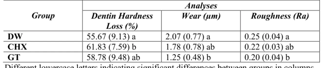
Table 1. Means and standard deviations of the loss of dentin hardness, wear and roughness values for all the treatments evaluated.
Group
Analyses Dentin Hardness
Loss (%)
Wear (µm) Roughness (Ra)
DW 55.67 (9.13) a 2.07 (0.77) a 0.25 (0.04) a
CHX 61.83 (7.59) b 1.78 (0.78) ab 0.22 (0.03) ab
GT 58.78 (9.48) ab 1.25 (0.48) b 0.20 (0.04) b
3.2 Capítulo 2
PROTECTIVE EFFECT OF MATRIX METALLOPROTEINASES INHIBITORS ON
RADICULAR DENTIN EROSION: AN IN VITRO STUDY
Maria Denise Rodrigues de-Moraes, University of Fortaleza, Fortaleza, Ceará, Brazil.
Graduate Program, Faculty of Pharmacy, Dentistry and Nursing, Federal University of Ceara,
Fortaleza, Ceara, Brazil. (85) 988965991; denisermoraes@gmail.com
Marcelo Victor Sidou Lemos, Graduate Program, Faculty of Pharmacy, Dentistry and
Nursing, Federal University of Ceara, Fortaleza, Ceara, Brazil. (85)996119893
Vanara Florêncio Passos, University of Fortaleza, Fortaleza, Ceara, Brazil. (85) 999882039;
vanarapassos@hotmail.com
Sérgio Lima Santiago, Department of Operative Dentistry, Faculty of Pharmacy, Dentistry
and Nursing, Federal University of Ceara, Fortaleza, Ceara, Brazil. (85) 988842704;
sergiosantiago@ufc.br
Address for correspondence:
Sérgio Lima Santiago
Rua Monsenhor Furtado, s/n – Rodolfo Teófilo
CEP 60430-355 – Fortaleza – CE – Brasil
PROTECTIVE EFFECT OF GREEN TEA INFUSION ON RADICULAR DENTIN
EROSION: AN IN VITRO STUDY
Abstract
Objective This study aims to evaluate the effect of green tea infusion in protecting tissue loss caused by erosion on human root dentin.
Materials and methods Root dentin blocks were immersed in human saliva (2h) and subsequently submitted to three-day-cycles of erosive challenge. The specimens (n=10) were
selected based on their surface hardness and submitted to erosive challenge-3 times/day (citric
acid 0.05 M, 1 min) and treatments (5 min): DW-Distilled water; CHX-0.12% Chlorhexidine
digluconate, and GT-Green tea infusion, with immersion in artificial saliva between (60 min)
and at the end of the cycles (overnight). At the end of each experimental day, measurements
were performed to verify surface wear, roughness and % of surface hardness loss (%SHL).
Scanning Electron microscopy (SEM) were qualitatively analyzed. Data were analyzed by
two-way repeated-measures ANOVA and Tukey test (α=0.05).
Results ANOVA two way repeated-measures revealed in the results of the wear difference between the groups and the time of experiment (p<0.05). Tukey’s test showed less wear to
GT than the DW (p<0.05). For the roughness, Tukey’s test showed the lowest mean value
was for the treatment with GT (p<0.05). In %SHL, it revealed significant differences for the
time and not for the groups. Tukey’s test showed on the third day the %SHL was higher.
Conclusion The green tea infusion has shown to be promising as a protective agent of root dentin in cyclic erosive challenge in vitro.
Clinical Relevance Root dentin treatment with green tea infusion can be considered effective against erosive tooth wear.
Introduction
Non-carious cervical lesions are associated with gingival recession exposing root
dentin [1]. It is difficult to diagnose these lesions in early stages, and in advanced stages the
dentine becomes more and more exposed [2]. In such cases, the deleterious effect of erosion
seems to be sharper due to a more rapid and extensive exposure of the dentin, because the
cementum layer overlying the surface of the root is thin and can be easily removed [3].
During demineralization, collagen fibrils present in the organic matrix are exposed4.
The exposed collagen fibrils are subject to degradation due to the action of a family of
collagenolytic enzymes known as matrix metalloproteinases (MMPs) [5,6]. MMPs are
endopeptidases, zinc and calcium dependent which contribute to the organization and
mineralization on the dentin matrix. Such enzymes are deposited in human dentin during
formation of the teeth and remain inactive, but when in acidic environments, as in caries and
erosion lesions, are activated [6,7]. Although, few studies evaluated the role of MMPs in
dental erosion, it can be speculated that such MMPs may be directly related to the degradation
of exposed collagen [4,8,9].
It was demonstrated that MMPs inhibitors reduce the loss of dentin substrate when it
is exposed to an erosive challenge [4,9-11]. Study has shown that chlorhexidine (CHX) has
MMP-2, -8 and -9 activity inhibiting potential. The effect of CHX to inhibit MMPs is
attributed due to a mechanism of zinc chelation, since the MMPs -2, -9 inhibition may be
avoided by adding calcium chloride to the CHX connections [12].
Besides CHX, fluorides and ferrous sulfate (FeSO4) have also been related as potential
anti-erosion agents [13], in relation to fluorides, its effects are mainly attributed to the
formation of a protective layer of calcium fluoride on dental tissue. This fact would make the
teeth more stable and less susceptible to demineralization, making a lower pH level necessary
to cause such effect [14-16]. In relation to ferrous sulfate, its action is attributed to the
replacement by cations Fe2+of the essential ions in the MMPs structure (Ca2+and Zn2+),
causing reversible inactivity[17].
The investigation of organic anti-erosion agents has gained increased importance.
Green tea polyphenols (Camellia sinensis) have been identified as promising agents in the strategy to preserve collagen fibrils exposed to collagenolytic degradation [9,10,18,19]. The
flavonoid epigallocatechin-3-gallate (EGCG) is the main polyphenol found in green tea, and
presents ability to inhibit the expression and activity of MMP-2 and MMP-9 [20-22]. Green
seems to interact with such enzymes via hydrogen bonds, which are responsible for the
change of its secondary structure and consequently inhibition [22].
The green tea extract is often used daily in diets and seems particularly attractive,
since this tea contains EGCG and others catechins that control dentin degradation. However,
there are still doubts concerning the action of treatment solutions to dentin erosion after
cyclical exposures of acids. Since the concentration and distribution of MMPs in the dentin
varies along different dentin depths [23], it seems valuable to evaluate whether using green
tea as a protease inhibitor would also protect loss of the root eroded dentin.
Therefore, the objective of this in vitro study was to evaluate the effect of green tea extract and chlorhexidine in protecting tissue loss of demineralized dentin by in vitro cyclic erosive protocol. The hypothesis is that the green tea infusion and 0.12% chlorhexidine
digluconate has a protective effect against wear of the demineralized dentin by short erosive
protocol of three-days.
Materials and methods
Experimental design
This is an in vitro, randomized and blind study with two factors under investigations: time (one, two and three days of the experimental cycle) and treatments DW: distilled water
(control), CHX: 0.12% chlorhexidine digluconate and GT: green tea infusion [GT- EGCG
(concentration 0.0014%)]. The specimens were randomly assigned into three groups (n=10) in
accordance with the treatment.
The protocol was approved by the local committee (#003438/2016). The response
variable used was the percentage of surface hardness loss (%SHL), evaluated by Knoop
microhardness (FM100 Future-Tech Corp., Kawasaki, Kanagawa, Japan), and dentin wear
and roughness, assessed by the contact profilometer Hommel Tester T1000 (Jenoptik,
Schwenningen, Germany). The dentin surface was qualitatively evaluated by Scanning
Electron microscopy (SEM).
Specimens preparation
Ninety recently extracted non-carious human third molars, which had been stored in a
from the roots with a diamond saw. Ninety dentin blocks (4x4x2 mm) from cervical third of
the roots were obtained using a longitudinal coupled double-sided diamond disk in a IsoMet
slow speed saw (Buehler, Lake Bluff, USA). Dentin blocks were mounted on acrylic
appliances and grounded in a water-cooled mechanical grinder (Arotec S.A., Cotia, Brazil)
using 400-, 600-, 800- and 1200-grit aluminum oxide abrasives, polished with felt paper and 1
µm diamond spray (Extec Corp., Enfield, USA) [18,24]. The initial values of surface Knoop
hardness were determined using a microhardness tester (FM100 Future-Tech Corp.,
Kawasaki, Kanagawa, Japan), which were five indentations with a load of 10 gf for 5 s,
distancing 100 micrometres from each other, taking as reference the center of the specimen.
Thirty root dentin blocks with an average of Knoop hardness (KH) of 61.96 ± 6.19 were
selected and randomly distributed in three experimental groups using a computer-generated
randomization list (Microsoft Excel 2007, EUA). Subsequently, a thin coating layer of
acid-resistant was applied on their polished surface, leaving the half area (2x2 mm) not protected
by the varnish and subjected to acid challenge [18].
Erosive Cycling
On each experimental day, five volunteers without erosion, salivary dysfunction or
active carious lesions donated fresh saliva samples. The secretion of saliva was stimulated by
chewing on paraffin wax for five minutes. Saliva from the first minute of chewing was
swallowed, and the rest was collected and deposited into a 50 mL centrifuge tubule. The
saliva samples were centrifuged for 10 min at 2000 rpm in a pre-cooled centrifuge (4°C)
NT-815 (Novatecnica, Piracicaba, Brazil). The clear fluid above the sediments was pooled and
used for pellicle formation [18,25]. The specimens from each group were immersed in
clarified saliva and incubated for two hours under stirring (oscillatory table TE143,
Piracicaba, SP, Brazil), at 37°C (Olidef CZ, Ribeirão Preto, SP, Brazil) in order to simulate
the formation of the acquired pellicle [25].
After the formation of the acquired pellicle, each block was immersed in a citric acid
solution at 0.05M (dehydrated citric acid, pH 3.75; Dinâmica®, Diadema, SP, Brazil) for 60
s. They were then washed with distilled water and subjected to respective treatments for 5
min, and subsequently immersed in artificial saliva for one hour, with all steps performed
under stirring at 100 rpm and kept at 37°C. This cycle was repeated for three days and three
times daily. The cycle time was defined by previous pilot study. At the end of each
[26]. After each experimental day analyses, the blocks were immersed in artificial saliva
under stirring and stored at 37 ° C overnight.
The cyclic erosive challenge comprised the following steps: formation of the acquired
pellicle, erosion, treatments and remineralization with artificial saliva, considering the
influence of variables such as stirring, temperature and time of exposure [27] (Fig.1). The
treatments were: 0.12% chlorhexidine digluconate (pH 5.5) prepared in a local compounding
pharmacy; the green tea (Dr Oetker, Jd. Do Lago, SP, Brazil) was prepared before each cycle
(200 mL), being 0.0014% the concentration of epigallocatechin-3-gallate (EGCG), obtained
after analysis in spectrophotometer, and pH of 5.45.
Fig. 1 Erosive cycle [18]
Surface Hardness Loss Percentage
Immediately after each experimental day, the blocks were analyzed in a
microdurometer, where five new indentations were made using the Knoop tip load of 10 gf /5
s (SH-after), being such indentations carried to 100 µm of the previous measurements. The percentage of SH loss (% SHL) was then calculated for each day, according to the following
equation: %SHL = [(SHbefore – SHafter) x100/ SHbefore] [18].
Measurement of wear and roughness
Before analysis, the nail varnish was carefully removed with acetone exposing the
untreated reference areas. These measurements were performed as described in De-Moraes et
al., (2016) [18]. Measurements of dentin surface loss were performed using the stylus
![Fig. 1 Erosive cycle [18]](https://thumb-eu.123doks.com/thumbv2/123dok_br/15364698.565857/44.892.180.520.424.659/fig-erosive-cycle.webp)