Professional flossing as a diagnostic
method for gingivitis in the primary
dentition
Abstract: The aim of this study was to evaluate lossing as a diagnostic method for interproximal gingival bleeding in children. For this cross-over study, 23 pre-schoolchildren presenting neither restorations nor ap-proximal carious cavities and with at least 15% of gingival bleeding sites were selected. Examinations were performed at three different moments (3-4 days interval). Examinations comprised repeated measurements of two gingival indices with a 10-minute interval in the following sequenc-es: the Ainamo & Bay Gingival Bleeding Index (GBI) followed by the Carter & Barnes lossing index (CBI); CBI followed by GBI; and GBI followed by GBI. Data analysis was performed only for the interproxi-mal sites, considering the GBI as the gold-standard. Agreement between indices, sensitivity (SE), speciicity (SP), positive (PPV) and negative pre-dictive values (NPV) were estimated. Percentage agreements in sequences GBI-CBI, CBI-GBI and GBI-GBI were 70.3%, 76.4% and 84.5%, re-spectively. Validation of lossing in the irst sequence (GBI-CBI) resulted in values of 0.61 (95%CI 0.53 – 0.68), 0.72 (95%CI 0.69 – 0.76), 0.33 (95%CI 0.28 – 0.39) and 0.89 (95%CI 0.86 – 0.92) respectively for SE, SP, PPV and NPV. It can be concluded that professional lossing is a use-ful tool in the diagnosis of interproximal gingival inlammatory status in children, especially in conditions of gingival health.
Descriptors: Gingivitis; Diagnosis; Dentition, primary; Dental plaque. Adriela Azevedo Souza Mariath(a)
Ana Eliza Lemes Bressani(a) Alex Nogueira Haas(b) Fernando Borba de Araujo(c) Cassiano Kuchenbecker Rösing(d)
(a)MScs, Department of Pediatric Dentistry;
(b)MSc, Professor, Department of Periodontology; (c)PhD, Professor, Department of Pediatric Dentistry; (d)PhD, Professor, Department of Periodontology – School of Dentistry, Federal University of Rio Grande do Sul, Porto Alegre, RS, Brazil.
Corresponding author:
Adriela Azevedo Souza Mariath Av. Alegrete, 305 - Ap. 401 - Petrópolis Porto Alegre - RS - Brazil
CEP: 90460-100
E-mail: [email protected]
Introduction
Evaluation of the periodontal status is fundamen-tal during oral health follow-ups of infant patients. The periodontal status is not limited to a periodon-tal diagnosis, but also provides an assessment of the quality and the routine of the mechanical plaque control performed. Studies have collected informa-tion on oral hygiene habits either by means of re-ported behavior or more directly by using plaque or gingival indices.1
It was demonstrated that prevalence, extent and severity of gingivitis increase with age, starting in the primary dentition and reaching their peak during adolescence.2,3 Gingivitis in children was also shown to be less severe compared to adults when similar amounts of plaque deposition are found. However, the same gingival indices have been used to diagnose the gingival status in children and adults.4,5
The most frequently used clinical indices for evaluation of gingival health have been the Plaque Index (PlI)6 and the Gingival Index (GI).7 The Plaque Index evaluates the presence of plaque attributing four scores according to the plaque volume present on dental surfaces. The Gingival Index associates vi-sual inspection with bleeding of the gingival margin for the diagnosis of the inlammatory condition of the gingiva.
Ainamo, Bay8 (1975) suggested the use of two di-chotomous indices. The irst, called Visible Plaque Index, aggregates PlI scores 0 and 1 as absence and scores 2 and 3 as presence of visible plaque.8 Simi-larly, a dichotomous index to evaluate gingival in-lammation was proposed (Gingival Bleeding Index – GBI). The rationale for these dichotomizations is the high subjectivity of quantiied classiications of plaque and gingival inlammation. Additionally, it was demonstrated that visual clinical manifesta-tions of gingivitis (edema and gingival redness) is preceded by bleeding of the gingival margin,9 con-sequently the sequence of GI scores would generate some misleading interpretation of the gingival in-lammatory condition. Moreover, gingival bleeding was related to histological inlammatory evaluations of the gingival tissues.10 Another important consid-eration related to dichotomous indices of gingivitis is their higher reproducibility.9,11
Speciic indices for evaluation of interproximal gingival inlammation have also been suggested.11-14 The Carter, Barnes11 (1974) gingival bleeding index (CBI) applies dental loss to mechanically stimulate the gingival sulcus aiming at evaluating the absence or presence of bleeding. The authors afirm there is no evidence of traumatic injury of the gingival mar-gin that could lead to a higher occurrence of bleed-ing.11 Loesche13 (1979) also introduced a different system, the papillary bleeding score (PBS), in which bleeding is evaluated after the insertion of a Stimu-dent® interdental cleaner. Tinoco, Gjermo15 (1992) observed that lossing was able to detect expected changes in gingival health after plaque control in children 4 to 6 years of age and demonstrated high-er sensitivity than GI and GBI to identify reductions in gingival inlammation.
Different indices are available to be applied in the evaluation of gingival inlammation. However, a method that could allow an evaluation of gingival inlammation combining control of dental plaque, oral hygiene instruction and motivation at the same time would be of great value. Such features, asso-ciated with easiness of assessment, are desirable characteristics of an index, mainly when applied in infant patients that can present dificult behavior during dental appointments. Thus, the aim of the present study was to evaluate the validity of dental loss as a diagnostic method for gingivitis in the de-ciduous dentition.
Material and Methods
Study design and sample
This research is in accordance with the Decla-ration of Helsinki and was approved by the Ethics Committee, School of Dentistry, Federal University of Rio Grande do Sul, Brazil. Patients were included in this study after parental acceptance and conir-mation through a signed Informed Consent.
Procedures
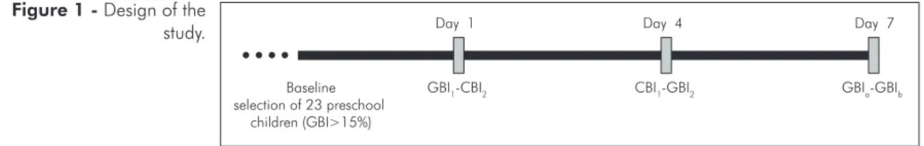
The study design consisted of three examination sequences performed in all children with an interval period of 3-4 days (Figure 1). These sequences com-prised assessment of two gingival indices with a 10-minute interval between them, as follows:
Sequence 1 (GBI1-CBI2) – Ainamo, Bay8 (1975) Gingival Bleeding Index (GBI1) recorded in four sites of all teeth present (mesial, buccal, distal, palatal/lingual) followed by Carter, Barnes11 (1974) Index (CBI2) recorded for all interproxi-mal surfaces;
Sequence 2 (CBI1-GBI2) – the same indices were recorded, however in an inverted sequence, CBI followed by GBI;
Sequence 3 (GBIa-GBIb) – GBI recording was re-peated after a 10-minute interval (Figure 1).
Gingival indices
For both indices, gingival bleeding was recorded as present in a period of 10 seconds after mechani-cal stimulation of the gingival margin. GBI was re-corded using a round sectioned Williams periodon-tal probe (Neumar, São Paulo, SP, Brazil) and CBI using a dental loss (Saniill, São Paulo, SP, Brazil).
All examinations were performed by one exam-iner at school, under natural/artiicial light source with the children laying down on class tables.
Examiner reproducibility
Before starting the study, the reliability for GBI was evaluated with duplicate recordings (3-day
in-•
•
•
terval) of the index in 10 non-participant children. Percentual agreement and kappa coeficient were 85% and 0.59, respectively.
Statistical analysis
Data analysis included only interproximal sites. Validation analysis of CBI was conducted consider-ing GBI as the gold-standard, includconsider-ing a total of 928 interproximal sites. Sensitivity (SE), speciicity (SP), positive (PPV) and negative (NPV) predictive values and their respective 95% conidence intervals (95%CI) were calculated. Agreement between the two indices in each of the examination sequences was evaluated calculating total percentage agree-ment. Wilcoxon signed rank tests were used to compare indices in the same examination sequence. Analysis of variance was conducted to compare the GBI recordings in the three different sequences. The alpha level was set at 5%.
Results
The mean percentage of interproximal gingival bleeding was 18.2% ± 10.6% in the irst examina-tion sequence (Table 1, GBI1). A signiicant increase in the percentage of bleeding sites was observed when CBI was recorded after GBI (sequence 1, GBI1 Figure 1 - Design of the
study. Day 1 Day 4 Day 7
Baseline selection of 23 preschool
children (GBI>15%)
CBI1-GBI2 GBIa-GBIb
GBI1-CBI2
Table 1 - Comparison between percentages of bleeding
sites in the three examination sequences (mean ± standard
deviation).
Sequences Percentage of
bleeding sites p (Wilcoxon)
1 GBI1 18.2 ± 10.7 0.001
CBI2 33.7 ± 18.3
2 CBI1 20.0 ± 14.6 0.881
GBI2 19.7 ± 12.7
3 GBIa 13.6 ± 9.8 0.118
18.2% ± 10.7% and CBI2 33.7% ± 18.3%). When CBI was recorded before GBI, the percentages of bleeding sites were very similar (20.0% ± 14.6% and 19.7% ± 12.7%, respectively) and there was no signiicant difference between the two indices. In the third sequence, there was a higher percent-age of interproximal bleeding sites after the second time GBI was recorded (GBIb 16.3% ± 11.6%) than after the irst recording (GBIa 13.6% ± 9.8%), with-out statistically signiicant difference between the two examinations. There was no signiicant differ-ence between the percentages of bleeding sites re-corded with GBI in the three examination sequences (ANOVA, p = 0.163).
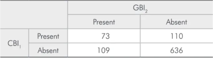
Tables 2 and 3 illustrate the frequency distribu-tion of gingival bleeding after GBI and CBI record-ing in examination sequences 1 and 2, respectively. Sensitivity, speciicity, positive and negative pre-dictive values were calculated and are expressed in Table 4. Speciicity of CBI recorded before (CBI1) and after (CBI2) GBI was 0.85 and 0.72,
respective-ly. Values of sensitivity for the respective sequences were 0.40 and 0.61. In relation to predictive values, the chances of a true result with the use of dental loss (CBI) in the absence of disease (absence of gingival inlammation detected by GBI) were 85% and 89% (negative predictive values presented in Table 4) when the dental loss was used before and after GBI had been recorded, respectively.
A perfect agreement between CBI and GBI was not observed, regardless of whether dental loss was used before (CBI1) or after (CBI2) GBI recording. Percentual agreement of the repeated recordings of GBI was 84.5% (Table 5).
Discussion
The present study tried to validate the use of dental loss (Carter, Barnes11 Index) for the diag-nosis of gingivitis in the deciduous dentition. One of the most dificult issues in validation studies is the choice of a gold-standard. There is no consen-sus in the literature about the gold-standard index for evaluation of gingival inlammation in children. The Gingival Index7 and the Gingival Bleeding Index8 are the indices mostly used in clinical and epidemiological studies.2,16 After considering the in-dices’ peculiarities, the GBI was chosen as the gold-standard in the present study because it is charac-terized by the diagnosis of gingival inlammation by stimulated bleeding, similarly to the Carter, Barnes11 (1974) Index.
Table 2 - Frequency distribution of bleeding sites with GBI followed by CBI.
GBI1
Present Absent
CBI2 Present 103 209
Absent 67 549
Table 3 - Frequency distribution of bleeding sites with CBI followed by GBI.
GBI2
Present Absent
CBI1 Present 73 110
Absent 109 636
Table 4 - Sensitivity, specificity, positive and negative predictive values for CBI recorded after (CBI2) and before (CBI1) GBI in sequences 1 and 2 (95% confidence interval).
Sensitivity Specificity Positive predictive value Negative predictive value
CBI2 0.61 (0.53 – 0.68) 0.72 (0.69 – 0.76) 0.33 (0.28 – 0.39) 0.89 (0.86 – 0.92)
CBI1 0.40 (0.33 – 0.48) 0.85 (0.83 – 0.88) 0.40 (0.33 – 0.47) 0.85 (0.83 – 0.88)
Table 5 - Percentual agreement between indices in the three sequences.
Sequences Percentual agreement
1 - GBI1/CBI2 70.3
2 - CBI1/GBI2 76.4
A higher percentage of gingival bleeding sites was observed when CBI was assessed after GBI. How-ever, this inding was not conirmed when CBI was assessed before GBI (Table 1). Two possible reasons for that can be speculated. First, the repetition of an index recording after a certain period of time (10 minutes in the present study) can represent a high-er occurrence of bleeding as a result of the trauma caused by the two consecutive mechanical stimula-tions.15 Although there was no difference when GBI was repeated (Table 1, sequence 3), it seems reason-able not to discard the possibility of trauma since bleeding after lossing is a result of a much more close contact with the papilla than that occurred after probing the gingival margin. Further investi-gations with appropriate experimental designs are needed to elucidate the role of repeated mechanical trauma in gingival bleeding overestimation.
Second, there are differences in the recording procedures for the two indices compared in the present investigation. CBI is assessed with a close contact between dental loss and interdental pa-pilla, reaching the interproximal area in a certain point not necessarily accessed by the probe during GBI recording, allowing the diagnosis of inlamma-tion at initial stages. Thilo et al.17 (1986) demon-strated that gingival inlammation in the interprox-imal area begins in the central area of the papilla. This region may not be accessible to the probe at initial stages of gingival inlammation, leading to false negative results.
There is some discussion in the literature about the viability of reproducing gingival indices. Marks et al.14 (1993) have evaluated the degree of repro-ducibility on the subject level of gingival indices and have observed a high variability across differ-ent examiners. Additionally, their results indicated that the Papillary Bleeding Index by Loesche dem-onstrated higher values of reproducibility compared to other indices. Besides the fact that marginal gin-gival bleeding upon probing is less subjective than gingival visual inspection,10 some variables can also be considered subjective when recording probing indices. Probing force and position, probe design and depth of insertion of the probe may be some examples of variables that are dificult to control
during assessment of gingival inlammation.18 These are some possible explanations for the complexity involved in reproducing gingival indices, including the repeated recordings of GBI performed in the present study in sequence 3. Thus, high kappa coef-icient values (> 0.61)19 are not expected for replicate recordings of GBI.
Duplicate measures of gingival inlammation also result in an increase in the percentage of gingi-val bleeding sites, as could be observed in sequences 1 and 3 (Table 1), similarly to the indings of Tinoco, Gjermo15 (1992) who observed an increase of 3.9% in gingival bleeding three days after the irst mea-surement. Besides, in sequence 2 (CBI1-GBI2) there was no such increase, a fact that reinforces the idea of dental loss reaching an inaccessible area for the probe. Therefore, a new mechanical stimulus pro-duced by the probe did not result in a signiicant in-crease of bleeding sites.
Professional lossing (CBI) was demonstrated to be accurate to evaluate the gingival inlammatory condition in the deciduous dentition. As shown in Table 4, speciicity and negative predictive value of CBI were satisfactory when it was recorded before and after GBI, while sensitivity and positive predic-tive value were lower. Therefore, assessing gingi-val inlammation with dental loss is more reliable in periodontal health than in disease. These lower values for sensitivity and positive predictive value are the result of a high occurrence of false-positives (209) (Table 2).
authors concluded that this modiied version of NBP (using dental loss to stimulate bleeding) seemed to be able to detect the expected change in gingi-val conditions more effectively than the GI and the dichotomized form. Thus, comparing to GBI and GI, they showed that only lossing was able to de-tect gingival health condition after some weeks of plaque control. These indings support that lossing is more precise or even more sensitive to detect early signs of gingival inlammation.
In addition to the indings of the present study, another favorable aspect of the professional lossing index is the possibility to combine patient motiva-tion and plaque control instrucmotiva-tion with
profession-al plaque removprofession-al. Motivationprofession-al factors are some of the greatest problems involved in preventing and treating behavioral diseases,20 such as dental caries and periodontal diseases. Thus, the control of peri-odontal and caries health-disease processes should start as early as possible, so children can learn and construct positive and lifelong habits.
Conclusions
It can be concluded that professional lossing, by means of assessment of the Carter, Barnes11 (1974) Index, is a useful tool for the diagnosis of inter-proximal gingival inlammatory status in children, mainly as an indicator of gingival health.
References
1. Harris R, Nicoll AD, Adair PM, Pine CM. Risk factors for dental caries in young children: a systematic review of the lit-erature. Community Dent Health. 2004;21(1 Suppl):71-85. 2. Jenkins WM, Papapanou PN. Epidemiology of
periodon-tal disease in children and adolescents. Periodontol 2000. 2001;26(1):16-32.
3. Oh TJ, Eber R, Wang HL. Periodontal diseases in the child and adolescent. J Clin Periodontol. 2002;29(5):400-10. 4. Feldens EG, Kramer PF, Feldens CA, Ferreira SH. Distribution
of plaque and gingivitis and associated factors in 3- to 5-year-old Brazilian children. J Dent Child (Chic). 2006;73(1):4-10.
5. Matsson L, Goldberg P. Gingival inflammatory reaction in children at different ages. J Clin Periodontol. 1985;12(2):98-103.
6. Silness J, Loe H. Periodontal Disease in Pregnancy. II. Cor-relation between Oral Hygiene and Periodontal Condition. Acta Odontol Scand. 1964;22(2):121-35.
7. Loe H, Silness J. Periodontal Disease in Pregnancy. I. Preva-lence and Severity. Acta Odontol Scand. 1963;21(5):533-51. 8. Ainamo J, Bay I. Problems and proposals for recording
gin-givitis and plaque. Int Dent J. 1975;25(4):229-35.
9. Muhlemann HR, Son S. Gingival sulcus bleeding - a lead-ing symptom in initial glead-ingivitis. Helv Odontol Acta. 1971;15(2):107-13.
10. Greenstein G. The role of bleeding upon probing in the diag-nosis of periodontal disease. A literature review. J Periodontol. 1984;55(12):684-8.
11. Carter HG, Barnes GP. The Gingival Bleeding Index. J Peri-odontol. 1974;45(11):801-5.
12. Abrams K, Caton J, Polson A. Histologic comparisons of in-terproximal gingival tissues related to the presence or absence of bleeding. J Periodontol. 1984;55(11):629-32.
13. Loesche WJ. Clinical and microbiological aspects of chemo-therapeutic agents used according to the specific plaque hy-pothesis. J Dent Res. 1979;58(12):2404-12.
14. Marks RG, Magnusson I, Taylor M, Clouser B, Maruniak J, Clark WB. Evaluation of reliability and reproducibility of dental indices. J Clin Periodontol. 1993;20(1):54-8. 15. Tinoco NM, Gjermo P. Comparison of the effectiveness of
three different methods in detection of changes in gingivitis in the primary dentition. Community Dent Oral Epidemiol. 1992;20(2):84-6.
16. Clerehugh V, Tugnait A. Periodontal diseases in children and adolescents: I. Aetiology and diagnosis. Dent Update. 2001;28(5):222-30, 232.
17. Thilo BE, Caton JG, Polson AM, Espeland MA. Cell popu-lations associated with interdental gingival bleeding. J Clin Periodontol. 1986;13(4):324-9.
18. Barnett ML. Suitability of gingival indices for use in thera-peutic trials. Is bleeding a sine qua non? J Clin Periodontol. 1996;23(6):582-6.
19. Landis JR, Koch GG. The measurement of observer agreement for categorical data. Biometrics. 1977;33(1):159-74. 20. Thomson WM, Poulton R, Milne BJ, Caspi A, Broughton