Carolina Bosso André(a) Thaiane Rodrigues Aguiar(a) Ana Paula Almeida Ayres(a) Glaucia Maria Bovi Ambrosano(b) Marcelo Giannini(a)
(a) Department of Restorative Dentistry, Piracicaba Dental School, State University of Campinas, Piracicaba, SP, Brazil.
(b) Department of Social Dentistry, Piracicaba Dental School, State University of Campinas, Piracicaba, SP, Brazil.
Corresponding Author:
Marcelo Giannini
E-mail: [email protected]
Bond strength of self-adhesive resin
cements to dry and moist dentin
Abstract: This study evaluated the effects of humidity conditions and evaluation times on the dentin bond strength (DBS) of two self-adhesive resin cements (RC). The RC used were: RelyX Unicem (3M ESPE) and Clearil SA Cement (Kuraray Med.). One hundred and twenty coronal portions of bovine incisors (n = 10) were used. Buccal surfaces were abraded in order to expose a lat dentinal surface (180-grit SiC) and to standardize the smear layer formation (600-grit SiC). The humidity con-ditions tested were: dry (air-dried for 10 s), slightly moist (water appli-cation with disposable applicator on dried dentin and water excess re-moved with absorbent paper), and moist (same application without water removal). The RC were used according to the manufacturers’ recommen-dations and were applied to prepolymerized resin discs (2 mm thick; Sin-fony, 3M ESPE), which were subsequently bonded to the dentin surfaces. After 24 h, half of the teeth were prepared for the microtensile bond strength test, while the other half were stored in water for 6 months and tested in tension (0.5 mm/min) until failure. A 3-way analysis of variance and the Tukey test were performed (preset alpha of 0.05). No RC showed any reduction of DBS after 6 months, and no signiicant difference was observed between them. The moist dentin increased the bond strength of Clearil SA Cement for both periods of time. Humidity conditions can change the DBS; however, the study’s results were product-dependent.
Descriptors: Dentin; Tensile Strength; Resin Cements; Humidity.
Introduction
The self-adhesive resin cements do not require acid conditioning of the tooth structure and application of the bonding agent before cementa-tion. These luting materials simplify the cementation procedures, because they save time and reduce the number of steps for cementation of indirect restorations and posts, when compared to conventional resin cements.1-3
The bonding to tooth structures is promoted by speciic functional monomers, which differ among different commercial products. Accord-ing to the manufacturers, the functional monomers are able to bond chemically the calcium from hydroxyapatite, which is one of the bonding mechanisms responsible for the retention of the restoration.4-6 However,
little information is available about these chemical reactions, and the durability of this bonding requires further investigation, since clinicians have switched from conventional resin cement to these new self-adhesive resin cements.7-9
Declaration of Interests: The authors certify that they have no commercial or associative interest that represents a conflict of interest in connection with the manuscript.
Submitted: Apr 07, 2013
The manufacturers’ instructions provide infor-mation regarding the proper use of these materials; however, some of these are unclear regarding the humidity conditions of mineralized tissues (enamel and dentin) before cementation. The purpose of this
in vitro study was to measure the bond strength of
pre-polymerized composite discs to underlying den-tin, using two self-adhesive resin-based cements un-der three humidity conditions. In addition, the bond failure site morphology was analyzed and compared among material types and humidity conditions. The research hypothesis tested was that the bond strength would be signiicantly higher when the res-in cement is applied to wet dentres-in than when it is used on dried surfaces.
Methodology
Two self-adhesive resin cements were selected:
• RelyX Unicem (3M ESPE, St. Paul, USA) and
• Clearil SA Cement (Kuraray Noritake Dental Inc., Kurashiki, Japan).
The composition and the batch number are pre-sented in Table 1. One hundred and twenty freshly extracted incisor bovine teeth (stored in a 0.05% thymol [LabSynth, Diadema, Brazil] solution at 5°C) were used and randomly divided into four groups (n = 10; Institutional Review Board proto-col #089/2009). Their buccal surfaces were wet and abraded with 180-grit silicon carbide paper (Carborundum Abrasivos, Vinhedo, Brazil) using a machine (APL-4, Arotec Ind. e Com. Ltda., Cotia, Brazil) to remove the enamel and to expose a lat dentin surface with a remaining dentin thickness ranging from 1.0 to 1.5 mm. Afterwards, the teeth were abraded with 600-grit silicon carbide paper (Carborundum Abrasivos, Vinhedo, Brazil) for 10 s to standardize the smear layer formation. The hu-midity conditions tested were:
1. Dry: air-dried for 10 s, 70 psi and a distance of 10 mm between the dentin surface and the tip of the air-syringe (Dabi Atlante, Ribeirão Preto, Brazil).
2. Slightly moist: water application with a Micro-brush disposable applicator (MicroMicro-brush Interna-tional, Grafton, USA) on dried dentin, and water
excess removed with absorbent paper (Kleenex, Kimberly-Clark, Mogi das Cruzes, Brazil).
3. Moist: the same application of condition 2 with-out the removal of water.
One hundred and twenty pre-polymerized (B2D shade; Sinfony, 3M ESPE, St. Paul, USA), light-cured composite resin discs, 2 mm thick and 10 mm in diameter, were prepared to simulate overlying laboratory-processed composite resin restorations. The surface of each disc to be bonded to the pre-pared tooth was airborne-particle abraded with 50 µm aluminum oxide (Danville Engineering Inc., San Ramon, USA) for 10 s (air pressure: 0.552 MPa; distance from the tip: 1.5 cm), and silanated using coupling agents (Ceramic Primer, 3M ESPE, St. Paul, USA or Clearil Ceramic Primer, Kuraray Noritake Dental Inc., Kurashiki, Japan), according to the manufacturer’s directions.10
All cements were manipulated and applied ac-cording to the manufacturers’ instructions. The mixed resin cement pastes were applied to the sand-blasted and silanated surface of the pre-polymerized composite resin disc, after which the disc was placed on the dentin surface. A 500 g load was applied for 5 min. For light-polymerized groups, the load was removed and the light-activating tip (XL 3000, 3M ESPE, St. Paul, USA) was positioned against the composite resin disc after loading, and the unit was activated for 40 s. To facilitate the specimen-grip-ping length while bond testing was being performed,
Table 1 - Composition and lot number of the materials tested in this study.
Resin
cement Composition (lot number) Clearfil SA
Cement Bis-GMA, TEGDMA, sodium fluoride, 10-methacryloyloxydecyl dihydrogen phosphate, hydrophobic aromatic dimethacrylate, hydrophobic aliphatic dimethacrylate, silanated barium glass filler, silanated colloidal silica, dl-camphorquinone, initiators, accelerators, catalysts, pigments (filler = 66 wt%; 45 vol%; avg. 2.5 µm) (0004AB) RelyX
sputter-coated with gold (MED 010, Balzers, Bal-zer, Liechtenstein) and examined by a single indi-vidual using a scanning electron microscope (volt-age 15 kV; VP 435, Leo, Cambridge, UK). Failure patterns were classiied as:
1. cohesive within the resin cement;
2. adhesive along the pre-polymerized composite overlay–resin cement interface;
3. adhesive along the dentin surface;
4. mixed when simultaneously exhibiting the den-tin surface and remnants of the resin cement; and
5. cohesive within the dentin.
Representative areas of the failure patterns were photographed at 400×.
Results
The mean bond strength values are presented in Table 2. Three-way ANOVA revealed statistically signiicant differences only for the factor, “humid-ity” (p = 0.0475). Conversely, the statistical analysis revealed no signiicant differences for the “material” (p = 0.3309) and “time” (p = 0.8859) factors. The double interactions between “material and humid-ity” and “material and time” factors were signii-cant (p = 0.0384 and p = 0.0072, respectively). The triple interaction was not statistically signiicant (p = 0.5440).
The Tukey-Kramer test revealed signiicant differences among humidity conditions only for Clearil SA Cement (p < 0.05). Analysis of data with respect to these differences in the humidity condi-tion showed that the Clearil SA Cement had a sig-niicantly lower bond strength when applied to the dried dentin surface than when applied to moist den-tin (p < 0.05). However, the dry and slightly moist a 3-mm-thick block of autopolymerizing composite
resin (Concise, 3M of Brazil, Sumaré, Brazil) was then added to the untreated, pre-polymerized com-posite resin surface.
The teeth were stored in distilled, deionized wa-ter at 37°C for 24 h, and were then vertically, seri-ally sectioned into several 1.0-mm-thick slabs using a cutting instrument (Isomet 1000, Buehler Ltd., Lake Bluff, USA). Each slab was further sectioned perpendicularly to produce bonded sticks approxi-mately 1.0 mm² in cross-section. Each bonded stick was attached to the grips of a microtensile testing device (Cometa, Piracicaba, Brazil) with cyanoacry-late glue (Super Bonder, Henkel/Loctite, Diadema, Brazil), and tested in tension in a universal testing machine (EZ Test, Shimazu, Kyoto, Japan) at a crosshead speed of 0.5 mm/min until failure. After testing, the specimens were removed carefully, and the cross-sectional area at the site of fracture was measured to the nearest 0.01 mm with a digital cali-per (727-6/150, Starret Ind. e Com. Ltda., Itu, Bra-zil). The specimen’s cross-sectional area was divided by the peak tensile load at failure to calculate the stress at fracture (MPa). A single failure stress value was then calculated for each tooth by averaging the values of 6 sticks from that tooth. A 3-way analy-sis of variance (ANOVA) (factors: material, humid-ity and time) was performed to determine the effect of these major factors on and their interaction with tensile strength. The Tukey-Kramer test was used to detect multiple comparisons among the experimen-tal groups. All statistical testing (SAS Institute Inc., Cary, USA) was performed at a preset alpha of 0.05.
Fractured surfaces of tested specimens were al-lowed to air-dry (Marconi Equip. Lab., Piracicaba, Brazil) overnight at 37°C, after which they were
Resin cement Evaluation time Humidity conditions
Dry Slightly moist Moist
Clearfil SA Cement
24 hours 12.7 (6.0) B 15.7 (6.3) AB 19.0 (8.4) A RelyX Unicem 18.9 (5.8) A 19.3 (3.6) A 19.7 (2.8) A
Clearfil SA Cement
6 months 14.2 (6.2) B 17.8 (7.0) AB 22.1 (4.6) A RelyX Unicem 18.3 (6.7) A 15.7 (2.9) A 16.5 (3.6) A No significant difference (p > 0.05) in bond strength was noted between resin cements and evaluation times (uppercase letters = row).
conditions did not differ from each other (p > 0.05). The storage time did not inluence the bond strength results (p > 0.05).
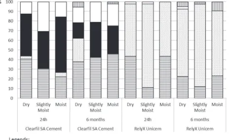
Figure 1 shows the proportional prevalence (%) of the failure patterns in all experimental groups. Rep-resentative images depicting the failure classiications are presented in Figures 2–6. All groups showed cohe-sive failure within the resin cement (type 1; Figure 2). RelyX Unicem presented a high incidence of adhesive
failure along the pre-polymerized composite overlay– resin cement interface (type 2; Figure 3), whereas ad-hesive failures along the dentin surface (type 3) and mixed fracture (type 4) were observed for Clearil SA Cement (Figures 4 and 5, respectively) at both evalu-ation times. Cohesive failures within the dentin (type 5) were observed for both luting materials only after the storage of specimens for 6 months (Figure 6).
Figure 1 - Distribution of failure modes among experimental groups.
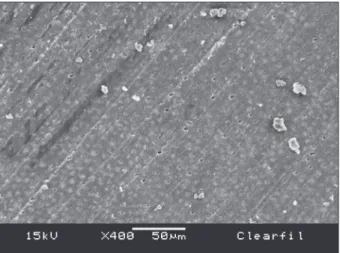
Figure 2 - Cohesive failure within the Clearfil SA Cement
Discussion
The functional monomer of RelyX Unicem is methacrylated phosphoric ester, which reacts chemi-cally with the hydroxyapatite, promoting the bond-ing to the dental structures and with iller particles that are the basic components of this self-adhesive resin cement. This second acid-base reaction is im-portant in neutralizing the acidic characteristic of the resin cement and reducing the hydrophilic-ity after mixing of the catalyst and base pastes.3,11-13
The bond strength of RelyX Unicem to dentin ob-tained in this study ranged from 15.7 ± 2.9 MPa to
19.7 ± 2.8 MPa. These values were higher than those obtained by Piwowarczyk et al.14 (6.2 MPa), Yang et
al.15 (8.2 MPa), Goracci et al.16 (6.8 MPa),
Holdereg-ger et al.17 (9.2 MPa), and Egilmez et al.18 (13 MPa).
On the other hand, similar results were found by De Munck et al.,11 Sarr et al.,19 Luhrs et al.,20 Mazzitelli
et al.,21 Ebert et al.,22 and Inukai et al.23
Although the hydrated dentin substrate can fa-cilitate ionization of the acid monomers, the results of this study suggested that the humidity conditions did not affect the bond strength of RelyX Unicem to dentin, i.e., no signiicant difference was observed when the self-adhesive was applied to surfaces with different humidity conditions. Conversely, when Guarda et al.24 did not over-dry the dentin surface
before cementation, a higher bond strength was ob-served for RelyX Unicem. Moreover, Mazzitelli et al.21 found that RelyX Unicem performed better
un-der simulated pulpar pressure, because the constant intrapulpar water perfusion changes the substrate wetness.
The functional monomer of Clearil SA Cement is the 10-methacryloyloxydecyl dihydrogen phos-phate (10-MDP), which is able to form a strong and stable ionic bond with calcium from hydroxyapap-tite.25 Besides this chemical interaction,26 Clearil
SA Cement can also provide micromechanical reten-tion, since its monomer is capable of iniltrating the dentinal substrate.27,28 The wetness of dentin was
Figure 6 - Cohesive failure within the dentin (type 5) after 6 months of water storage with RelyX Unicem (original mag-nification 400×).
Figure 4 - Adhesive failure along the dentin surface (type 3) with absence of the self-adhesive resin (Clearfil SA Cement) over the dentin surface (original magnification 400×).
important for increasing the bond strength of this resin cement. In the presence of water, the acidic monomer of Clearil SA resin cement was ionized, acid-etched the dentin, and interacted with the den-tinal substrate. Thus, the extrinsic denden-tinal wetness may have optimized these acid-base reactions, al-lowing for better setting and bonding.
In comparison with RelyX Unicem, few studies have investigated the bond strength of Clearil SA Cement to dentin. In this study, the bond strength re-sults for Clearil SA Cement ranged from 12.7 ± 6.0 MPa in dry dentin to 22.1 ± 4.6 MPa in moist den-tin, which were signiicantly different. The bond strength results for Clearil SA Cement from Ebert et al.,22 Inukai et al.23 and Ilday et al.29 corroborated
our data, while Egilmez et al.18 showed lower bond
strength values than those obtained in this study. The self-adhesive resin cements (RelyX Unicem and Clearil SA Cement) tested in this study showed no signiicant difference between materials in terms of bond strength. Although they present different compositions, these commercial formulations did not inluence the bonding to dentin. In addition, the storage in water for 6 months did not alter the bond strength to dentin for any of the resin cements tested, independent of the humidity conditions of dentin. The chemical reaction between 10-MDP and cal-cium from hydroxyapatite is considerably stable,25-28
which explains the absence of bond strength reduc-tion for Clearil SA Cement. The setting reacreduc-tion of RelyX Unicem increases the pH value, changing the
monomer nature from hydrophilic to hydrophobic, and this neutralization reaction is important for the long-term stability of RelyX Unicem cement regard-ing bond strength to dentin.3,11-13
Although no change was observed in bond strength, the failure pattern was modiied after stor-age of the bonded beam specimens for 6 months in water. The specimens stored in water for 6 months induced some cohesive failures within dentin, which did not occur for the specimens stored for 24 hours. The storage of bonded beams instead of restored teeth and without a peripheral composite-enamel bond30 seemed to accelerate the degradation rate
of the exposed dentin, resulting in cohesive failures within dentin during the tensile test.
Conclusion
The storage in water for 6 months does not de-crease the bond strength of self-adhesive resin ce-ments to dentin. Conversely, the humidity con-ditions can change the bond strength to dentin; however, these results are product-dependent.
Acknowledgements
This study was supported by grants from PI-BIC/CNPq (Programa Institucional de Bolsas de Iniciação Cientíica / Conselho Nacional de
De-senvolvimento Cientíico e Tecnológico) and
FA-PESP (Fundação de Amparo à Pesquisa do Estado
de São Paulo), processes nos. 2009/51674-6 and
2010/13599-0, Brazil.
References
1. Radovic I, Monticelli F, Goracci C, Vulicevic ZR, Ferrari M. Self-adhesive resin cements: a literature review. J Adhes Dent. 2008 Aug;10(4):251-8.
2. Burgess JO, Ghuman T, Cakir D. Self-adhesive resin cements. J Esthet Restor Dent. 2010 Dec;22(6):412-9.
3. Ferracane JL, Stansbury JW, Burke FJ. Self-adhesive resin cements - chemistry, properties and clinical considerations. J Oral Rehabil. 2011Apr;38(4):295-314.
4. Aguiar TR, Di Francescantonio M, Ambrosano GMB, Gi-annini M. Effect of curing mode on bond strength of self-adhesive resin luting cements to dentin. J Biomed Mater Res B Appl Biomater. 2010 Apr;93(1):122-7.
5. Pisani-Proença J, Erhardt MC, Amaral R, Valandro LF, Bot-tino MA, Del Castillo-Salmerón R. Influence of different
sur-face conditioning protocols on microtensile bond strength of self-adhesive resin cements to dentin. J Prosthet Dent. 2011 Apr;105(4):227-35.
6. Vaz RR, Hipólito VD, D’Alpino PH, Goes MF. Bond strength and interfacial micromorphology of etch-and-rinse and self-adhesive resin cements to dentin. J Prosthodont. 2012 Feb;21(2):101-11.
9. Hooshmand T, Mohajerfar M, Keshvad A, Motahhary P. Mi-croleakage and marginal gap of adhesive cements for noble al-loy full cast crowns. Oper Dent. 2011 May-Jun;36(3):258-65. 10. Soares CJ, Giannini M, Oliveira MT, Paulillo LAMS, Martins
LRM. Effect of surface treatments of laboratory-fabricated composites on the microtensile bond strength to a luting resin cement. J Appl Oral Sci. 2004 Mar;12(1):45-50.
11. De Munck J, Vargas M, Van Landuyt K, Hikita K, Lambrechts P, Van Meerbeek B. Bonding of an auto-adhesive luting mate-rial to enamel and dentin. Dent Mater. 2004 Dec;20(10):963-71.
12. Gerth HUB, Dammaschke T, Zuchner H, Schafer E. Chemi-cal analysis and bonding reaction of RelyX Unicem and Bifix composites - a comparative study. Dent Mater. 2006 Oct;22(10):934-41.
13. Al-Assaf K, Chakmakchi M, Palaghias G, Karanika-Kouma A, Eliades G. Interfacial characteristics of adhesive luting resins and composites with dentine. Dent Mater. 2007 Jul;23(7):829-39.
14. Piwowarczyk A, Bender R, Ottl P, Lauer HC. Long-term bond between dual-polymerizing cementing agents and human hard dental tissue. Dent Mater. 2007 Feb;23(2):211-7.
15. Yang B, Ludwig K, Adelung R, Kern M. Micro-tensile bond strength of three luting resins to human regional dentin. Dent Mater. 2006 Jan;22(1):45-56.
16. Goracci C, Cury AH, Cantoro A, Papacchini F, Tay FR, Fer-rari M. Microtensile bond strength and interfacial properties of self-etching and self-adhesive resin cements used to lute composite onlays under different seating forces. J Adhes Dent. 2006 Oct;8(5):327-35.
17. Holderegger C, Sailer I, Schuhmacher C, Schläpfer R, Häm-merle C, Fischer J. Shear bond strength of resin cements to human dentin. Dent Mater. 2008 Jul;24(7):944-50. 18. Egilmez F, Ergun G, Cekic-Nagas I, Vallittu PK, Lassila LV.
Bond strength of self-adhesive resin cements to dentin after antibacterial and chelating solution treatment. Acta Odontol Scand. 2013 Jan;71(1):22-31.
19. Sarr M, Mine A, De Munck J, Cardoso MV, Kane AW, Vreven J, et al. Immediate bonding effectiveness of contem-porary composite cements to dentin. Clin Oral Investig. 2010 Oct;14(5):569-77.
20. Luhrs AK, Guhr S, Gunay H, Geurtsen W. Shear bond strength of self-adhesive resins compared to resin cements
with etch and rinse adhesives to enamel and dentin in vitro. Clin Oral Investig. 2010 Apr;14(2):193-9.
21. Mazzitelli C, Monticelli F, Osorio R, Casuccia A, Toledano M, Ferrari M. Effect of simulated pulpal pressure on self-adhesive cements bonding to dentin. Dent Mater. 2008 Sep;24(9):1156-63.
22. Ebert J, Leyer A, Gunther O, Lohbauer U, Petschelt A, Fran-kenberger R, et al. Bond strength of adhesive cements to root canal dentin tested with a novel pull-out approach. J Endod. 2011 Nov;37(11):1558-61.
23. Inukai T, Abe T, Ito Y, Pilecki P, Wilson RF, Watson TF, et al. Adhesion of indirect MOD resin composite inlays luted with self-adhesive and self-etching resin cements. Oper Dent. 2012 Sep-Oct;37(5):474-84.
24. Guarda GB, Gonçalves LS, Correr AB, Moraes RR, Sinhoreti MAC, Correr-Sobrinho L. Luting glass ceramic restorations using a self-adhesive resin cement under different dentin condi-tions. J Appl Oral Sci. 2010 May-Jun;18(3):244-8.
25. Yoshida Y, Nagakane K, Fukuda R, Nakayama Y, Okazaki M, Shintani H, et al. Comparative study on adhesive performance of functional monomers. J Dent Res. 2004 Jun;83(6):454-8. 26. Fujita K, Ma S, Aida M, Maeda T, Ikemi T, Hirata M, et al.
Effect of reacted acidic monomer with calcium on bonding performance. J Dent Res. 2011 May;90(5):607-12.
27. Reis AF, Arrais CAG, Novaes PD, Carvalho RM, De Goes MF, Giannini M. Ultramorphological analysis of resin–dentin interfaces produced with water-based single-step and two-step adhesives: nanoleakage expression. J Biomed Mater Res B Appl Biomater. 2004 Oct;71(1):90-8.
28. Reis AF, Giannini M, Pereira PNR. Long-term TEM anal-ysis of the nanoleakage patterns in resin–dentin interfaces produced by different bonding strategies. Dent Mater. 2007 Sep;23(9):1164-72.
29. Ilday N, Gungor H, Duymus ZY. Dentine treatment effects on bonding strength of adhesive resin cements. Mater Res Innov. 2011 Jun;15(3):202-7.