Matheus Melo PITHON(a) Rogério LACERDA-SANTOS(b) Danillo Lyrio de OLIVEIRA(a) Janaína Viana ALVES(a) Joyce Pereira BRITTO(a) Eduardo da Silva SOUZA(a) Leandro Pereira ALVES(a) George BARBOSA(c)
Raildo da Silva COqUEIRO(d) Alex Ferreira dos SANTOS(c)
(a)Universidade Estadual do Sudoeste da Bahia – UESB, Curso de Odontologia, Departamento de Ortodontia, Jequié, BA, Brazil.
(b)Universidade Federal de Campina Grande – UFCG, Curso de Odontologia, Departamento de Clínica Infantil, Patos, PB, Brazil.
(c)Universidade Estadual do Sudoeste da Bahia – UESB, Curso de Sistema da Informação, Departamento de Ciência da Computação, Jequié, BA, Brazil.
(d)Universidade Estadual do Sudoeste da Bahia – UESB, Curso de Odontologia, Departamento de Epidemiologia, Jequié, BA, Brazil.
Esthetic perception of facial profile after
treatment with the Thurow appliance
Abstract: The objective of this article was to investigate the perception
of esthetic changes in the facial proile of bilateral Class II patients treated with the Modiied Thurow Appliance for extraoral treatment. Silhouettes were traced of proiles of patients who initially presented a bilateral Class II molar relationship and who, post-treatment, presented molars in a Class I relationship. Three groups were formed: the irst composed of patients with maxillary protrusion (SNA >84°), the second with maxillary retrusion (SNA <80°), and the third with a well-positioned maxilla (SNA 80-84°). A panel of 200 lay evaluators judged the proile esthetics by a randomized drawing of the silhouettes. The multiple analysis results showed that the proile esthetic scores for the three positions of the maxilla were greatly inluenced by signiicant interactions with the characteristics (like sex and age) of the evaluators:
retrusive maxilla (Score *Age Group, p < 0.001), normal maxilla (Score
*Sex, p = 0.024; Score *Age Group, p = 0.050) and protrusive maxilla
(Score *Age Group, p < 0.001). It was observed that the proile of Class
II patients with protrusion, normal relationship and retrusion of the maxilla, improved in their esthetic post-treatment result; however, the evaluators showed greater satisfaction with the groups of protrusion and normal position of the maxilla. The Modiied Thurow Appliance provided signiicant improvements in the esthetics of the proile of patients who presented protrusion and normal position of the maxilla. However, its use was not the best treatment option for patients with maxillary retrusion.
Keywords: Perception; Face; Malocclusion.
Introduction
Angle’s Class II malocclusion is characterized by a dental discrepancy in which the mesial groove of the lower irst permanent molar articulates posterior to the mesiobuccal cusp of the irst permanent maxillary molar. It can also be characterized by a maxillomandibular skeletal discrepancy in the anteroposterior direction, by maxillary protrusion, by mandibular retrusion or by a combination of these factors. The incidence of this malocclusion ranges from 35% to 42%, and may attain up to 50% of the
clinical cases treated by orthodontists.1,2
An ininite variety of appliances are available for the orthopedic correction of Class II malocclusion; these include both intraoral and
extraoral appliances.3,4,5,6 Among the intraoral orthopedic appliances,
Declaration of Interests: The authors certify that they have no commercial or associative interest that represents a conflict of interest in connection with the manuscript.
Corresponding Author: Matheus Melo Pithon
E-mail: [email protected]
DOI: 10.1590/1807-3107BOR-2015.vol29.0043
Submitted: May 25, 2014
those of Herbst,7Bionator,8 Planas9 and other types
are mentioned in the literature. In regard to extraoral orthopedic appliances, there are those made with an outer facial arch attached to rings cemented to the molars, and there is also the Thurow Appliance, where the outer facial arch is attached to acrylic resin
adapted to the occlusal surfaces of the teeth.10
Treatment with the Thurow Appliance is made by restricting anterior growth of the maxilla, or redirecting facial growth, and is indicated precisely when the malocclusion is determined predominantly
by maxillary protrusion.10,11 However, these devices
are also used with skeletal Malocclusion Class II in growing individuals presenting a well-positioned or
retropositioned maxilla.12 However, it is interesting
to query what the esthetic repercussions would be of using these appliances in different maxillary positions?
In this sense, the authors’ purpose in the present study was to evaluate the perception of laypersons with respect to silhouettes of Class II patients with different maxillary positions, treated with the Modiied Thurow Appliance.
Methodology
Research was conducted in the archives of the orthodontic documentation of the Pediatric Dental Clinic of the dentistry course at the State University of Southeast Bahia, in order to identify the records of patients treated with the Modiied Thurow Appliance,
who presented different anteroposterior positions of
the maxilla. The inclusion criteria for the sample were: patients treated with the Modiied Thurow Appliance, treated with pre- and post-treatment orthodontic models, and having pre- and post-treatment lateral cephalometric radiographs that came from the same radiology center. The accepted patients had a Class II molar relationship bilaterally (< 5 mm) on pre-treatment, and a good Class I molar relationship
on post-treatment, with angle SN.GoGn ≤ 35 on pre-
and post-treatment, similar skeletal maturation (hand-wrist radiographic evaluation) on pre- and treatment, mixed dentition on pre- and post-treatment, and absence of open bite and crossbite on pre- and post-treatment. All the patients were treated without extractions and without using a ixed orthodontic appliance afterwards.
The research participants were divided into three
groups, based on the anteroposterior position of the maxilla. Ten patients were identiied as initially having maxillary protrusion (SNA > 84°), ten initially having maxillary retrusion (SNA < 80°), and ten fitting into the normal group (SNA 80-84°). The Modiied Thurow Appliance components included an occlusal self-polymerizing acrylic resin (OrtoCril, Sao Paulo, Brazil) base plate covering the entire palate up to contact of the occlusal surfaces of the primary
molars and the permanent 1st molar. The appliance
also had an expander screw (Morelli Ortodontia, São Paulo, Brazil) centralized in the palate at the level of the primary second molars, and Adams’ cribs, for retention, on both upper irst permanent molars. In addition, high traction and cervical traction were used on the patients, with a high or low mandibular plane, respectively.
The sample size was calculated considering the minimal difference between the means of treatment of 1 mm for any of the linear distances (Class II right or Class II left) and a standard deviation of 0.5 mm. Considering a one-sided signiicance level of 0.01 and a power of 85%, at least 10 patients per group were required. This experiment was approved by the Human Research Ethics Committee, CEP/ CAAE: 0154.0.454.000-11.
Table 1. Pre-treatment and post-treatment linear and angular measurements of patients used for constructing the profile silhouettes.
Variable Patients with maxillary protrusion Patients with normal maxilla Patients with maxillary retrusion
Mean Mean Mean
SNA (º)
Pre-treatment 87 82 75.5
Post-treatment 82.5 80 74.5
Difference -4.5 -2 -1
SNB (º)
Pre-treatment 79.5 77.5 70.5
Post-treatment 79.5 77.5 71
Difference 0 0 0.5
ANB (º)
Pre-treatment 7.5 4.5 5
Post-treatment 3 2.5 3.5
Difference -4.5 -2 -1.5
SnGoGn (º)
Pre-treatment 34 33 33.5
Post-treatment 34 33 33.5
Difference 0 0 0
1.NA (º)
Pre-treatment 33.5 30 28.5
Post-treatment 24.5 23 21
Difference -9 -7 -7.5
Overjet
Pre-treatment 7 6.5 5.5
Post-treatment 2.5 3 2
Difference -4.5 -3.5 -3.5
Overbite
Pre-treatment 6 9.5 6.5
Post-treatment 2.5 2.5 2
Difference -3.5 -7.0 -4.5
Class II right (mm)
Pre-treatment 3 3 2.5
Post-treatment 0 0 0
Difference -3 -3 -2.5
Class II left (mm)
Pre-treatment 3 3 3
Post-treatment 0 0 0
considering the degree of magniication recorded for all the radiographs, i.e., 8%.
According to the previously described methodology, pre- and post-treatment lateral cephalometric tracings were used to produce dark silhouettes, identiied only by numbers. The tracings were scanned by the GT
2400 appliance (Hewlett-PackardLtd., Tokyo, Japan),
deined for the gray scale with a resolution of 500 dpi, and saved as TIFF images in the Adobe Photoshop CS3 program (Adobe Systems, San Jose, USA). Each traced line was rotated so that the Horizontal Frankfurt (FH) Plane would be parallel to the ground.
For the purpose of creating the profile presentation for the judges, the silhouettes were transferred to the PowerPoint program, version 2010 (Microsoft Corporation, Redmond, USA) in random order. Each image of the silhouette was classiied with a sequential number to help the evaluators, and these numbers were crossed with the speciic initial or inal proile, at intervals of 10 seconds for each silhouette proile to be evaluated (Figure 1).
The panel of evaluators consisted of 200 laypersons (Table 2). The ages assessed represented the majority of patients who initially seek orthodontic treatment. The groups were stratiied to allow full identiication with a group of like individuals,13 in order to maintain
the cognitive speciicities of the age groups and quality of results.14 The evaluators were invited to assess each
silhouette proile and attribute an attractiveness score, using the Likert scale from 0 (not very attractive) to 7 (very attractive).
The analysis of variance (ANOVA) for the mixed model of repeated measures was used to determine the differences in the evaluations of the pre- and
post-treatment esthetic proile scores for the three clinical proiles assessed: maxillary protrusion, maxilla in a normal position and maxillary retrusion. ANOVA was also used to evaluate the effect of the evaluator’s characteristics, such as sex and age group. The level of significance adopted was 5%. All the analyses were performed with the SPSS 15.0 for Windows (SPSS Inc., Chicago, USA) statistical software program.
Results
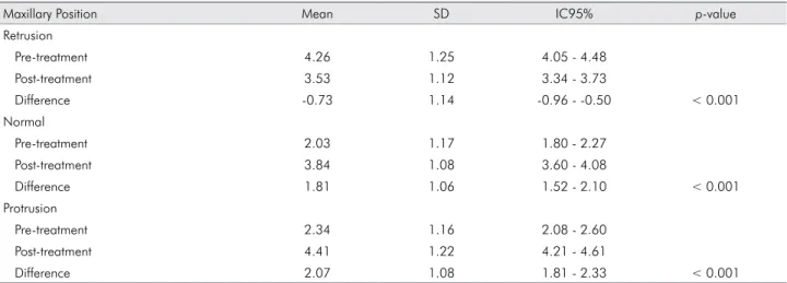
In the case of the retrusive maxilla, the esthetic profile evaluation scores were lower for the post-treatment silhouettes, indicating the evaluators’ dissatisfaction with the inal result of the treatment. In contrast, in the case of the well-positioned and the protrusive maxilla, the esthetic proile evaluation scores were higher for the post-treatment silhouettes, indicating that the evaluators considered the inal result of the treatment as attractive (Table 3).
Multiple analysis showed that the esthetic profile scores for the three maxilla positions were inluenced by signiicant interactions with the evaluators’ characteristics (like sex and age):
retrusive maxilla (Score *Age Group, p < 0.001),
normal maxilla (Score *Sex, p = 0.024; Score *Age
Group, p = 0.05) and protrusive maxilla (Score *Age
Group, p < 0.001).
The results indicated a signiicant main effect of sex in combination with the evaluators’ age group on the scores of the esthetic proile for the maxilla in a position of retrusion (Table 4). The interaction between sex and age group showed that among the evaluators from 8 to 12 and from 18 to 22 years of age, the participants of the female sex gave higher
scores. However, among the evaluators from 13 to 17 and from 23 to 27 years of age, participants of the male sex gave higher scores.
The evaluations varied in respect to the maxilla in the normal position, when considering sex and age group (Table 4). In general, the highest scores were given by evaluators of the male sex and age group from 8 to 12 years. There was no statistical difference among the scores of the evaluators of the
three older age groups (13 to 17 years, 18 to 22 years and 23 to 27 years).
The evaluations in respect to the maxilla in the protrusion position varied only when considering the age group (Table 4). According to the previously described methodology, pre- and post-treatment lateral cephalometric tracings were used to produce dark silhouettes, identiied only by numbers. There was no statistical difference among the scores given by the evaluators of the three older age groups (13 to 17 years, 18 to 22 years and 23 to 27 years).
Discussion
Recently, it has been observed that much attention is being given to the perception of laypersons, as well as of dental professionals, in regard to esthetic evaluations.15,16 The results of this study indicate that the
individuals who evaluated the silhouettes presented to them were capable of observing the differences existing among these images, as demonstrated by
their assessment scores.17
Table 2. Description of the evaluators.
Variables n %
Sex
Male 100 50.0
Female 100 50.0
Age Groups
8 to 12 years 50 25.0
13 to 17 years 50 25.0
18 to 22 years 50 25.0
23 to 27 years 50 25.0
Table 3. Differences in the evaluations between the pre-treatment and post-treatment silhouettes.
Maxillary Position Mean SD IC95% p-value
Retrusion
Pre-treatment 4.26 1.25 4.05 - 4.48
Post-treatment 3.53 1.12 3.34 - 3.73
Difference -0.73 1.14 -0.96 - -0.50 < 0.001
Normal
Pre-treatment 2.03 1.17 1.80 - 2.27
Post-treatment 3.84 1.08 3.60 - 4.08
Difference 1.81 1.06 1.52 - 2.10 < 0.001
Protrusion
Pre-treatment 2.34 1.16 2.08 - 2.60
Post-treatment 4.41 1.22 4.21 - 4.61
Difference 2.07 1.08 1.81 - 2.33 < 0.001
SD: standard deviation; IC95%: interval of confidence of 95% of the mean.
Table 4. Effects of evaluators’ characteristics on evaluations of the silhouettes.
Characteristics Maxillary Position (p-values)
Retrusion Normal Protrusion
Sex 0.396 < 0.05 0.164
Age Group 0.691 < 0.001 < 0.001
Clinically, orthodontists diverge in regard to the use of photographs, silhouettes and cephalometric tracings to evaluate the esthetics of the facial proile. Nevertheless, by observing silhouettes, one is able to eliminate factors that inluence attractiveness in the eye of the observer, such as sex, age, skin color, shape and color of hair, and style and color of eyes, factors
that may be noted when using photographs.15,18,19,20
However, by eliminating attributes like sex, other features of the face may be excessively emphasized, for example, the size and shape of the nose, or difference in the interlabial gap.15
Variants such as the sex and age group of the evaluators indicated a signiicant effect on the esthetic proile scores given in the study. The evaluators of the female sex in the age range from 8 to 12 years and from 18 to 22 years were the ones who attributed the highest attractiveness scores during the evaluation of maxillary retrusion. In contrast, the evaluators of the male sex in the age range from 8 to 12 years attributed higher attractiveness scores when assessing the maxilla in the normal position. Note that there was no statistical difference for the other age groups, i.e., from 13 to 17, 18 to 22 and 23 to 27 years of age. This is in contrast to a study in which men were more
critical than women and attributed lower scores.21
There was variation in regard to age group only for maxillary protrusion, where the higher scores were given by the evaluators from 8 to 12 years old. Previous studies in which esthetic proiles relating to maxillary protrusion were evaluated also detected no
signiicant differences between men and women.15,22
The maxillary anteroposterior position is directly related to the esthetic proile. Normally, North Americans prefer straight proiles to concave or convex proiles.23,24,25
In patients with extreme protrusion or retrusion, the maxillary position has a negative inluence on esthetics, particularly because it will affect the balance between the maxillae when seen in proile. Therefore, surgical
correction may be an option.23
According to the data found, differences between the pre- and post-treatment evaluations occurred in the three proile silhouettes assessed; however, the evaluation was more signiicant for the patients with maxillary protrusion, who obtained the highest
attractiveness scores in the assessment, similar to the group of patients with normal maxillary position.
In the treatment of Class II patients with maxillary retrusion, the attractiveness scores were lower in the post-treatment assessment, indicating that the evaluators were dissatisied with the inal esthetic result, and that the treatment given was not the best treatment option for these cases. This shows that the decision of what treatment should be given must also be based on the individual characteristics of the patient, the diagnosis and the desired result, and not only on the esthetic results. The results obtained were found to differ from those of a previous study, in which the use of the Thurow extraoral appliance was successful in enhancing the proile esthetics of Class II patients
with different maxillary anteroposterior positions.23
The esthetic improvements in the facial proile of the groups with protrusion and normal maxillary position, treated with the Modiied Thurow Appliance, normally included better chin projection, reduction in the interlabial gap, and reduction in the depth of the labiomental sulcus, factors also observed in the treatment of patients with the same malocclusion and maxillary anteroposterior positions as those treated
with the extraoral appliance.3,23
In view of the results found, the Modiied Thurow Appliance can be considered a good option in the treatment of Class II malocclusions with protrusive and well-positioned maxillae, making this an important ally for the orthodontist, because it is a low cost and easy-to-fabricate device.
Conclusions
1. Hodge TM, Boyd PT, Munyombwe T, Littlewood SJ. Orthodontists’ perceptions of the need for orthognathic surgery in patients with Class II Division 1 malocclusion based on extraoral examinations. Am J Orthod Dentofacial Orthop. 2012 Jul;142(1):52-9.
2. Millett DT, Cunningham SJ, O’Brien KD, Benson PE, Oliveira CM. Treatment and stability of class II division 2 malocclusion in children and adolescents: a systematic review. Am J Orthod Dentofacial Orthop. 2012 Aug;142(2):159-69. 3. Lima Filho RM, Lima AL, Ruellas ACO. Mandibular
changes in skeletal class II patients treated with Kloehn cervical headgear. Am J Orthod Dentofacial Orthop. 2003 Jul;124(1):83-90.
4. Nayak KU, Goyal V, Malviya N. Two-phase treatment of class II malocclusion in young growing patient. Contemp Clin Dent. 2011 Oct-Dec;2(4):376-80.
5. Tripathi NB, Patil SN. Treatment of class II division 1 malocclusion with myofunctional trainer system in early mixed dentition period. J Contemp Dent Pract. 2011 Nov;12(6):497-500.
6. Janson G, Valarelli DP, Valarelli FP, Freitas MR. Treatment times of Class II malocclusion: four premolar and non-extraction protocols. Eur J Orthod. 2012 Apr;34(2):182-7. 7. Wieslander L. Intensive treatment of severe Class II
malocclusions with a headgear-Herbst appliance in the early mixed dentition. Am J Orthod. 1984 Jul;86(1):1-13.
8. Almeida MR, Henriques JF, Almeida RR, Almeida-Pedrin RR, Ursi W. Treatment effects produced by the Bionator appliance. Comparison with an untreated Class II sample. Eur J Orthod. 2004 Feb;26(1):65-72.
9. Simoes WA, Petrovic A, Stutzmann J. Modus operandi of Planas’ appliance. J Clin Pediatr Dent. 1992 Winter;16(2):79-85. 10. Thurow RC. Craniomaxillary orthopedic correction with en
masse dental control. Am J Orthod. 1975 Dec;68(6):601-24. 11. Stuani MB, Stuani AS. Modified Thurow appliance: a clinical
alternative for correcting skeletal open bite. Am J Orthod Dentofacial Orthop. 2005 Jul;128(1):118-25.
12. Pithon MM, Santos RL, Sampaio GAM, Meneses IHC, Coqueiro RS. Anteroposterior and vertical changes in skeletal Class II patients treated with modified Thurow Appliance. Braz Dent J. 2014 Apr,25(2):170-4.
13. Aberastury A, Knobel M. Normal Adolescence. 9th ed. Porto Alegre: Artes Médicas; 1991. 92 p.
14. Marx RG, Menezes A, Horovitz L, Jones EC, Warren RF. A comparison of two time intervals for test-retest reliability of health status instruments. J Clin Epidemiol. 2003 Aug;56(8):730–5. 15. Ng D, Silva RK, Smit R, Silva H, Farella M. Facial attractiveness
of skeletal Class II patients before and after mandibular advancement surgery as perceived by people with different backgrounds. Eur J Orthod. 2013 Aug;35(4):515-20.
16. Pithon MM, Santos AM, Couto FS, Coqueiro RS, Freitas LM, Souza RA, et al. Perception of the esthetic impact of mandibular incisor extraction treatment on laypersons, dental professionals, and dental students. Angle Orthod 2012 Jul;82(4):732-8.
17. Pithon MM, Santos AM, Couto FS, Freitas LM, Coqueiro RS. Comparative evaluation of esthetic perception of black spaces in patients with mandibular incisor extraction. Angle Orthod. 2012 Sep;82(5):806-11.
18. Sloss EA, Southard KA, Qian F, Stock SE, Mann KR, Meyer DL, et al. Comparison of soft-tissue profiles after treatment with headgear or Herbst appliance. Am J Orthod Dentofacial Orthop. 2008 Apr;133(4):509-14.
19. Mergen JL, Southard KA, Dawson DV, Fogle LL, Casko JS, Southard TE. Treatment outcomes of growing Class II Division 1 patients with varying degrees of anteroposterior and vertical dysplasias, Part 2. Profile silhouette evaluation. Am J Orthod Dentofacial Orthop. 2004 Apr;125(4):457-62. 20. Shelly AD, Southard TE, Southard KA, Casko JS, Jakobsen JR,
Fridrich KL, et al. Evaluation of profile esthetic change with mandibular advancement surgery. Am J Orthod Dentofacial Orthop. 2000 Jun;117(6):630-7.
21. Zange SE, Ramos AL, Cuoghi OA, Mendonca MR, Suguino R. Perceptions of laypersons and orthodontists regarding the buccal corridor in long- and short-face individuals. Angle Orthod. 2011 Jan;81(1):86-90.
22. Abu Alhaija ES, Al-Shamsi NO, Al-Khateeb S. Perceptions of Jordanian laypersons and dental professionals to altered smile aesthetics. Eur J Orthod. 2011Aug;33(4):450-6. 23. Mann KR, Marshall SD, Qian F, Southard KA, Southard
TE. Effect of maxillary anteroposterior position on profile esthetics in headgear-treated patients. Am J Orthod Dentofacial Orthop. 2011 Feb;139(2):228-34.
24. De Smit A, Dermaut L. Soft-tissue profile preference. Am J Orthod. 1984 Jul;86(1):67-73.
25. Kerr WJ, O’Donnell JM. Panel perception of facial attractiveness. Br J Orthod. 1990 Nov;17(4):299-304.