Cephalometric effects of the use of 10-hour Force Theory for Class II treatment
Marise de Castro Cabrera1, Carlos Alberto Gregório Cabrera2, Karina Maria Salvatore de Freitas3, Marcos Roberto de Freitas4,
Guilherme Janson4, Laura de Castro Cabrera5
Objective: This study aimed to evaluate the cephalometric effects promoted by the orthodontic treatment of Class
II malocclusion patients with the use of the 10-Hour Force Theory, that consists in the use of fixed appliances with 8 hours a day using a cervical headgear appliance and 16 hours a day using Class II elastics, 8 hours on the first man-dibular molar and 8 hours in the second manman-dibular molar. Methods: Sample comprised 31 patients with mean initial age of 14.90 years, final mean age of 17.25 years and mean treatment time of 2.35 years. The lateral cephalo-grams in pre-treatment and post-treatment stages were evaluated. Evaluation of cephalometric changes between initial and final treatment phases was performed by paired t test. Results: The cases treated with the 10-Hour Force Theory presented a slight restriction of anterior displacement of the maxilla, increase in the effective length of the mandible, significant improvement of the maxillomandibular relationship, significant increase in anterior lower face height, distal tipping of the maxillary premolar crowns, extrusion and distal tipping of the roots of maxil-lary molars, significant proclination and protrusion of mandibular incisors, significant extrusion and mesialization of mandibular molars, besides a significant correction of the molar relationship, overjet and overbite. Conclusion: The use of the 10-Hour Force Theory in treatment of Class II malocclusion provided satisfactory results.
Keywords: Angle Class II malocclusion. Corrective orthodontics. Extraoral traction appliances.
How to cite this article: Cabrera MC, Cabrera CAG, Freitas KMS, Freitas MR, Jan-son G, Cabrera LC. Cephalometric effects of the use of 10-hour Force Theory for Class II treatment. Dental Press J Orthod. 2012 Nov-Dec;17(6):31-40.
Submitted: August 14, 2008 - Revised and accepted: September 08, 2008
Contact address: Karina Maria Salvatore de Freitas Rua Jamil Gebara 1-25, Apto 111 – Bauru/SP – Brazil CEP: 17017-150 – E-mail: [email protected]
1 Coordinator of the Specialization Course in Orthodontics, Herrero Faculty. 2 Professor of the Specialization Course in Orthodontics, Herrero Faculty. 3 Coordinator of Master Course in Dentistry/Orthodontics, UNINGÁ. 4 Professor of the Department of Pediatric Dentistry, Orthodontics and Social
Health, FOB-USP.
5 Student of the Specialization Course in Orthodontics, Herrero Faculty.
» The author reports no commercial, proprietary or financial interest in the products or companies described in this article.
Objetivo: esse estudo objetivou avaliar os efeitos cefalométricos promovidos pelo tratamento ortodôntico de
pa-cientes com má oclusão de Classe II com o uso da Teoria de Força das 10 Horas, que consiste no uso de aparelho ortodôntico fixo, 8 horas diárias de uso de aparelho extrabucal cervical e 16 horas de uso de elásticos de Classe II, sendo 8 horas com apoio no primeiro molar inferior e 8 horas com apoio no segundo molar inferior. Métodos: a amostra consistiu de 31 pacientes, com idade média inicial de 14,90 anos, idade média final de 17,25 anos e tempo médio de tratamento de 2,35 anos. Foram avaliadas as telerradiografias em norma lateral nas fases pré e pós-tra-tamento ortodôntico. Para avaliação das alterações cefalométricas entre as fases inicial e final de trapós-tra-tamento, foi utilizado o teste t dependente. Resultados: os casos tratados com a Teoria de Força das 10 Horas apresentaram uma suave restrição do deslocamento anterior da maxila, aumento do comprimento efetivo da mandíbula, melhora significativa da relação maxilomandibular, aumento significativa da altura facial anteroinferior, inclinação para distal da coroa dos pré-molares superiores, extrusão e inclinação para distal da raiz dos molares superiores, inclinação para vestibular e protrusão significativa dos incisivos inferiores, extrusão e mesialização significativos dos molares inferio-res, além de correção significativa da relação molar e dos trespasses horizontal e vertical. Conclusão: o uso da Teoria de Força das 10 Horas no tratamento da má oclusão de Classe II proporcionou resultados satisfatórios.
INTRODUCTION
The goal of orthodontic treatment is to restore the normal occlusion characteristics. The Six Keys to the Perfect Occlusion advocated by the North American orthodontist Andrews1 provide safe therapeutic goals,
so we can identify deviations and seek perfect finishing of orthodontic treatment. The maxillary arch should match the mandibular arch containing it, so that there is a perfect intercuspation and thus promoting maxi-mum function with minimaxi-mum effort considering the longevity of the stomatognathic system.22,33,34
The Class II malocclusion represents a chal-lenge in modern orthodontics. It represents one of the most comprehensive malocclusion in amount of treatment protocols and diversity of appliances for its treatment. The diagnosis of the Class II malocclu-sion is crucial to determine the treatment planning. Silva Filho, Freitas and Cavassan36 reported that 42%
of the malocclusions are Class II, being 15% skeletal and 27% dental, which has no facial involvement and shows a good relationship between the apical bases. The treatment planning for Class II associated to dental protrusion can include dental extractions or distalization of the molars.
The treatment protocol most adequate for the suc-cess of orthodontic treatment of the Class II maloc-clusion, seeking better results, remains a problem for the orthodontist. The headgear with cervical anchor-age is an effective therapeutic option used in ortho-dontics for maxillary molar distalization during the craniofacial growth and development stage,26 but its
efficiency is closely associated with patient coopera-tion since this appliance is removable, presents nega-tive social impact and external elements, hindering the cooperation by teenagers. Given this, many intra-oral devices have been in evidence, providing satisfac-tory results and requiring minimal patient collabora-tion.10,19,21,23,28,29,35 It is known through researches that
these devices have limitations and can cause unwant-ed tooth movement.
An important contribution to the treatment of molar distalization, comes from the concept of An-drews’3,4 so called “Ten-Hour Force Theory.” This
concept is based on a biological theory which explains that a tooth only initiates its process of orthodontic movement after being submitted to force application for a continuous period of 10 hours, i.e., when applying
a force with the aim of promoting orthodontic move-ment, the osteoclasts responsible for bone resorption and osteoblasts responsible for bone apposition, only promote conditions for movement after 10 hours of continuous use of force. When the force ceases, the tooth movement stops immediately. After a period of approximately 30 minutes at rest, it will be necessary to apply more 10 hours of continuous force to restart the orthodontic movement.
It is a way to generate a non-reciprocal force. Based on this theory, the professional can alternate the use of intermaxillary elastic with the use of the extraoral headgear to obtain a distalization of the maxillary mo-lars, so that forces are imposed on those teeth for a pe-riod of approximately 24 hours a day. “Teeth that do not require movement, can be used for up to ten hours, as non reciprocal anchorage for teeth that need move-ment, or as anchors for the application of orthopedic force”.3 This treatment protocol can be used in two
stages during orthodontic treatment: 1st. Phase — dis-talization of maxillary molars; and 2nd. Phase — Re-traction or reduction of the overjet. Each phase has three stages.
As there were no publications on the Ten-Hour Force Theory or reports of experimental or micro-scopic studies, Cuoghi11 analyzed and quantified
(macro-and microscopically), the first moments of the induced tooth movement in the teeth of five young adult monkeys. For the conditions of continuous dis-sipating force, the tooth movement after 5, 10, 15 and 20 hours was analyzed and for continuous dissipat-ing, but intermittent force, the periods of 10hF/5hR (hF=hours of force; hR=hours of rest), 10hF/10hR, 10hF/5hR/5hF and 10hF/5hR/10hF. Based on the methodology used and considering the inherent limi-tations, the results obtained allowed the author to verify that for a favorable tooth movement, the forces must be applied continuously throughout the day. The rest periods should be minimal and do not reach 5 hours. In the first moments, the interruption in hours of continuous dissipating force application does not favor the efficiency of the induced tooth movement. The author concluded from this study that the An-drews’4 hypothesis on the amount of movement is
The absence of studies published adopting this methodology reinforces the relevance of this research.
PROPOSITION
This study aimed to evaluate the cephalometric ef-fects promoted by orthodontic treatment of patients with Class II malocclusion using the 10-Hour Force Theory, which consist of the use of fixed orthodontic appliances with 8 hours daily use of cervical headgear and 16 hours use of Class II elastics, 8 hours applied to the first mandibular molar and 8 hours applied to the second mandibular molar.
MATERIAL AND METHODS Material
The sample used for this study consisted of 62 lat-eral cephalometric radiographs, 31 pre-treatment and 31 post-treatment, of a group of patients with initial Class II malocclusion who were treated orthodonti-cally with fixed appliances and the use of the Ten-Hour Force Theory. The criteria for sample selection were based on the following characteristics: 1- bilat-eral Class II molar relationship, 2 - Exclusion of cases with absence or loss of permanent teeth.
The objective at the end of treatment was to achieve the Six Keys to the Perfect Occlusion.1,2
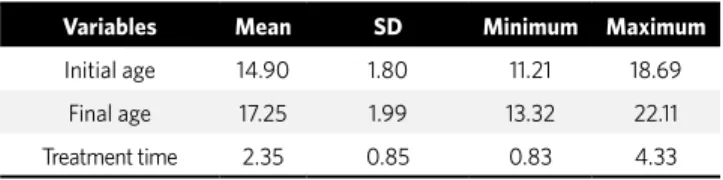
The sample consisted of 31 patients with mean initial age of 14.90 years and final age of 17.25 years (Table 1), 22 female and 9 male. Patients’ records were obtained from the archives of Cabrera & Cabrera orth-odontic clinic and were treated by them. The Class II malocclusion was corrected by using the Ten-Hour Force Theory associated to fixed orthodontic appli-ances with Straight Wire technique (A’ Company). The mean treatment time was 2.35 years.
Methods
10-Hour Force Theory
The protocol for the Ten-Hour Force Theory used for molar distalization began with banding and solder-ing double tubes to the maxillary first molars (A ‘Com-pany) and bonding of Straight Wire Andrews brackets (A’ Company), 0.022 x 0.025-in slot, with the excep-tion of the maxillary premolars.
The fixed orthodontic treatment was performed conventionally, with teeth leveling and alignment until reaching the 0.016-in stainless steel wire, when
the preparation was started for the first stage of the Ten-Hour Force Theory (T10), which aims to distalize maxillary molars. Initially, the extraoral headgear ap-pliance was made and then the sliding jig for the use of intermaxillary Class II elastics. A hook made of 0.8 mm brass wire was soldered to the telescopic tube by holding the hook with a Mathieu plier at the end the telescopic tube of 0.07 mm in diameter and by apply-ing solderapply-ing flux to the base of the hook. The brass wire was cut 7 mm above the solder and after finish-ing, the hook was made facing mesially and cervically.
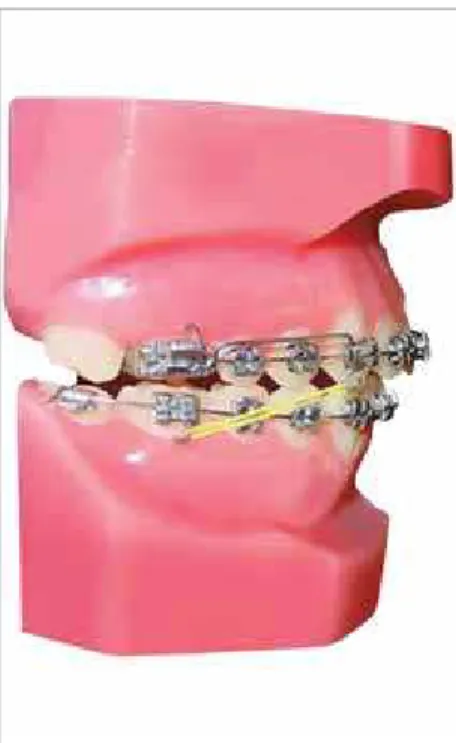
With the maxillary arch previously prepared, without premolar bonding to receive the T10, the tance between the upper tube entrance and the dis-tal surface of the canine bracket was measured with the telescope tube directly in the oral cavity, marking with a pen, and the excess of the telescopic tube was cut with a carborundum disc. The procedures were repeated on the opposite side. The telescopic tubes were attached to the leveling arch with the hooks fac-ing mesially and tied to the bracket slots usfac-ing metal-lic ligature (Fig 1). The procedures were repeated on the opposite side.
1st Phase: Distalization of the first molars
This phase is divided into 3 distinct stages:
» 1st stage – headgear appliance with cervical trac-tion, with a force intensity of 400 to 500 cN/side, used at night, only for 8 hours a day (Fig 2).
» 2nd stage – Class II intermaxillary elastics that should be attached from the first molar tube hook to the telescopic tube hook, bilaterally (Fig 3). The pa-tient is instructed to use it for 8 hours daily.
» 3rd stage – Class II intermaxillary elastics which should be attached from second mandibular molar tube hook to the telescopic tube hook, bilaterally (Fig 4). The patient is instructed to use it for 8 hours a day.
Variables Mean SD Minimum Maximum
Initial age 14.90 1.80 11.21 18.69
Final age 17.25 1.99 13.32 22.11
Treatment time 2.35 0.85 0.83 4.33
Table 1 - Descriptive statistics (mean, standard deviation, minimum and
Figure 1 - Upper and lower arches prepared to re-ceive the T10.
Figure 3 - T10 second stage: Use of Class II
elastics, anchored on the lower first molar.
Figure 4 - T10 third stage: Use of the headgear. Figure 5 - Sliding jig substituting the
tele-scopic tube for the use of Class II elastics in T10.
Figure 2 - T10 first stage: Use of the headgear.
The magnitude of elastic force should be between 200 and 250 cN, measured with a dynamometer. Elas-tics should be changed daily for both sides and they should only be removed during meals for a period of approximately 30 minutes.
Thus, the maxillary first molars receive 24 hours of continuous force per day, favoring its distaliza-tion, and the mandibular teeth are not moved since the force is removed upon completion of eight hours of application, not reaching the 10-hour period neces-sary for the orthodontic movement to be initiated.
This phase ends when the anteroposterior cor-rection of the maxillary first molar is complete (Class I molar relationship). In this situation, the brackets of the maxillary premolars can be bonded. Orthodontic treatment follows conventionally until the achievement of the six keys to normal occlusion recommended by Andrews,1 when it should end with
the removal of the fixed orthodontic appliance and placement of retainers, for the maxillary arch a modi-fied Hawley plate and a fixed retainer bonded from ca-nine to caca-nine for the lower arch.
The telescopic tube used during the applica-tion of the Ten-Hour Force Theory, can be re-placed by a sliding jig made of stainless steel wire
with 0.7 mm diameter and with this device the fixed appliance can be fully bonded, since the jig is placed above the brackets (Fig 5).
The use of the Ten-Hour Force Theory can be used for retraction or reduction of the overjet. This phase is divided into three distinct stages of 8 hours. The force application is performed by intramaxillary elas-tics attached from the maxillary first molar tube to the rectangular wire hook associated to the headgear for 8 hours. After this period, intermaxillary Class II elastics are tied to the rectangular wire hook alternat-ing between two mandibular posterior teeth on each side for every 8 hours during the day. The use of the Ten-Hour Force Theory, can also be applied for the purpose of anchorage loss, always using the elastics in 3 different stages.
Cephalometric method
measurements were performed automatically by the software. All radiographs had the magnification factor (6%, 7.9% and 9.8%) corrected by the software. Cepha-lometric variables used are shown in Figures 6 to 9.
Error of the method
To assess intra-examiner error, 20 radiographs were randomly selected, and the measurements were repeated in the Dolphin software after an interval of one month. The formula proposed by Dahlberg12
(Se2=Sd2/2n) was applied to estimate the magnitude of
casual errors, while systematic errors were evaluated by paired t test, according to Houston.20
Statistical analysis
To evaluate the cephalometric changes that oc-curred between initial and final treatment stages, the dependent t test was used. All tests were performed
in the Statistica software (Statistica for Windows, version 6.0, StatSoft Inc.), adopting a significance level of 5% (p < 0.05).
RESULTS
Systematic errors were found for only two vari-ables: ANB and 4-PTV. The greater random errors found were 1.79° for ANB and 1.25 mm for 4-PTV (Table 2).Table 3 shows the results of the intragroup comparison of cephalometric changes.
DISCUSSION Method
To assess maxillary and mandibular dental compo-nents, points were marked at the first molars, first pre-molars and central incisors. The point located in the center of clinical crown (centroid), exactly at the mid-point of the mesiodistal distance, was chosen due to
3
4
6 2
7
8
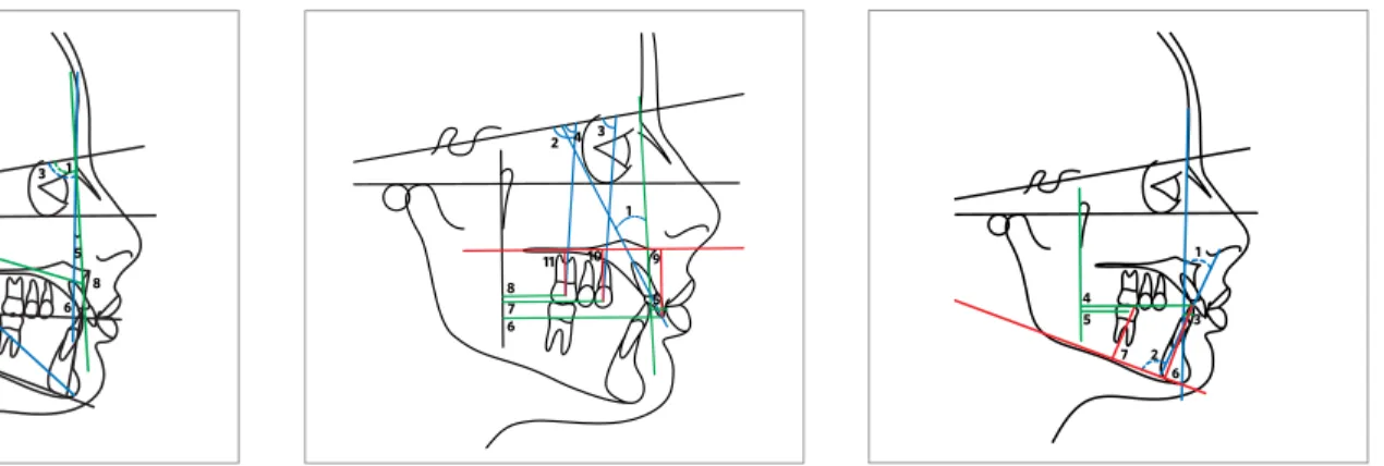
Figure 6 - Skeletal cephalometric measurements:
1) SNA; 2) Co-A; 3) SNB; 4) Co-Gn; 5) ANB; 6) Wits; 7) FMA; 8) LIFH.
Figure 7 - Upper dental cephalometric
mea-surements: 1) 1.NA; 2) 1.SN; 3) 6.SN; 4) 4.SN; 5) 1-NA; 6) 1-PTV; 7) 4-PTV; 8) 6-PTV; 9) 1-PP; 10) 4-PP; 11) 6-PP.
Figure 9 - Dental measurements: 1) Molar Relationship (MR); 2) Overjet (OJ); 3) Overbite (OB). The
mesial surface of the upper first molar should be 3 mm distal to the mesial surface of the lower first molar to describe a Class I molar relationship (negative value = -3 mm). The example demonstrates a -0.5 mm for the molar relationship.
3
Figure 8 - Lower dental cephalometric
mea-surements; 1) 1.NB; 2) IMPA; 3) 1-NB; 4) 1-PTV; 5) 6-PTV; 6) 1-GoMe; 7) 6-GoMe.
1 2 3
5 1
1 2 4 3
5 6
7 8
9 10
11 1
2 4 5
Variables
1st. Measurement 2nd. Measurement
Dahlberg p
n = 20 n = 20
Mean SD Mean SD
Maxillary component
SNA (degrees) 81.26 3.12 81.98 3.74 1.26 0.512
Co-A (mm) 91.92 4.93 92.15 5.51 0.98 0.890
Mandibular component
SNB (degrees) 77.65 2.96 78.25 3.37 1.57 0.553
Co-Gn (mm) 116.68 6.45 117.94 6.00 0.86 0.526
Maxillomandibular relationship
ANB (degrees) 3.97 1.29 3.22 1.01 1.79 0.047*
Wits (mm) -0.53 1.62 -0.14 1.95 0.52 0.495
Vertical component
FMA (degrees) 25.53 3.08 26.72 3.85 1.38 0.287
AFAI (mm) 68.77 4.03 67.61 4.38 0.73 0.388
Maxillary dental component
1.NA (degrees) 25.43 4.87 24.31 4.60 1.06 0.459
1.SN (degrees) 104.69 4.01 106.18 3.58 1.29 0.222
1-NA (mm) 6.34 1.82 6.68 2.09 0.84 0.586
1-PP (mm) 29.01 1.85 29.91 1.24 0.77 0.078
1-PTV (mm) 59.97 2.75 60.31 2.70 1.10 0.695
4.SN (degrees) 75.65 2.51 76.08 3.13 1.68 0.634
4-PP (mm) 23.44 1.00 22.61 1.70 0.92 0.067
4-PTV (mm) 40.37 2.35 41.95 1.77 1.25 0.021*
6.SN (degrees) 69.65 3.34 70.63 3.65 1.20 0.381
6-PP (mm) 20.68 1.87 19.55 1.81 1.01 0.059
6-PTV (mm) 24.44 1.46 23.81 1.10 0.96 0.131
Mandibular dental component
1.NB (degrees) 29.22 4.61 31.67 3.03 1.34 0.054
IMPA (degrees) 96.75 3.70 97.02 3.90 1.62 0.823
1-NB (mm) 7.10 1.35 6.75 1.14 0.86 0.381
1-GoMe (mm) 41.27 1.29 40.33 1.64 0.79 0.051
1-PTV (mm) 56.36 2.73 57.93 2.50 0.63 0.065
6-GoMe (mm) 30.24 1.53 29.36 1.60 0.89 0.083
6-PTV (mm) 24.78 2.11 25.39 1.58 0.99 0.307
Dental relationship
Molar Relationship (mm) -0.39 0.85 0.10 0.97 0.27 0.097
Overjet (mm) 3.41 1.27 4.13 1.15 0.87 0.067
Overbite (mm) 2.56 1.44 3.05 1.69 0.45 0.329
Table 2 - Results of dependent t test and the Dahlberg’s12 formula, applied to the studied variables, to estimate systematic and casual errors,
respectively.
the claim of several authors that this point accurate-ly represents dental changes, when compared with points located in the mesial and distal surfaces, which can show excessive changes when considering distal tooth angulation, not reflecting the actual movement of the maxillary molar and superestimating extrusion or intrusion of the assessed teeth.15,31
For the evaluation of the linear dental changes, the pterigomaxillary vertical line was used. This measure is considered a very reliable reference, due to the fact that it does not change significantly in the anteroposterior direction during the craniofa-cial growth and for being consistently used in the literature.5,8,15
Among the 29 variables evaluated, only two sys-tematic errors were noted for the following variables: ANB, 4-PTV (Table 2). For the casual errors, values lower than 1 mm for linear variables and values lower than 1.5° for the angular variables are considered ac-ceptable. The greater casual errors found were 1.79° for the ANB and 1.25 mm for the measure 4-PVT.
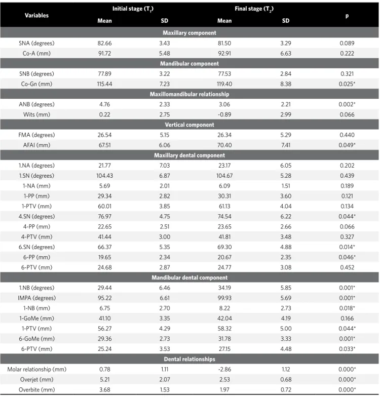
Table 3 - Results of the intragroup comparison of the cephalometric changes between the initial and final treatment stages (dependent t test) (n = 31).
Variables
Initial stage (T1) Final stage (T2)
p
Mean SD Mean SD
Maxillary component
SNA (degrees) 82.66 3.43 81.50 3.29 0.089
Co-A (mm) 91.72 5.48 92.91 6.63 0.222
Mandibular component
SNB (degrees) 77.89 3.22 77.53 2.84 0.321
Co-Gn (mm) 115.44 7.23 119.40 8.38 0.025*
Maxillomandibular relationship
ANB (degrees) 4.76 2.33 3.06 2.21 0.002*
Wits (mm) 0.22 2.75 -0.89 2.99 0.066
Vertical component
FMA (degrees) 26.54 5.15 26.34 5.29 0.440
AFAI (mm) 67.51 6.06 70.40 7.41 0.049*
Maxillary dental component
1.NA (degrees) 21.77 7.03 23.17 6.05 0.202
1.SN (degrees) 104.43 6.87 104.67 5.28 0.439
1-NA (mm) 5.69 2.01 6.09 1.51 0.189
1-PP (mm) 29.34 2.82 30.31 3.60 0.121
1-PTV (mm) 60.01 3.85 61.13 4.04 0.134
4.SN (degrees) 76.97 4.75 74.54 6.22 0.044*
4-PP (mm) 22.65 2.51 23.65 2.66 0.066
4-PTV (mm) 41.44 3.00 41.81 3.48 0.327
6.SN (degrees) 66.37 5.35 69.30 4.88 0.014*
6-PP (mm) 19.65 2.34 20.67 2.35 0.046*
6-PTV (mm) 24.68 2.87 24.77 3.08 0.452
Mandibular dental component
1.NB (degrees) 29.44 6.46 34.19 5.85 0.001*
IMPA (degrees) 95.22 6.61 99.93 5.69 0.001*
1-NB (mm) 6.75 2.70 8.22 2.73 0.018*
1-GoMe (mm) 41.10 3.35 42.04 4.19 0.166
1-PTV (mm) 56.27 4.29 58.32 5.00 0.044*
6-GoMe (mm) 29.36 2.73 31.78 3.33 0.001*
6-PTV (mm) 25.24 3.53 27.15 4.48 0.033*
Dental relationships
Molar relationship (mm) 0.78 1.11 -2.86 1.12 0.000*
Overjet (mm) 5.21 2.07 2.53 0.68 0.000*
Overbite (mm) 3.68 1.53 1.97 0.72 0.000*
* Statistically significant for p < 0.05.
Changes with treatment
Intragroup comparison of cephalometric chang-es between the initial and final treatment stagchang-es are shown in Table 3.
There was a slight restriction, but not significant, of the anterior development of the maxilla (SNA, Table 3), probably due to the use of headgear only 8 hours a day. The redirection of maxillary growth, with anterior growth restriction in young patients who used extraoral headgear is widely reported in the literature.9,14,17,32
There was a significant increase in the effective length of the mandible (Co-Gn, Table 3). This prob-ably occurred because patients still were at the final growth phase.24,25
There was an improvement in the anteroposte-rior discrepancy of the apical bases (ANB, Table 3). This improvement was already expected, since there was a slight restriction of the anterior development of the maxilla and a significant increase of the effective length of the mandible. In addition, this improvement is reported in cases treated with extraoral headgear and Class II elastics.16,25,27
A significant increase can be observed in low-er antlow-erior facial height (LAFH) with treatment, probably due to the extrusion of the maxillary first molars that occurred with the use of the extraoral headgear (Table 3). The extrusion and increase of the LAFH has been previously reported in the lit-erature using this mechanics for correction of the Class II malocclusion.6,7,9,18,37
There was no significant change in the position of the maxillary incisors with treatment (Table 3). The maxillary first premolars showed a significant distal tipping of the crown, and the maxillary molars showed a significant distal tipping of the root and extrusion, with treatment (Table 3).
The maxillary first molar did not experience a sig-nificant distalization, as reported by some authors.6,37
This must have been due to the minor use of the head-gear and also due to the lack of restriction of the max-illary displacement observed in this study.
The mandibular incisors were significantly buc-cally tipped and protruded with treatment (Table 3). This effect was caused by the use of Class II inter-maxillary elastics, and probably due to the initial crowding exhibited by patients.
The mandibular molars presented significant ex-trusion and mesial movement during treatment (Ta-ble 3). This result agrees with some reports in the lit-erature,5,13,30 showing mesial movement of mandibular
molars and buccal inclination of mandibular incisors. Papaioannou-Maragou and Papaioannou32 also
ob-served the same effect in the mandibular incisors us-ing Class II elastics without headgear, agreeus-ing with Ellen, Schneider and Sellke13 who observed a buccal
inclination of the mandibular incisors, extrusion and mesial movement of the mandibular molars. Nelson, Hansen and Hägg30 also observed mesial movement of
the mandibular molars.
The Class II molar relationship was corrected satisfactorily and significantly during treatment (Table 3). As the molar remained stable, with no dis-talization, the correction of the molar relationship was probably due to the mesial movement of the mandibular molar. The overjet and overbite showed significant decrease with treatment, demonstrating that they were corrected with treatment. This im-provement in the horizontal direction is mainly due to buccal inclination and protrusion of the mandibu-lar incisors, correcting the overjet, and the overbite correction is probably due to the extrusion of the maxillary and mandibular molars.
Clinical considerations
The correction of the Class II molar relationship evidenced by the protocol of the 10-Hour Force Theo-ry associated with the use of fixed appliances, showed that the treatment time was as planned because there was no need for further retraction of anterior teeth, since there was no anchorage loss.
There are no references in the literature of this type of protocol, so there is no possibility of compari-son. There are other studies comparing the extraoral headgear to other intraoral device using intermaxil-lary elastics.5,32
patient’s profile, is a good reason for cooperating with the use of elastics for a few months.
The protocol using the T10 resource, did not pro-vide orthopedic effects, but resulted in satisfactory dental effects such as the satisfactory molar rela-tionship and adequate incisal guidance. Although it requires patient cooperation for the use of the head-gear at night and exchange of intermaxillary elastics, since it is an easy resource to understand and estheti-cally pleasing to the patient, it is possible to obtain a successful treatment.
These results are evidence that the 10-Hour Force Theory would be an option for the treatment of the Class II molar correction. Since Orthodontics is based on scientific evidence, more researches are needed to truly prove the validity of this theory, as the comparison of theory with the use of conven-tional Class II elastics applied on the mandibular molars for 24 hours or the use of extraoral headgear with continuous use of Class II elastics; in addition,
it can be emphasized the importance of studies eval-uating the stability of the results obtained through the use of the 10-Hour Force Theory for molar dis-talization.
CONCLUSIONS
1. Andrews LF. The six keys to normal occlusion. Am J Orthod. 1972;62(3):296-309.
2. Andrews LF. Straight Wire: o conceito e o aparelho. 2ª ed. Curitiba:
Interativas; 1997.
3. Andrews LF. The ten-hour force theory. San Diego: [S.l.]; 1975. 4. Andrews LF. The ten-hour force theory. 5th ed. 1996.
5. Angelieri F. Comparação dos efeitos cefalométricos promovidos pelos
aparelhos extrabucal cervical e Pendulum [tese]. Bauru (SP): Universidade de São Paulo; 2005.
6. Baumrind S, Molthen R, West EE, Miller DM. Distal displacement of the maxilla and
the upper first molar. Am J Orthod. 1979;75(6):630-40.
7. Blueher WA. Cephalometric analysis of treatment with cervical anchorage. Angle
Orthod. 1959;29(1):45-53.
8. Brickman CD, Sinha PK, Nanda RS. Evaluation of the Jones Jig appliance for distal molar movement. Am J Orthod Dentofacial Orthop. 2000;118(5):526-34. 9. Cangialosi TJ, Meistrell ME Jr, Leung MA, Ko JY. A cephalometric appraisal of
edgewise Class II nonextraction treatment with extraoral force. Am J Orthod Dentofacial Orthop. 1988;93(4):315-24.
10. Carano A, Testa M. The distal jet for upper molar distalization. J Clin Orthod. 1996;30(7):374-80.
11. Cuoghi OA. Avaliação dos primeiros momentos da movimentação dentária
induzida: estudo microscópico em macacos da espécie Cebus apella [tese]. Bauru (SP): Universidade de São Paulo; 1996.
12. Dahlberg G. Statistical methods for medical and biological students. New York: Interscience; 1940.
13. Ellen EK, Schneider BJ, Sellke T. A comparative study of anchorage in bioprogressive versus standard edgewise treatment in Class II correction with intermaxillary elastic force. Am J Orthod Dentofacial Orthop. 1998;114(4):430-6. 14. Gandini MRS, Gandini LG Jr, Martins JCR, Del Santo M Jr. Effects of cervical
headgear and edgewise appliances on growing patients. Am J Orthod Dentofacial Orthop. 2001;119(5):531-8; discussion 538-9.
15. Ghosh J, Nanda RS. Evaluation of an intraoral maxillary molar distalization technique. Am J Orthod Dentofacial Orthop. 1996;110(6):639-46. 16. Gianelly AA, Valentini V. The role of “orthopedics” and orthodontics in the
treatment of Class II, division 1 malocclusions. Am J Orthod. 1976;69(6):668-78.
17. Henriques JFC. Estudo cefalométrico comparativo de três tipos de ancoragem
extrabucal sobre as estruturas dentoesqueléticas, em pacientes com Classe II, 1ª divisão [tese]. Bauru (SP): Universidade de São Paulo; 1993.
18. Henriques JFC, Martins DC, Pinzan, A. Estudo cefalométrico da ação da
ancoragem extrabucal cervical, na dentadura mista, sobre a maxila, mandíbula e dentes, em pacientes com Classe II, divisão 1. Ortodontia. 1979;12(2):76-86. 19. Hilgers J. The Pendulum appliance for Class II non-compliance therapy. J Clin
Orthod. 1992;26(11):706-14.
REFERENCES
20. Houston WJB. The analysis of errors in orthodontic measurements. Am J Orthod. 1983;83(5):382-90.
21. Itoh T, Tokuda T, Kiyosue S, Hirose T, Matsumoto M, Chaconas SJ. Molar
distalization with repelling magnets. J Clin Orthod. 1991;25(10):611-7. 22. Janson GRP, Martins DR, Henriques JFC, Freitas MR, Pinzan A, Almeida RR.
Oclusão funcional e ajuste oclusal. In: Viazis SD. Atlas de Ortodontia avançado. São Paulo: Ed. Santos; 1999. p. 203-14.
23. Jones RD, White JM. Rapid Class II molar correction with an open-coil jig. J Clin Orthod. 1992;26(10):661-4.
24. Keeling SD, Wheeler TT, King GJ, Garvan CW, Cohen DA, Cabassa S, et al. Anteroposterior skeletal and dental changes after early Class II treatment with bionators and headgear. Am J Orthod Dentofacial Orthop. 1998;113(1):40-50. 25. Kim KR, Muhl ZF. Changes in mandibular growth direction during and after cervical
headgear treatment. Am J Orthod Dentofacial Orthop. 2001;119(5):522-30. 26. Kloehn SJ. Guiding alveolar growth and eruption of teeth to reduce treatment time
and produce a more balanced denture and face. Angle Orthod 1947;17(1-2):10-33. 27. Lima Filho R, Lima AL, Ruellas ACO. Estudo longitudinal das alterações no ângulo
ANB em pacientes Classe II esquelética, tratados com aparelho extra-oral de Kloehn. Rev Dental Press Ortod Ortop Facial. 2003;8(2):21-9.
28. Locatelli R, Bednar J, Dietz VS, Gianelly AA. Molar distalization with superelastic NiTi wire. J Clin Orthod. 1992;26(5):277-9.
29. Muse DS, Fillman MJ, Emmerson WJ, Mitchell RD. Molar and incisor changes with Wilson rapid molar distalization. Am J Orthod Dentofacial Orthop. 1993;104(6):556-65.
30. Nelson B, Hansen K, Hägg U. Class II correction in patients treated with Class II elastics and with fixed functional appliances: a comparative study. Am J Orthod Dentofacial Orthop. 2000;118(2):142-9.
31. Ngantung V, Nanda RS, Bowman SJ. Posttreatment evaluation of the distal jet appliance. Am J Orthod Dentofacial Orthop. 2001;120(2):178-85.
32. Papaioannou-Maragou O, Papaioannou A. Comparison of treatment results with the edgewise and the Begg approach. J Clin Pediatr Dent. 1994;19(1):27-30. 33. Roth RH. Functional occlusion for the orthodontist. Part I. J Clin Orthod.
1981;15(1):32-51.
34. Roth RH. Functional occlusion for the orthodontist. Part III. J Clin Orthod. 1981;15(3):174-98.
35. Silva E, Gasque CA, Vieira AM. Ertty system: um novo conceito na distalização de molares. Rev Clín Ortod Dental Press. 2003;2(3):45-60.
36. Silva Filho OG, Freitas SF, Cavassan AO. Prevalência de oclusão normal e má oclusão em escolares da cidade de Bauru (São Paulo). Parte I: relação sagital. Rev Odontol Univ São Paulo. 1990;4(2):130-7.