In a previous study in dogs, the early removal of expanded polytetrafluoroethylene (ePTFE) membrane (2 weeks after placement) showed histomorphometric results (of new bone, cementum and periodontal ligament) similar to that obtained with membrane removal at 4 weeks after placement. This study evaluated the influence of early removal of an ePTFE membrane on the treatment of Class II furcation defects. Twelve patients who provided 12 pairs of mandibular furcation defects were recruited for the study. Baseline clinical measurements were recorded: plaque index (PI), gingival index (GI), bleeding on probing (BOP), probing depth (PD), gingival margin position (GMP) and relative clinical attachment level (RCAL). Full flaps were elevated and hard tissue measurements were performed during the surgery: relative vertical (RVBL) and horizontal (RHBL) bone level. The ePTFE membranes were adapted and sutured to their correspondent tooth and removed at 2 weeks in the test group (TGr) and at 4 weeks in the control group (CGr). After 1 year all sites were re-entered, and soft and hard tissue measurements were recorded. There were no statistically significant differences between TGr and CGr for any baseline measurement. After 12 months, there were no statistically significant differences between TGr and CGr in the PD (p=0.74), GMP (p=0.76) and RCAL (p=0.44) values. However, the RHBL resolution was significant for both groups (CGr p=0.01 and TGr p=0.02), without difference between groups (p=0.39). Early removal of membranes did not affect the outcome on the treatment of Class II furcation defects.
Effect of Early Membrane Removal on
the Treatment of Mandibular Class II
Furcation Defects - A Controlled Clinical
Trial with Re-entry after 12 Months
Alexandre C. Leite, Rafael R. de Oliveira, Arthur B. Novaes Jr., Patrícia A. O’Connell, Márcio F. M. Grisi, Mário Taba Jr., Daniela B. Palioto, Sérgio L. S. Souza
Department of Surgery, Oral and Maxillofacial Traumatology and Periodontology, Dental School of Ribeirão Preto, University of São Paulo, Ribeirão Preto, SP, Brazil
Correspondence: Prof. Dr. Sérgio Luís Scombatti de Souza, Avenida do Café s/n, 14040-904, Ribeirão Preto, SP, Brasil. Tel: +55-16-3602-4141. e-mail: [email protected]
Key Words: furcation defects; guided tissue regeneration; polytetrafluoroethylene membrane.
Introduction
The ultimate goal of periodontal therapy is to prevent further attachment loss and predictably restore the periodontal supporting structures that were lost due to the progression of the periodontal disease in a way that the architecture and function of the lost structures may be reestablished (1). Histological and clinical studies have reported the potential of guided tissue regeneration (GTR) to regenerate periodontal ligament, cementum and alveolar bone (2,3), and the technical reasons for the success of this technique are well described in literature (4). In GTR, a barrier is inserted between the root surface and the gingival tissues to inhibit the apical migration of the epithelium and gingival connective tissue of the flap, allowing the granulation tissue derived from the periodontal ligament and osseous tissues to repopulate the space adjacent to the denuded root surface. These cells have the ability to differentiate into osteoblast-like cells and cementoblast-like cells, as well as new periodontal ligament fibroblasts and thereby promote tissue regeneration.
Expanded polytetrafluoroethylene (ePTFE) membrane is a non-resorbable material extensively investigated for the
treatment of periodontal defects, with significant gains in new attachment (5). The period for membrane removal varies from 4 to 6 weeks after placement (6); a previous study has shown the benefit of ePTFE membrane retrieval at this time (7), and longer periods did not provide additional benefits (8). The exposure of the membrane in the early healing phase has been negatively correlated with the amount of newly formed tissues under the barrier (9), which occurs due to the bacterial contamination of the membrane. As the length of exposure time increases, the risk of contamination and infection also increases (10). Therefore, the membrane should be retrieved as soon as the healing clot results in a tissue with sufficient maturity to resist the surgical trauma of the re-entry procedure, inhibiting the apical down growth of gingival epithelium along the root surface.
Early membrane removal on GTR in humans of new bone, cementum and periodontal ligament) similar
to the membrane removal at 4 weeks after placement, leading to the conclusion that the early removal of e-PTFE membranes did not influence the GTR results (12). Then, it should be of interest to test the hypothesis that early removal of ePTFE membranes in humans does not affect clinically the healing process. Therefore, the purpose of this study was to evaluate if tissue formed under an ePTFE membrane retrieved 2 weeks postoperatively promotes similar clinical results to the healing tissue formed under the same type of membrane retrieved 4 weeks after surgery.
Material and Methods
Study Population and Experimental Design
The study was designed as a randomized, controlled clinical trial performed using the split-mouth design. It was conducted according to the guidelines of the Helsinki Declaration, after approval of the research protocol by the institutional Ethics Committee (Process 2006.1.409.58.5). The participants signed and informed consent form after receiving explanation of the procedure, its associated risks and benefits, and the need for documentation and re-entry surgery. The patients were treated from that date on a sequential basis.
The inclusion criteria were: adult subjects with clinically detectable mandibular buccal or lingual Class II furcation
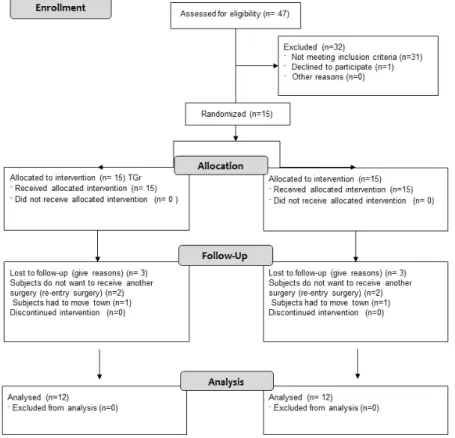
defects with ≥5 mm attachment loss and with ≥3 mm of horizontal probing depth (13), with stable occlusion, presenting an unremarkable general health according to medical history and clinical judgment, and not taking any medications for ≥6 months before the beginning of the study. Subjects with aggressive forms of periodontitis, smokers and subjects with significant systemic diseases (e.g., cancer, acquired immunodeficiency syndrome and diabetes) were not included in the study. The following additional exclusion criteria included: mandibular Class III furcation defects, presence of apical radiolucency and previous lack of cooperation with the maintenance program or use of antibiotics for 12 months before the study baseline measurements were recorded. The study population consisted of subjects who had previously been treated non-surgically for advanced chronic periodontitis and adhered to maintenance care ≥1 year before the beginning of the study. Treatment included scaling and root planing and plaque control measures. Baseline full-mouth plaque and bleeding scores were recorded. The examined sites were isolated furcations that did not respond adequately after a comprehensive initial therapy phase. The study flowchart is shown in Figure 1.
Twelve adult patients (5 women and 7 men, age range: 37 to 60 years; mean age: 46.01 years) meeting the inclusion criteria were treated within a 1-year period at a Graduate
404
A.C. Leite et al.
Program Clinic of Ribeirão Preto School of Dentistry, University of São Paulo, Brazil. Twelve pairs of mandibular Class II furcation defects (20 buccal and 4 lingual) found in 18 second molars (9 pairs), 4 first molars (pairs) and 2 third molars 1 pair) were included.
Basic periodontal therapy was performed with detailed full-mouth scaling and root planing under local anesthesia. Four weeks after the completion of the basic therapy, baseline examination was performed with an automated periodontal probe (Florida Probe; Florida Probe Corporation, Gainesville, FL, USA) at three sites per tooth (mesial, central and distal) using a customized acrylic stent as reference to establish the exact site and angle of the measurement, ensuring reproducibility during the examinations.
The following clinical parameters were recorded at baseline and 12 months after surgery: plaque index (PI) (14), gingival index (GI) (15), bleeding on probing (BOP) (16), probing depth (PD), relative clinical attachment level (RCAL) and gingival margin position (GMP). Additionally, hard tissue measurements were undertaken during membrane placement surgery and at re-entry in order to determine the relative vertical (RVBL) and horizontal (RHBL) bone level, defined as primary study variables. Surgical procedures and measurements were done by the same non-masked examiners.
Examiner Calibration
Five patients, each showing 2 pairs of contralateral mandibular molars with probing depths ≥6 mm on at least one aspect of each tooth, were used to calibrate the examiner. The examiner evaluated the patients on 2 separate occasions, 48 h apart. Calibration was accepted if measurements at baseline and after 48 h were similar
to the millimeter at ≥90% level.
Surgical Procedures
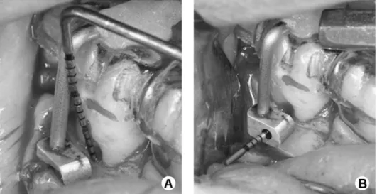
The bilateral surgeries were carried out at the same appointment, following administration of local anesthesia. Intrasulcular and vertical releasing incisions were performed and mucoperiosteal flaps were reflected. The inner aspect of the flaps was curetted and complete debridement of the osseous defects and thorough scaling and root planing using Gracey curettes (Hu-Friedy, Chicago, IL, USA) were performed. After root conditioning with EDTA 24% for 2 min and irrigation with physiologic saline solution, intrasurgical measurements were recorded. The vertical osseous component of the furcation (RVBL) was recorded using the acrylic stent at the pre-surgical midline reference points. Vertical measurements were obtained from the base of the stent to the bottom of the defect (Fig. 2A). The horizontal component of the defect (RHBL) was measured from a line tangential to the buccal root surfaces extending horizontally to the deepest portion of the defect (Fig. 2B). The measurements were recorded to the nearest millimeter using a color probe (PCPUNC; Hu-Friedy, Chicago, IL, USA).
Next, the ePTFE membranes (Gore-Tex membrane; W.L. Gore & Associates, Flagstaff, AZ, USA) were sutured with ePTFE sutures around the teeth, covering the buccal furcations. Flaps were sutured with the same material, completely covering the membranes. For each surgery, 500 mg of amoxicillin plus clavulanic acid was prescribed for every 8 h during 10 days, starting 24 h before the procedure. Patients were instructed to refrain from mechanical biofilm control in the areas where surgery had been preformed and to use a 0.2% chlorhexidine solution topically until membrane retrieval. On one side the membranes were
Early membrane removal on GTR in humans removed after 2 weeks (test group [TGr]), and the remaining
membranes were removed after 4 weeks (control group [CGr]) by careful elevation of the flap to gain access to the membranes (Fig. 3A and B). The designation of which side was TGr or CGr was randomly determined by a coin toss. Sutures were removed after 15 days. Both sides had a healing period of 12 weeks after removal of membranes and, during this period, patients were maintained on a plaque control program once a week for the first month, every two weeks for the second and third months, and then once a month until the re-entry procedure (17).
Twelve months after surgery, the soft tissue changes were evaluated and measurements were made using the same acrylic stent and reference points as for the initial examination. Intrasulcular and vertical releasing incisions and mucoperiosteal flaps were elevated at all sites to gain access to the treated furcations (Fig. 3C and D). Changes in the vertical or horizontal morphology of the bone defects were assessed and the osseous measurements were made using the same stent as in the initial surgery.
Statistical Analysis
Mean values and standard deviation were calculated. The Mann-Whitney U test was performed to determine if both groups had similar clinical measurements at baseline,
and if one treatment produced better clinical results after the 12-month follow-up. The Friedman test was used to analyze whether clinical measurements differed before and after treatment. For all statistical analyses, a significance level of 5% was used. A software package (SPSS version 16.0.0; SPSS, Chicago, IL, USA) was used for all calculations.
Results
All patients showed good compliance throughout the study and the healing period was uneventful for both treatment groups, without signs of infection or complications. In addition, there were no membrane exposures during the period of observation.
The overall results including baseline recordings as well as the final outcome of both treatments are presented in Tables 1 to 4. Initially, the PI was 1.0 ± 0.5 in both groups. At 12 months, plaque scores were markedly reduced and no statistically significant differences were observed between plaque scores of surfaces treated by both therapies (Table 1). The GI was significantly reduced in both groups at the 12-month follow-up evaluation compared to baseline (p<0.05), with no statistically significant differences between groups (Table 1). At the baseline, both groups presented similar mean values for PD, GMP, RCAL, RVBL and RHBL (p>0.05) as shown in Tables 1-3. After 12 months,
406
A.C. Leite et al.
there were no statistically significant differences (Mann Whitney test) between TGr and CGr in the values of PD (p=0.74), GMP (p=0.76) and RCAL (p=0.44).
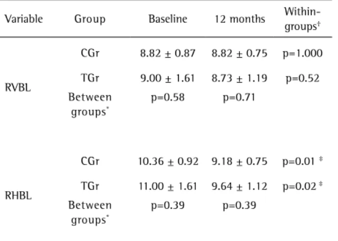
Regarding the surgical parameters, both groups showed similar mean values for RVBL at baseline; after 12 months, in the re-entry procedure, no statistical significant differences were observed by the analysis between groups (p=0.71) (Table 3). The RHBL was reduced in both groups at the 12-month evaluation compared to baseline, and these differences were statistically significant (p=0.02 for TGr and p=0.001 for CGr). In the analysis between groups,
no statistically significant differences were observed for baseline values (p=0.397) and for the 12-month re-entry values (p=0.39) (Table 3). Additionally, the mean changes of clinical and surgical parameters (baseline to 12 months) are expressed in Table 4 and also no statistically significant differences were found between TGr and CGr.
Table 1. Plaque index (PI), gingival index (GI) and bleeding on probing (BOP): mean scores (±SD, N=12 patients) at baseline and 12 months
Index/treatment Baseline (±SD) 12 months (±SD) p value
PI
TGr 1.00 ± 0.50 0.6 ± 0.4 n.s.
Cgr 1.00 ± 0.50 0.4 ± 0.4 n.s.
p value n.s. n.s.
GI
TGr 1.70 ± 0.60 0.70 ± 0.40 *
Cgr 1.80 ± 0.70 0.60 ± 0.50 *
p value n.s. n.s.
BOP
TGr 47% 19% *
CGr 50% 21% *
p value n.s. n.s.
Significance of differences within (Wilcoxon signed rank test, non-parametric test) and between (Mann-Whitney U test, non-non-parametric test) the groups. (*p<0.05; n.s: not significant)
Table 2. Probing depth (PD), gingival margin position (GMP) and relative clinical attachment level (RCAL). mean scores (±SD; N=12 patients) at baseline and 12 months
Variable Group Baseline 12 months
Within-groups†
PD
CGr 3.43 ± 1.20 3.00 ± 0.60 p=0.28
TGr 3.47 ± 1.32 3.10 ± 0.88 p=0.08
Between-groups* p=0.645 p=0.740
GMP
CGr 4.73 ± 1.03 3.80 ± 1.22 p=0.07
TGr 4.62 ± 1.06 4.24 ± 1.06 p=0.52
Between-groups* p=0.974 p=0.766
RCAL
CGr 9.09 ± 1.19 8.85 ± 1.09 p=0.51
TGr 9.27 ± 1.21 9.25 ± 1.34 p=0.35 Between-groups* p=0.42 p=0.44
† Friedman test.
Table 3. Relative vertical bone level (RVBL) and Relative horizontal bone level (RHBL) mean scores (±SD; N=12 patients) at baseline and 12 months
Variable Group Baseline 12 months
Within-groups†
RVBL
CGr 8.82 ± 0.87 8.82 ± 0.75 p=1.000
TGr 9.00 ± 1.61 8.73 ± 1.19 p=0.52 Between
groups*
p=0.58 p=0.71
RHBL
CGr 10.36 ± 0.92 9.18 ± 0.75 p=0.01 ‡
TGr 11.00 ± 1.61 9.64 ± 1.12 p=0.02 ‡ Between
groups*
p=0.39 p=0.39
‡ Statistical significance based on p<0.05.
Table 4. Mean changes (baseline – 12 months) ± SD (mm) and p values of hard and soft tissue parameters
Variable Group Baseline - 12 months
RHBL
CGr 1.18 ± 1.08
TGr 1.36 ± 1.57
Between-groups* p=0.83
RVBL
CGr 0 ± 0.92
TGr 0.27 ± 1.44
Between-groups* p=0.64
PD
CGr 0.43 ± 0.84
TGr 0.37 ± 1.03
Between-groups* p=0.91
GMP
CGr 0.93 ± 1.18
TGr 0.38 ± 1.09
Between-groups* p=0.11
RCAL
CGr 0.24 ± 1.15
TGr 0.02 ± 1.32
Between-groups* p=0.72
Early membrane removal on GTR in humans
Discussion
Experimental studies demonstrated the regenerative potential of GTR to promote periodontal regeneration (3,5). The clinical efficacy of this procedure for Class II furcation regeneration was established in a meta-analysis carried out by Kinaia et al. (18). This study indicated that both resorbable and non-resorbable membranes were more effective than open flap debridement for reducing vertical probing depths, gaining vertical attachment levels and gaining vertical and horizontal bone in class II furcation regeneration (18).
ePTFE membranes have also demonstrated an adequate clinical performance, being able of inhibiting the down growth of epithelium, promoting the isolation of the gingival connective tissue, and protection of the blood clot (8,19).Replacement of the blood clot eventually leads to the formation of new periodontal tissues. Non-resorbable membranes are usually removed after 4 to 6 weeks, exposing the immature tissue to the tissues that were excluded from the early healing phases. Experimental studies demonstrated that this tissue is able to inhibit the down growth of the epithelium and be replaced by new periodontal ligament, cementum and bone (19).
The present clinical trial evaluated the effect of early ePTFE membrane removal (2 weeks after placement) in the periodontal regeneration of Class II furcation defects. The results demonstrated that this procedure leads to outcomes similar to the control group (4 weeks after placement). Previous studies demonstrated the dynamics of periodontal tissue formation in animals, reporting histologic findings in less than 4 weeks of healing. According to Wikesjö et al. (20), 7 days after periodontal tissue injury, it may be observed the formation of collagen fibers in the connective tissue, free from inflammatory cells and in contact with dentin. After 14 days, the tissue can present new collagen fibers, with an organization indicative of attachment to the dentin. Describing the tissues formed at 2 weeks of wound healing after GTR, Matsuura et al. (21) reported that the defect was occupied predominantly by granulation tissue composed of numerous inflammatory cells and blood vessels, as well as formation of a fine layer of connective tissue between the granulation tissue and the root surface. At this stage, there is no significant bone or cementum formation. Macedo et al. (12) evaluated in dogs the effect of early e-PTFE membrane removal (2 weeks after placement) in the periodontal regeneration of class II furcation defects. The results demonstrated that this procedure promoted the formation of new bone, cementum and periodontal ligament similar to the control group (membrane removal at 4 weeks after placement). The authors concluded that the early removal of the e-PTFE membranes in dogs did not influence the GTR results. Thus, the healing tissues formed
under this membrane within 2 weeks seem to have physical and structural characteristics that inhibit the down growth of epithelium and regenerate the periodontal tissues. In different experimental trials, several histological aspects of GTR have been highlighted (12,19,20). Unfortunately, the animals used in these studies showed metabolic rates that were at least more than twice those of humans. Furthermore, more parameters can be kept constant in animal models, which could never be possible in a clinical trial. Therefore, caution should be exercised when extrapolating histological results to humans.
It is relevant to mention that no exposure of the ePTFE membrane was observed in this trial. Careful surgical technique and a strict protocol of biofilm control may have contributed to this. A previous study demonstrated that the membranes can be a retentive factor for bacterial colonization, and that membrane exposure to the oral cavity was negatively correlated with the results of the regenerative therapy (22).
Diverse clinical methodologies have been used to determine hard tissue measurements in order to evaluate the outcome of therapies at re-entry. Horizontal defects present at least two variables affecting measurements: the horizontal component and the vertical intrabony component. Studies that define a predetermined point to measure the horizontal component of the furcation at the most coronal aspect of the furcation entrance or furcation dome level could underestimate the residual osseous defect. This is the reason why the present study used the method advocated by Kenney et al. (23) to obtain the bone measurements in the furcation area. The primary response variable in the treatment of furcation defects is the attachment level in horizontal direction, and both groups resulted in similar horizontal defect fill. The intergroup comparison revealed no statistically significant differences observed for baseline values (p=0.397) and for the 12-month re-entry values (p=0.399) (Table 1). In addition, the mean changes of RHBL (baseline to 12 months) were 1.18 ± 1.08 mm (CGr) and 1.36 ± 1.57 mm (TGr) and no statistically significant differences were found (p=0.839) (Table 4).
In the analysis of the vertical component (RVBL) no statistically significant differences were observed compared to the baseline measurements with the values obtained at the re-entry for TGr (p=0.527) and for CGr (p=1.000). Additionally, no significant differences were observed comparing TGr and CGr in baseline measurements (p=0.397) and re-entry measurements (p=0.399) (Table 3).
408
A.C. Leite et al.
reports (6,22). Most probably these differences are due to local factors that influence the outcomes of regeneration in class II furcations. Indeed, furcation involvement is probably the most difficult type of defect to standardize. Along with the variables associated with the osseous defect itself, the aspects associated with the tooth, and more specifically with furcation morphology, obviously play a significant role in the outcome of GTR. According to Bowers et al. (25), the distance between the roof of furcation and crest of bone, roof of furcation and base of defect, depth of horizontal defect, and divergence of roots at the crest of bone can influence the results of treatment.
Although the present study indicates a similarity in the results regardless of the time of membrane retrieval (2 and 4 weeks), there is some controversy in the literature. The optimal time for membrane removal was evaluated in clinical studies, which showed that prevention of purulence is related to the timely removal of the material within 4 to 6 weeks, and that the early removal of membranes may have a negative influence on the amount of regeneration in sites that received a graft (7).
The period of membrane removal varies from 4 to 6 weeks after placement (6) and longer time periods do not provide additional benefits (8). The exposure of the membrane in the early healing phase has been negatively correlated with the amount of newly formed tissues under the barrier (9), which occurs due to bacterial contamination of the membrane. Therefore, the membrane should be retrieved as soon as the healing clot results in a tissue with sufficient maturity to resist the surgical trauma of the re-entry procedure, inhibiting the apical down growth of gingival epithelium along the root surface and regenerating the periodontal tissues. Previous studies demonstrated that early healing tissues formed in less than 4 weeks may show an arrangement indicative of physical attachment to the dentin and are able to resist tensile forces on the root surface-gingival flap interface during this period (11,12).
According to the present results, early membrane removal at 2 weeks leads to similar clinical outcomes to those observed in the group where the membranes remained for 4 weeks. However, the possibility of a Type II error (no difference between TGr and CGr when a true difference exists) should be considered in the results, mainly due to the sample size (post-hoc power calculation = 27.8%). This is an important limitation of the present study, which should be considered as a preliminary report. The power calculation showed that, for a power level of 80%, the ideal sample must have at least 60 patients per group (60 for TGr and 60 for CGr) to detect a minimum clinically significant difference in bone level of 0.5 mm, using a significance level of 5%. Thereby, future studies with large sample sizes should support or contrast these findings.
The early removal has advantages, such as a shorter treatment time, reducing the period of antimicrobial coverage, as well as lower risk of membrane exposure. Therefore, in periodontal clinics where many patients undergo this treatment, the risk of membrane exposure increases and thus early membrane removal could be beneficial.
It may be concluded that the early removal of ePTFE membranes in this trial did not influence significantly the GTR outcomes. Thus, the healing tissues formed under these membranes within 2 weeks seemed to have characteristics that could inhibit the down growth of epithelium and regenerate periodontal tissues. Both groups presented limited bone regeneration. This study is a preliminary report, and further studies with larger sample sizes are required to confirm the obtained results.
Resumo
Em estudo prévio, em cães, a remoção precoce da membrana de politetrafluoretileno expandido (PTFE-e), 2 semanas após a colocação, mostrou resultados histomorfométricos (formação de novo osso, cemento e ligamento periodontal) similares aos de remoção da membrana 4 semanas após a sua colocação. Este estudo avaliou a influência da remoção precoce de uma membrana de PTFE-e no tratamento de defeitos de bifurcação classe II. Foram selecionados para o estudo 12 pacientes, com 12 pares de defeitos de bifurcação mandibulares. Foram feitas as seguintes medidas clínicas iniciais: índice de placa (IP), índice gingival (IG), sangramento à sondagem (SAS), profundidade de sondagem (PS), posição da margem gingival (PMG) e nível relativo da inserção clínica (NRIC). Foram elevados retalhos totais e as medidas do tecido ósseo foram tomadas transcirurgicamente: níveis ósseos vertical (NOV) e horizontal (NOH). Membranas de PTFE-e foram adaptadas e suturadas aos dentes correspondentes e removidas após 2 semanas no grupos teste (GT) ou quatro semanas no grupo controle (GC). Após 1 ano, em todos os sites foi realizada reentrada cirúrgica e medidas clínicas e ósseas foram novamente feitas. Não houve diferenças estatisticamente significantes entre GT e GC para nenhuma das medidas iniciais avaliadas. Após 12 meses, não houve diferenças estatisticamente significantes entre GT e GC para os valores de PS (p=0,74), PMG (p=0,76) e NRIC (p=0,44). Entretanto, a resolução do nível ósseo horizontal foi significante para ambos os grupos (GC: p=0,01 e GT: p=0,02), sem diferenças entre grupos (p=0,39). A remoção precoce da membrana não afetou os resultados do tratamento de defeitos de bifurcação Classe II.
Acknowledgements
This study was supported by grants from the Coordination of Improvement of Higher Education Personnel (CAPES), Brasília, DF, Brazil. The authors state that there is no conflict of interest with any financial organization regarding the material discussed in this manuscript.
References
1. Villar CC, Cochran DL. Regeneration of periodontal tissues: guided tissue regeneration. Dent Clin North Am 2010;54:73-92.
2. de Andrade PF, de Souza SL, de Oliveira Macedo G, Novaes Jr. AB, de Moraes Grisi MF, Taba Jr. M, et al.. Acellular dermal matrix as a membrane for guided tissue regeneration in the treatment of Class II furcation lesions: a histometric and clinical study in dogs. J Periodontol 2007;78:1288-1299.
Early membrane removal on GTR in humans
biological foundation and preclinical evidence: a systematic review. J Clin Periodontol 2008;35:106-116. Review
4. Novaes Jr AB, Palioto DB, Andrade PF, Marchesan JT. Regeneration of class II furcation defects: determinants of increased success.Braz Dent J 2005;16: 87-97.
5. Queiroz AC, Nóbrega PB, Oliveira FS, Novaes AB Jr, Taba M Jr, Palioto DB, et al.. Treatment of intrabony defects with anorganic bone matrix/p-15 or guided tissue regeneration in patients with aggressive periodontitis. Braz Dent J 2013;24:204-212.
6. Machtei EE. The effect of membrane exposure on the outcome of regenerative procedures in humans: A meta-analysis. J Periodontol 2001;72:512-516.
7. Murphy KG. Postoperative healing complications associated with Gore-Tex periodontal material. Part I. Incidence and characterization. Int J Periodontics Restorative Dent 1995;15:363-375.
8. Caffesse RG, Smith BA, Castelli WA, Nasjleti CE. New attachment achieved by guided tissue regeneration in beagle dogs. J Periodontol 1988;59:589-594.
9. Souza SLS, Novaes Jr. AB, Pontes CC, Taba Jr. M, Grisi MFM, Silveira e Souza AMM. Guided bone regeneration with intentionally exposed membranes and its implications for implant dentistry. A 6 months re-entry randomized clinical trial. J Osseointegration 2010;2:1-7. 10. Polson AM, Caton J. Factors influencing periodontal repair and
regeneration. J Periodontol1982;53:617-625.
11. Werfully S, Areibi G, Toner M, Bergquist J, Walker J, Renvert S, et al.. Tensile strength, histological and immunohistochemical observations of periodontal wound healing in the dog. J Periodontal Res 2002;37:366-374.
12. Macedo GO, Souza SL, Novaes AB Jr, Grisi MF, Taba M Jr, Palioto DB. The effect of early membrane removal on the regeneration of class II furcation defects in dogs. J Periodontol2006; 77:46-53
13. Hamp SE, Nyman S, Lindhe J. Periodontal treatment of multirooted teeth. Results after 5 years. J Clin Periodontol 1975;2:126-135. 14. Silness J, Löe H. Periodontal disease in pregnancy. II. Correlation
between oral hygiene and periodontal condition. Acta Odontol Scand 1964;22:112-135.
15. Löe H, Silness J. Periodontal disease in pregnancy. I. Prevalence and severity. Acta Odontol Scand 1963;21:533-551.
16. Schwarz F, Berakdar M, Georg T, Reich E, Sculean A. Clinical evaluation
of an Er:YAG laser combined with scaling and root planning for non-surgical periodontal treatment. A controlled, prospective clinical study. J Clin Periodontol 2003;30:26-34.
17. Villaça JH, Rodrigues DC, Novaes AB Jr, Taba M Jr, Souza SL, Grisi MF. Root trunk concavities as a risk factor for regenerative procedures of class II furcation lesions in humans. J Periodontol 2004;75:1493-1499. 18. Kinaia BM, Steiger J, Neely AL, Shah M, Bhola M. Treatment of Class
II molar furcation involvement: meta-analyses of reentry results. J Periodontol 2011;82:413-428.
19. Machtei EE, Dunford RG, Norderyd OM, Zambon JJ, Genco RJ. Guided tissue regeneration and antiinfective therapy in the treatment of Class II furcation defects. J Periodontol 1993;64:968-973.
20. Wikesjö UM, Nilveus RE, Selvig KA. Significance of early healing events on periodontal repair: A review. J Periodontol 1992;63:158-165. 21. Matsuura M, Herr Y, Han KY, Lin WL, Genco RJ, Cho MI.
Immunohistochemical expression of extracellular matrix components of normal and healing periodontal tissues in the beagle dog. J Periodontol 1995;66:579-593 (erratum 1995;66:905-914).
22. Nowzari H, Matian F, Slots J. Periodontal pathogens on polytetrafluoroethylene membrane for guided tissue regeneration inhibit healing. J Clin Periodontol 1995;22:469-474.
23. Kenney EB, Lekovic V, Elbaz JJ, Kovacvic K, Carranza FA Jr, Takei HH. The use of a porous hydroxylapatite implant in periodontal defects. II. Treatment of Class II furcation lesions in lower molars. J Periodontol 1988;59:67-72.
24. Yukna RA, Evans GH, Aichelmann-Reidy MB, Mayer ET. Clinical comparison of bioactive glass bone replacement graft material and expanded polytetrafluoroethylene barrier membrane in treating human mandibular molar class II furcations. J Periodontol 2001;72:125-133. 25. Bowers GM, Schallhorn RG, McClain PK, Morrison GM, Morgan R,
Reynolds MA. Factors influencing the outcome of regenerative therapy in mandibular Class II furcations: Part I. J Periodontol 2003;74:1255-1268.