Treatment of gingival recession with
collagen membrane and DFDBA: a
histometric study in dogs
Abstract: In a previous study, we evaluated the indings related to the use of resorbable collagen membranes in humans along with DFDBA (demin-eralized freeze-dried bone allograft). The aim of this subsequent study was to histometrically evaluate in dogs, the healing response of gingival recessions treated with collagen membrane + DFDBA (Guided Tissue Re-generation, GTR) compared to a coronally positioned lap (CPF). Two types of treatment were randomly carried out in a split-mouth study. Group 1 was considered as test (GTR: collagen membrane + DFDBA), whereas Group 2 stood for the control (only CPF). The dogs were given chemical bacterial plaque control with 0.2% chlorhexidine digluconate during a 90-day repair period. Afterwards, the animals were killed to ob-tain biopsies and histometric evaluation of the process of cementum and bone formation, epithelial migration and gingival level. A statistically sig-niicant difference was found between groups with a larger extension of neoformed cementum (GTR = 32.72%; CPF = 18.82%; p = 0.0004), new bone (GTR = 23.20%; CPF = 09.90%; p = 0.0401) and with a smaller area of residual gingival recession in the test group (GTR = 50.69%; CPF = 59.73%; p = 0.0055) compared to the control group. The only item assessed that showed no statistical difference was epithelial prolifer-ation on the root surface, with means of 15.14% for the GTR group and 20.34% for the CPF group (p = 0.0890). Within the limits of this study we concluded that the treatment of gingival recession defects with GTR, associating collagen membrane with DFDBA, showed better outcomes in terms of a larger extension of neoformed cementum and bone, as well as in terms of a smaller proportion of residual recessions.
Descriptors: Gingival recession; Guided tissue regeneration; Reconstructive surgical procedures; Histology, comparative. Elizabeth Pimentel Rosetti(a)
Rosemary Adriana Chiérici Marcantonio(b)
Joni Augusto Cirelli(b) Elizangela Partata Zuza(a) Elcio Marcantonio Jr(b)
(a) PhD, Professor of Periodontics, School
of Dentistry, Educational Foundation of Barretos (UNIFEB), Barretos, SP, Brazil.
(b) PhD, Professor of Periodontics, School of
Dentistry of Araraquara, São Paulo State University (UNESP), Araraquara, SP, Brazil.
Corresponding author:
Elizangela Partata Zuza
Rua Buarque, 67, Campos Eliseos Ribeirão Preto - SP - Brazil CEP: 14080-530
E-mail: [email protected]
Introduction
Physical barriers (membranes) have been used in periodontal defects in order to provide Guided Tis-sue Regeneration (GTR). The GTR technique has shown to be a viable alternative when compared to other root coverage techniques used for gingival re-cessions (free gingival graft, repositioned laps, and connective tissue grafts). This technique does not use donor tissue from other areas of the mouth,1 and it has been demonstrating an ability for histological regeneration.2 Several studies have been conducted to evaluate the eficiency of the GTR procedure to treat gingival recessions.3-10 Some other studies have also assessed histological periodontal regeneration, using or not resorbable membranes as barriers.2,11-18
Some clinical studies evaluated the combined use of membrane and bone graft in dehiscences;19-23 however, there is still a lack of histological studies that evaluate the regenerative outcomes of the GTR procedure with the use of both membrane and bone graft.13 Rosetti et al.22 (2000) evaluated the clinical results of the association between membrane and DFDBA (demineralized freeze-dried bone allograft) in humans. Thus, the purpose of this subsequent study was to evaluate the healing response of gin-gival recession treated with collagen membrane and DFDBA by means of histometrical assessment in dogs.
Material and Methods
This study was approved by the Ethics Com-mittee in Animal Research of Araraquara Dental School - “Júlio de Mesquita Filho”. Five male dogs were used with mean ages between 1.5 and 3 years, and weighing between 12 and 18 kg. The animals were healthy and showed no periodontal disease or gingival recession in the initial clinical and radio-graphic examination. Using a split-mouth design, two maxillary cuspids (canines) were randomly cho-sen from each animal, totaling 10 teeth. Thus, one of the sides received the test treatment, and the op-posite side served as the control treatment.
Defect creation and chronification
The anesthetic procedure included previous seda-tion with intramuscular injecseda-tion of dihydrothiazine
(4 mg/kg), followed by general anesthesia induction through intravenous injection of sodium thiopental (12 mg/kg). The local anesthesia was carried out through iniltrative technique with 2% lidocaine (1:100.000 of noradrenalin).
Mucoperiosteal laps were performed and the bone tissue was removed with rotary instruments and Ochsenbein’s microchisel in order to create de-fects and buccal recessions as described by
Cortelli-ni et al.4 (1991). Hence, the defects showed a vertical
height of 7 mm (from the cementoenamel junction to the bone crest), and a horizontal width of 5 mm (in the cementoenamel junction).
The roots were scaled with Gracey 7-8 periodon-tal curettes (Neumar, São Paulo, SP, Brazil) in order to remove periodontal ligament ibers and cemen-tum. Next, a metallic matrix was ixed on the root for defect chroniication.24 The laps were sutured with 4.0 silk (Silk suture, Johnson & Johnson, São Paulo, SP, Brazil), which was removed after 7 days under general anesthesia. During the 60-day chroni-ication period, the dogs were fed only with water and soft rations in order to provide increased plaque accumulation and to develop chronic inlammatory response.
After the chroniication period the matrixes were removed, the areas scaled, and prophylaxis was carried out with a rubber cup. Chemical bacterial plaque control was performed daily (5 times/week) with topical application of a 0.2% chlorhexidine solution (Periogard, Colgate-Palmolive, São Paulo, SP, Brazil).
Defect treatment
at a concentration of 125 mg/ml. The solution was rubbed for 3 minutes with cotton pellets, which were changed every 30 seconds. Then, the roots were irrigated with distilled water for 20 seconds.
At this stage, the defects were randomly treated according to the following groups:
GTR:
• the teeth received bioabsorbable collagen membrane (Universidade de São Paulo, São Car-los, SP, Brazil)25 and demineralized freeze-dried bone allograft (DFDBA, Dembone, Paciic Coast Tissue Bank, 93-195/254-48). The membranes overlapped the bone defects at least 3 mm and were sutured to the tooth. DFDBAs were used to maintain the space between the root and the membrane. The laps were positioned coronally and sutured with an interrupted sling suture with 4.0 silk (Silk suture, Johnson & Johnson, São Paulo, SP, Brazil).
CPF:
• Coronally positioned lap (CPF) was used alone.
At the end of the surgeries, the dogs received intramuscular injection of antibiotic (Penicillin G benzatin - 40.000 UI/kg) and analgesic (Dipyrone - 2 ml/10 kg) as post-operative medications. After 10 days, the animals were anesthetized to remove the sutures.
The dogs were observed for a period of 90 days, and, throughout this time, they were fed with water and soft rations in order to minimize mechanical in-juries to the soft tissues. The dogs also received dai-ly a chemical bacterial plaque control through topi-cal application of 0.2% chlorhexidine gel (Farmácia Santa Paula, Araraquara, SP, Brazil).
Animal killing and histological processing After a repair period (90 days), the dogs were sedated with dihydrothiazine and killed with a So-dium Thiopental overdose (Abbott Laboratórios do Brazil Ltda., São Paulo, SP, Brazil). The biop-sies were harvested, ixed in 10% formalin and de-calciied in Morse solution (50% formic acid and 20% sodium citrate, ratio 1:1) for approximately 4 months (solution changed every 2 days). Routine histological processing and parafin immersion were performed. Series-sections with thickness of 5 µm were cut along the bucco-lingual plane using a Jung
Supercut 2065 automatic microtome (Leica Instru-ments Gmbh, Heldelberg, Germany). Three sections of each tooth were selected to be assessed under op-tical microscopy.
Histometric assessment
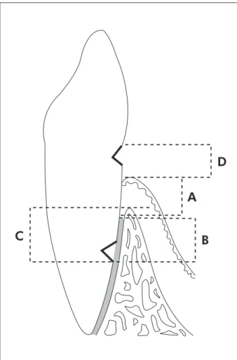
The sections were stained with Hematoxilin - Eo-sin (H&E) and Massom tricrome for the histomet-ric evaluation, and they were codiied in regards to the experimental groups. A computer with a system of image assessment was used, and an optical mi-croscope Diastar (Cambridge Instruments, Buffalo, NY, USA) was coupled to a video camera DXC-107A/107AP (Sony Eletronics Inc., Tokyo, Japan), which was connected to a microcomputer with image assessment software (Sigma Proscan, Jandel Scientif-ic, San Rafael, CA, USA). The sections of the images selected through optical microscopy were captured by the video camera and digitized to the microcom-puter. Thus, linear measurements were performed. The following variables were analyzed (Figure 1):
Level of epithelium: distance from the gingiva
A.
crest until the apical region of the epithelial tis-sue (sulcular + junctional).
Cementum Formation: distance from the apical
B.
notch to the coronary region of the neoformed cementum.
Bone Formation: distance from the apical notch
C.
to the coronary region of neoformed tissue. Residual Gingiva Recession: linear extension of
D.
roots covered by no tissue, measured from the gingiva margin to the coronal notch.
After the measurements of the ive sections, a mean was derived for each parameter. To obtain means of each parameter, the measurements were carried out at three different moments. The statis-tical analysis used Mann-Whitney’s test. The null hypothesis was considered when there was no dif-ference between the two treatment modalities and the rejection of this hypothesis was accepted when p < 0.05.
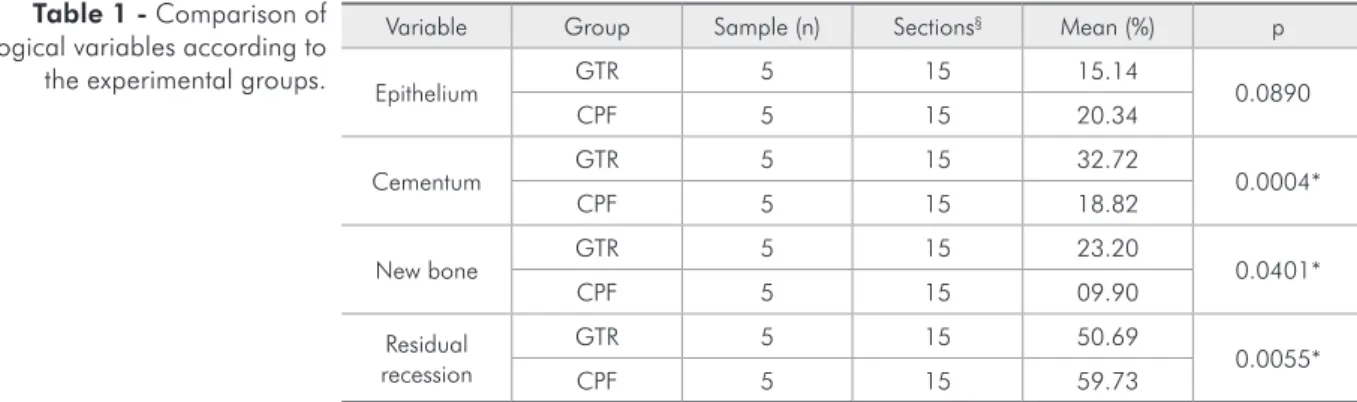
Results
CPF = 18.82%; p = 0.0004), new bone (GTR = 23.20%; CPF = 9.90%; p = 0.0401) and a smaller extension of residual gingival recession in the test group (GTR = 50.69%) when compared to the control group (CPF = 59.73%), with p = 0.0055 (Table 1).
The only item that showed no statistical differ-ence during the assessment was the proliferation of the epithelium (sulcular and junctional) on the root surface, with averages of 15.14% for the GTR group and 20.34% for the CPF group (p = 0.0890).
The neoformation of bone occurred closer to the apical notch due to osteoblastic cell migration to-wards this region.
Discussion
Kimble et al.21 (2004) showed no signiicantly different clinical results between the test (membrane
and DFDBA) and control (membrane alone) groups; nevertheless, other studies19,23 reported that the use of this combination can bring additional clinical beneits, such as higher percentage of root coverage, increased clinical insertion and keratinized gingiva.
The literature presents clinical studies on gin-gival recessions that do or do not associate the use of bioabsorbable membranes with bone grafts;19-23 however, most of the histological research evaluated the use of membranes alone.2,11-18 To provide further information on the clinical beneits mentioned in previous studies22 the association of collagen mem-branes with DFDBA was chosen in order to assess the histometrical healing response and because there is a lack of research evaluating the regenera-tive process by means of microscopy, evaluating fac-tors such as epithelial proliferation, cementum and bone formation, as well as residual recessions of the gingival margin.
According to Bowers et al.11 (1989), the chances of regeneration are improved when DFDBA is com-bined with GTR treatment. These authors have observed that periodontal defects treated with ho-mogenous bone showed a greater bone illing and cementogenesis when compared to the defects that were only scaled. Our indings conirm that the GTR technique (with bioabsorbable collagen membrane and DFDBA) positively inluences periodontal regen-eration, but it was not possible to isolate the effect of the membrane or DFDBA because there was not a control group with membrane or DFDBA alone.
The averages of new cementum formation were statistically signiicant for GTR (32.72%) when compared to CPF (18.82%), as well as bone neofor-mation with averages of 23.20% (GTR) and 9.90% (CPF). This may have occurred due to the exclusion of epithelial cells provided by the membrane, which was eficient in allowing repopulation of the non-differentiated mesenchymal cells in the region of the periodontal ligament, leading to a considerable re-generation of the tissues lost.26
Bone and cementum neoformation was also ob-served by Lee et al.15 (2002), who found that there was a greater bone and cementum formation in the GTR than in the CPF group throughout 16 weeks, although they were not statistically signiicant. Figure 1 - Linear measurement settings (cross-sectional
plane).
B
D
A
It must be pointed out that the authors used only membranes without bone graft.
Considering the root coverage, the average of residual recession was 50.69% for the GTR group and 59.73% for the CPF group, with a statistically signiicant difference (Mann-Whitney). Although less residual recessions have been observed when the GTR technique was used, the study by Leknes et al.6 (2005) showed that the use of bioabsorbable mem-branes does not seem to improve short- and long-term clinical outcomes, and that CPF offers a sim-pler treatment approach for Class I and II recessions with similar results compared to GTR. Our results concerning the residual recessions showed discrep-ancies in relation to other studies because we ob-tained 50.69% for the GTR group while Rosetti et al.22 (2000) found 15.8% and Paolantonio7 (2002) 18.99%.
The only assessed item that showed no statisti-cal difference during the assessment was epithelial proliferation on the root surface, with averages of
15.14% for the GTR group and 20.34% for the CPF group. Nonetheless, even though this item showed no statistical difference, there was a larger epithe-lial migration on the root surface for the CPF group than for the GTR group, which conirms the barrier property of the membrane.
Conclusion
Within the limits of this study, it was possible to conclude that the treatment of gingival recession with collagen membrane and DFDBA showed better results than the treatment with Coronally Positioned Flap alone, which was demonstrated by the larger extension of neoformed cementum and bone, as well as by a smaller proportion of residual recessions.
Acknowledgements
The authors would like to express their gratitude to Prof. Carlos Rossa Junior for his assistance dur-ing the experimental stages of this study.
Variable Group Sample (n) Sections§ Mean (%) p
Epithelium GTR 5 15 15.14 0.0890
CPF 5 15 20.34
Cementum GTR 5 15 32.72 0.0004*
CPF 5 15 18.82
New bone GTR 5 15 23.20 0.0401*
CPF 5 15 09.90
Residual recession
GTR 5 15 50.69
0.0055*
CPF 5 15 59.73
GTR: guided tissue regeneration; CPF: coronally positioned flap; * statistically significant value; § Three
sec-tions for each tooth.
Table 1 - Comparison of histological variables according to the experimental groups.
References
1. Sedon CL, Breault LG, Covington LL, Bishop BG. The subepi-thelial connective tissue graft: part II. Histologic healing and clinical root coverage. J Contemp Dent Pract. 2005;6(2):139-50.
2. Cortellini P, Clauser C, Pini Prato G. Histologic assessment of new attachment following the treatment of a human buccal recession by means of a guided tissue regeneration procedure. J Periodontol. 1993;64(5):387-91.
3. Borghetti A, Glise JM, Monnet-Corti V, Dejou J. Comparative clinical study of a bioabsorbable membrane and subepithelial
connective tissue graft in the treatment of human gingival recession. J Periodontol. 1999;70(2):123-30.
4. Cortellini P, DeSanctis M, Pini Prato G, Baldi C, Clauser C. Guided tissue regeneration procedure using a fibrin-fibro-nectin system in surgically induced recession in dogs. Int J Periodontics Restorative Dent. 1991;11(2):150-63.
inci-sions. Results of a series of consecutive cases. J Periodontol. 1998;69(12):1426-34.
6. Leknes KN, Amarante ES, Price DE, Boe OE, Skavland RJ, Lie T. Coronally positioned flap procedures with or without a biodegradable membrane in the treatment of human gingi-val recession. A 6-year follow-up study. J Clin Periodontol. 2005;32(5):518-29.
7. Paolantonio M. Treatment of gingival recessions by combined periodontal regenerative technique, guided tissue regenera-tion, and subpedicle connective tissue graft. A comparative clinical study. J Periodontol. 2002;73(1):53-62.
8. Trombelli L, Scabbia A, Tatakis DN, Calura G. Subpedicle connective tissue graft versus guided tissue regeneration with bioabsorbable membrane in the treatment of human gingival recession defects. J Periodontol. 1998;69(11):1271-7. 9. Trombelli L, Tatakis DN, Scabbia A, Zimmerman GJ.
Com-parison of mucogingival changes following treatment with coronally positioned flap and guided tissue regeneration pro-cedures. Int J Periodontics Restorative Dent. 1997;17(5):448-55.
10. Wang HL, Bunyaratavej P, Labadie M, Shyr Y, MacNeil RL. Comparison of 2 clinical techniques for treatment of gingival recession. J Periodontol. 2001;72(10):1301-11.
11. Bowers GM, Chadroff B, Carnevale R, Mellonig J, Corio R, Emerson J et al. Histologic evaluation of new attach-ment apparatus formation in humans. Part I. J Periodontol. 1989;60(12):664-74.
12. da Silva Pereira SL, Sallum AW, Casati MZ, Caffesse RG, Weng D, Nociti Jr FH et al. Comparison of bioabsorbable and non-resorbable membranes in the treatment of dehis-cence-type defects. A histomorphometric study in dogs. J Periodontol. 2000;71(8):1306-14.
13. Harris RJ. Human histologic evaluation of a bone graft combined with GTR in the treatment of osseous dehiscence defects: a case report. Int J Periodontics Restorative Dent. 2000;20(5):510-9.
14. Harris RJ. Histologic evaluation of root coverage obtained with GTR in humans: a case report. Int J Periodontics Re-storative Dent. 2001;21(3):240-51.
15. Lee EJ, Meraw SJ, Oh TJ, Giannobile WV, Wang HL. Com-parative histologic analysis of coronally advanced flap with and without collagen membrane for root coverage. J Periodon-tol. 2002;73(7):779-88.
16. Parma-Benfenati S, Tinti C. Histologic evaluation of a new at-tachment utilizing a titanium-reinforced barrier membrane in
a mucogingival recession defect. A case report. J Periodontol. 1998;69(7):834-9.
17. Sallum EA, Pimentel SP, Saldanha JB, Nogueira-Filho GR, Casati MZ, Nociti FH et al. Enamel matrix derivative and guided tissue regeneration in the treatment of dehiscence-type defects: a histomorphometric study in dogs. J Periodontol. 2004;75(10):1357-63.
18. Weng D, Hurzeler MB, Quinones CR, Pechstadt B, Mota L, Caffesse RG. Healing patterns in recession defects treated with ePTFE membranes and with free connective tissue grafts. A histologic and histometric study in the beagle dog. J Clin Periodontol. 1998;25(3):238-45.
19. Dodge JR, Greenwell H, Drisko C, Wittwer JW, Yancey J, Rebitski G. Improved bone regeneration and root coverage using a resorbable membrane with physically assisted cell migration and DFDBA. Int J Periodontics Restorative Dent. 2000;20(4):398-411.
20. Duval BT, Maynard JG, Gunsolley JC, Waldrop TC. Treat-ment of human mucogingival defects utilizing a bioabsorbable membrane with and without a demineralized freeze-dried bone allograft. J Periodontol. 2000;71(11):1687-92. 21. Kimble KM, Eber RM, Soehren S, Shyr Y, Wang HL.
Treat-ment of gingival recession using a collagen membrane with or without the use of demineralized freeze-dried bone allograft for space maintenance. J Periodontol. 2004;75(2):210-20. 22. Rosetti EP, Marcantonio RAC, Rossa Jr C, Chaves ES, Goissis
G, Marcantonio Jr E. Treatment of gingival recession: compar-ative study between subepithelial connective tissue graft and guided tissue regeneration. J Periodontol. 2000;71(9):1441-7. 23. Wang HL, Kimble K, Eber R. Use of bone grafts for the en-hancement of a GTR-based root coverage procedure: a pilot case study. Int J Periodontics Restorative Dent. 2002;22(2):119-27.
24. de Oliveira CA, Spolidorio LC, Cirelli JA, Marcantonio RA. Acellular dermal matrix allograft used alone and in com-bination with enamel matrix protein in gingival recession: histologic study in dogs. Int J Periodontics Restorative Dent. 2005;25(6):595-603.
25. Cirelli JA, Marcantonio Jr E, Marcantonio RAC, Lia RCC, Goissis G, Rossa C Jr. Evaluation of anionic collagen mem-branes in the treatment of class II furcation lesions: a histo-metric analysis in dogs. Biomaterials. 1997;18(18):1227-34. 26. Pitaru S, Tal H, Soldinger M, Azar AO, Noff M. Collagen