Virulence Factor Critical for
Borrelia burgdorferi
Persistence in Mice
Tisha Choudhury Ellis1, Sunny Jain1, Angelika K. Linowski1, Kelli Rike1, Aaron Bestor2, Patricia A. Rosa2, Micah Halpern1, Stephanie Kurhanewicz1, Mollie W. Jewett1*
1Burnett School of Biomedical Sciences, University of Central Florida College of Medicine, Orlando, Florida, United States of America,2Laboratory of Zoonotic Pathogens, Rocky Mountain Laboratories, National Institute of Allergy and Infectious Diseases, National Institutes of Health, Hamilton, Montana, United States of America
Abstract
Analysis of the transcriptome ofBorrelia burgdorferi, the causative agent of Lyme disease, during infection has proven difficult due to the low spirochete loads in the mammalian tissues. To overcome this challenge, we have developed anIn VivoExpression Technology (IVET) system for identification of B. burgdorferi genes expressed during an active murine infection. Spirochetes lacking linear plasmid (lp) 25 are non-infectious yet highly transformable. Mouse infection can be restored to these spirochetes by expression of the essential lp25-encoded pncAgene alone. Therefore, this IVET-based approach selects forin vivo-expressed promoters that drive expression ofpncA resulting in the recovery of infectious spirochetes lacking lp25 following a three week infection in mice. Screening of approximately 15,000 clones in mice identified 289 uniquein vivo-expressed DNA fragments from across all 22 replicons of theB. burgdorferiB31 genome. Thein vivo-expressed candidate genes putatively encode proteins in various functional categories including antigenicity, metabolism, motility, nutrient transport and unknown functions. Candidate genebbk46on essential virulence plasmid lp36 was found to be highly inducedin vivoand to be RpoS-independent. Immunocompetent mice inoculated with spirochetes lackingbbk46seroconverted but no spirochetes were recovered from mouse tissues three weeks post inoculation. However, thebbk46gene was not required forB. burgdorferi infection of immunodeficient mice. Therefore, through an initial IVET screen inB. burgdorferiwe have identified a novelin vivo-induced virulence factor critical for the ability of the spirochete to evade the humoral immune response and persistently infect mice.
Citation:Ellis TC, Jain S, Linowski AK, Rike K, Bestor A, et al. (2013)In VivoExpression Technology Identifies a Novel Virulence Factor Critical forBorrelia burgdorferi Persistence in Mice. PLoS Pathog 9(8): e1003567. doi:10.1371/journal.ppat.1003567
Editor:Jenifer Coburn, Medical College of Wisconsin, United States of America
ReceivedFebruary 27, 2013;AcceptedJuly 1, 2013;PublishedAugust 29, 2013
This is an open-access article, free of all copyright, and may be freely reproduced, distributed, transmitted, modified, built upon, or otherwise used by anyone for any lawful purpose. The work is made available under the Creative Commons CC0 public domain dedication.
Funding:Research reported in this publication was supported by the National Institute of Allergy and Infectious Diseases of the National Institutes of Health under award numbers K22AI081730 (MWJ) and R01AI099094 (MWJ), the Intramural Research Program, NIAID/NIH (PAR), the National Research Fund for Tick-Borne Diseases 2012–2013 pilot grant (MWJ) and UCF startup funds (MWJ). The funders had no role in study design, data collection and analysis, decision to publish, or preparation of the manuscript.
Competing Interests:The authors have declared that no competing interests exist.
* E-mail: [email protected]
Introduction
Lyme disease is a multi-stage inflammatory disease caused by the pathogenic spirocheteBorrelia burgdorferi, which is transmitted by the bite of an infected tick [1].B. burgdorferihas an enzootic life cycle that requires persistence in two disparate environments, the arthropod vector and the mammalian host.B. burgdorferi is well adapted to modulate its expression profile in response to the different conditions encountered throughout its infectious cycle [2]. Although the specific environmental signals that induce changes in spirochete gene expression are not fully defined, it has been reported that changes in temperature, pH, the presence or absence of mammalian blood, as well as changes in bacterial growth rate, can affect patterns of gene expression [2–8]. DNA microarray analysis and proteomics have been used to examine changes in the global expression profile ofB. burgdorferigrown underin vitroconditions that partially mimic the tick and mouse environments [3–5]. A rat dialysis membrane chamber (DMC) implant model, together with microarray technol-ogy, has been used to help identifyB. burgdorferigenes expressed in response to mammalian host-specific signals [7–10]. Although the
data reported in these studies provide insight into the molecular mechanisms of gene regulation, they may not fully reflect the patterns of B. burgdorferi gene expression during an active mammalian infection. Furthermore, transcriptome analysis ofB. burgdorferi during murine infection has proven difficult given that spirochete loads in the blood and tissues are too low to recover sufficient spirochete RNA for direct microarray analysis [11].
15]. IVET is a sensitive and versatile method for identification ofin vivo-expressed genes that has been used with pathogenic bacteria and fungi in a wide variety of host environments and has identified a number of previously uncharacterized virulence genes [14–18]. Using IVET we have developed and applied a genome-wide genetic screening approach to identifyB. burgdorferigenes that are expressed during an active murine infection. This is the first time that an IVET strategy has been applied toB. burgdorferi. Moreover, we have identified a novel gene on virulence plasmid lp36 that is strongly induced in vivo and required for B. burgdorferi persistent infection in the mouse.
Results
Mammalian host-adapted spirochetes demonstrate a 100-fold decrease in ID50relative toin vitrogrown spirochetes
It is clear thatB. burgdorferimodulates its gene expression profile at different stages of the infectious cycle [2]. The infectious dose of
wild-typeB. burgdorferivaries depending on the environment from which the spirochetes are derived. For example, the 50% infectious dose (ID50) of spirochetes derived from partially fed ticks has been found to be two orders of magnitude lower than that of log phase in vitro grown B. burgdorferi [19]. In order to quantitatively assess the impact that adaptation to the mammalian environment has on B. burgdorferi infectivity the 50% infectious dose (ID50) of spirochetes derived directly from the mammalian host was determined and compared to that of log phase in vitro grown spirochetes.B. burgdorferiare only transiently present in the blood of immunocompetent mice [20], whereas spirochetes persist longer in the blood of immunocompromised mice [21]. Therefore, the blood of severe combined immunodeficiency (scid) mice infected with B. burgdorferi was used as a source of spirochetes adapted to the mammalian environment. Strikingly, an inoculum containing approximately eightin vivo-derived spirochetes was able to infect five out of six mice, whereas, 5,000 in vitro grown spirochetes were required to obtain this level of infectivity (Table 1). The ID50 forin vivo-derived spirochetes was found to be less than eight organisms. In contrast, the ID50forin vitrogrown spirochetes was calculated to be 660 organisms. These data indicate that mammalian host-adapted spirochetes are 100-fold more infectious than in vitro grown spirochetes, likely due to appropriate coordinate expression of in vivo-expressed genes important for murine infectivity.
TheB. burgdorferiIVET system is a robust method for selection ofB. burgdorferisequences that are expressed during murine infection
We have developed a genome-wide genetic screening method to identify B. burgdorferi genes that are expressed during mouse infection using anin vivoexpression technology (IVET) approach [12,13]. The in vivo expression technology vector, pBbIVET, carries the B. burgdorferi bmpB Rho-independent transcription terminator sequence [22] repeated in triplicate (3XTT), to prevent any read-through promoter activity from the pBSV2* Borrelia shuttle vector backbone, followed by the promoter-less in vivo-essentialpncAgene (Figure 1) [23,24]. Spirochetes lacking linear plasmid (lp) 25 are non-infectious in mice and severely compro-mised in the tick vector [25–31]. ThepncAgene, located on lp25, encodes a nicotinamidase that is sufficient to restore murine infectivity to B. burgdorferi lacking the entire lp25 plasmid [24]. Genetic transformation of low-passage, infectious B. burgdorferi occurs at low frequency and efficiency hampering introduction of
Table 1.In vivo-adaptedB. burgdorferiare highly infectious.
In vivogrown spirochetes In vitrogrown spirochetes
Spirochete dose
Number of mice infecteda/number
of mice analyzed Spirochete dose
Number of mice infecteda/number of mice analyzed
86102 6/6 56104 6/6
86101 6/6 56103 6/6
86100 5/6 56102 2/6
56101 1/6
56100 0/6
ID50b ,8 spirochetes ID50b 660 spirochetes
aMouse infection was determined 3 weeks post inoculation by serological response toB. burgdorferiproteins and reisolation of spirochetes from ear, bladder and joint tissues.
bThe ID
50was calculated according to method of Reed and Muench [83]. doi:10.1371/journal.ppat.1003567.t001
Author Summary
Lyme disease is caused by tick-bite transmission of the pathogenic spirochete Borrelia burgdorferi. An increased understanding of how B. burgdorferisurvives throughout its infectious cycle is critical for the development of innovative diagnostic and therapeutic protocols to reduce the incidence of Lyme disease. One of the major difficulties blocking this effort has been genome-wide identification of the B. burgdorferi genes that are expressed in the mammalian host environment. Using in vivo expression technology (IVET) in B. burgdorferi for the first time, we have identified B. burgdorferi genes that are expressed during an active murine infection. We demonstrate that candidate genebbk46, encoded on essential linear plasmid 36, is highly expressed in vivo and, unlike some other knownB. burgdorferi in vivo-induced genes, is not RpoS regulated. Spirochetes lackingbbk46establish an infection in mice and elicit an antibody response but are undetect-able in mouse tissues three weeks post inoculation. The
a complex DNA library into an infectious background [32,33]. Because B. burgdorferi clones lacking lp25 and lp56 demonstrate increased transformability [34], we isolated a clonal derivative of the low-passage infectious clone A3 that lacks both lp25 and lp56. This clone was designated A3 68-1 [35]. Clones A3 and A3 68-1 were transformed by electroporation with 20mg of a Borrelia shuttle vector. The transformation frequency and efficiency of A3 68-1 was determined relative to that of the A3 parent. As expected, genetic transformation of A3 68-1 occurred at a high frequency and efficiency. We recovered approximately 2,000 transformants/ ml in clone A3 68-1, whereas no transformants were recovered with a parallel transformation of clone A3 (data not shown). In order to test the function of the B. burgdorferi IVET system, the promoter for thein vivoessentialospCgene [36], was cloned in front of the promoter-less pncA gene in pBbIVET (Figure 1), creating plasmid pBbIVET-ospCp. This plasmid, along with pBbIVET alone, was transformed into the non-infectious, low-passage, highly transformableB. burgdorfericlone A3 68-1. All clones were tested for their abilities to infect groups of 6 C3H/HeN mice at an infectious dose 100 (ID100) of 16104spirochetes [37], indicating the presence or absence of an active promoter sufficient to drive expression ofpncA thereby restoring infectivity. Spirochetes were reisolated from the ear, bladder and joint tissues of 5/6 mice infected with B. burgdorferi harboring pBbIVET-ospCp. No spirochetes were reisolated from mice (0/6) infected with B. burgdorferi carrying the promoter-less pBbIVET alone. Together these data demonstrated that our promoter trap system functioned with a knownin vivoactive promoter.
Screening forB. burgdorferigenes expressed during murine infection
AB. burgdorferi genomic DNA library using an average DNA fragment size of approximately 200 bps was constructed upstream of the promoter-lesspncAgene (Figure 1) in the pBbIVET vector in E. coli, yielding approximately 30,000 independent clones. A small subset of individuals from the 30,000 clone library inE. coliwas
analyzed by PCR and DNA sequencing and determined to carry non-identicalB. burgdorferi DNA fragments. The strategy used to construct the pBbIVET library allowed the DNA fragments to be cloned in either the forward or reverse orientation relative to the pncAgene. Therefore, a library of 30,000 clones each harboring a unique 200 bp DNA fragment represented approximately 26 coverage of the 1.5 Mb genome of B. burgdorferi. Although the initial analysis of the transformation efficiency ofB. burgdorfericlone A3 68-1 demonstrated that each transformation of 20mg of a single purified plasmid into this genetic background yielded approximately 10,000 transformants, this transformation efficiency was not achieved when 20mg of complex library plasmid DNA was transformed into A3 68-1. Forty four transformations of the library plasmid DNA resulted in recovery of approximately 15,000 individual clones inB. burgdorferiA3 68-1, representing an IVET library in B. burgdorferi with approximately 16coverage of the spirochete genome. As described for the pBbIVET library inE. coli, a subset of individuals from 15,000 clone library in B. burgdorferi were analyzed and found to carry non-identical B. burgdorferiDNA fragments.
Like the BbIVET system described here, many IVET strategies are based upon complementation of auxotrophy. For microor-ganisms other than B. burgdorferi these strategies have allowed negative selection against ‘‘promoter-less’’ clones in minimal medium in which the auxotroph mutants are unable to grow [38].B. burgdorferilackingpncAare not attenuated for growth in the complex, undefinedB. burgdorferimedium, BSKII. Moreover, there is currently no minimal medium available that supports the growth of wild-typeB. burgdorferi. Therefore, the BbIVET system did not include negative selection against ‘‘promoter-less’’ clonesin vitro. 179 mice were infected with pools from theB. burgdorferi IVET library of approximately 100 clones each, with each clone at a dose of 16104spirochetes resulting in a total dose of 16106spirochetes per mouse. Three weeks post inoculation, mice were sacrificed and ear, heart, bladder and joint tissues were harvested for reisolation of infectious spirochetes. 175 out of 179 mice became infected with B. burgdorferias determined by reisolation of spirochetes from at least two or more of the tissue sites analyzed (Table 2). However, due to the potentially stochastic nature of the kinetics of infection [39] and/or tissue-specific promoter activity of distinct B. burgdorferigenomic fragments not all four tissue sites from all 175 infected mice were found to be positive for spirochete reisolation. Nonetheless, the recovery of live spirochetes from infected mouse tissues suggested that these spirochetes harbored in vivo active promoter(s) in the pBbIVET plasmid sufficient to drive expression of the in vivo-essential pncA gene to restore spirochete mouse infectivity. Total genomic DNA was isolated from each pool of reisolated spirochetes from each of the four mouse tissues and the pBbIVET plasmid DNA rescued in E. coli. Colony PCR using primers targeting the genomic DNA insert region of the pBbIVET vector was performed on 24 of the resultingE. colicolonies from each plasmid rescue transformation. No reisolated spirochetes were found to harbor a pBbIVET plasmid lacking a genomic DNA fragment insert. The amplified inserts were analyzed by restriction digest using a cocktail of the A/T-rich restriction enzymes to identify those DNA fragments with distinct restriction patterns, suggesting that these fragments represent differentin vivo active promoters. Up to eleven non-identical restriction digest patterns were detected for every subset of 24E. colitransformants carrying pBbIVET DNA that were analyzed (Figure S1). The DNA fragments corresponding to each distinct restriction digest pattern were further analyzed by DNA sequencing and the identities of the sequences determined by microbial genome BLAST analysis. Screening of approximately 15,000 BbIVET
Figure 1. Schematic representation of the pBbIVET vector.
Features of this vector include: 3XTT, the transcriptional terminator sequence forbmpB[22] repeated in triplicate;pncA, promoterlesspncA gene; flgBpkan, kanamycin resistance cassette; zeo, zeocin resistance
marker; ColE1,E. coliorigin of replication; ORFs 1, 2, 3,B. burgdorfericp9 replication machinery. The EcoRI restriction site was used to clone theB. burgdorfericontrolin vivo-expressed promoter,ospCp, as well as theB.
burgdorferi(Bb) gDNA library, in front of the promoterlesspncAgene. The pBbIVET vector was derived from theB. burgdorferishuttle vector pBSV2* [80].
clones through mice resulted in the identification of 289 non-identicalB. burgdorferi in vivo-expressed (Bbive) DNA fragments from across the chromosome and all 21 plasmid replicons of the B. burgdorferi B31 segmented genome (Table 2). Although the 1:1 molar ratio of insert to vector used to generate the pBbIVET library did not preclude insertion of more than one fragment into each clone, only 20 out of the 289 clones were found to harbor two distinct DNA fragments. Of these clones the 39 DNA fragment, proximal to thepncAORF, was assumed to be the active promoter and was included in the subsequent analyses.
B. burgdorferi in vivoexpressed promoters map to distinct classes of putative regulatory sequences across the genome
Genomic mapping of the 289 uniqueBbivepromoters identified in this genetic screen demonstrated that 67% of the sequences mapped to sense DNA in the same direction as annotated open reading frames, 27% mapped to antisense DNA in the opposite direction to annotated open reading frames and 6% mapped to intergenic regions lacking annotated open reading frames. Of the large percentage of sense sequences, 41%, which represented 28% of the total Bbive sequences, mapped to regions just upstream of and in the same orientation to annotated open reading frames, suggesting that these sequences are promoters for the associated open reading frames and that these open reading frames are candidatein vivo-expressed genes. The remaining 59% of the sense sequences, which represented 39% of the totalBbive sequences, mapped within annotated open reading frames, suggesting the possibility for promoter elements within B. burgdorferigenes. Similar findings of putative transcriptional start sites within genes and operons have been reported for other bacterial pathogens [40,41]. The sequences that mapped to putative promoter locations in the genome and the genes associated with these promoters were prioritized for further analysis. Among the list of 80 sequences, 9 promoter regions were represented by two overlapping genomic DNA fragments. Five of these overlapping sequence pairs shared the same 39 end, suggesting that the sequences belonging to each pair contained the same promoter. Whereas, the other four overlapping sequence pairs harbored distinct 39 ends, suggesting that each sequence contained a unique promoter. The 71in vivo-expressed candidate genes have been annotated to encode proteins in various functional categories including: cell division, cell enve-lope, replication, metabolism, motility, protein synthesis, trans-port and unknown functions (Table 3).
IVET identified candidate genebbk46on virulence plasmid lp36
Linear plasmid 36 is required forB. burgdorferimouse infection; however, the genetic elements on lp36 that contribute to this
phenotype have not been fully defined [37]. IVET identified a candidate in vivo-expressed promoter sequence, Bbive162, which mapped to lp36. This sequence was found to be 60 bp long, with 48 bp immediately upstream of and in the same direction as the BBK46 open reading frame (Figure 2), suggesting that thebbk46 gene may be expressed during mammalian infection and may contribute to the essential role of lp36 inB. burgdorferiinfectivity. Therefore, thebbk46gene was selected for further analysis.
Expression of thebbk46gene is induced during murine infection
Our BbIVET screen identified genebbk46as a putativein vivo-expressed gene. The BbIVET screen was designed to identifyB. burgdorferiDNA fragments that are expressedin vivo and did not discriminate between those promoters that are specifically induced in vivoand those promoters that are expressed bothin vitroandin vivo. Therefore, quantitative reverse transcription PCR (qRT-PCR) was used to validate the expression ofbbk46 in vivoand to determine whether bbk46 expression was upregulated in vivo compared toin vitro. Total RNA was isolated from bladder tissue collected from mice infected with 16105 wild-type B. burgdorferi three weeks post-inoculation as well as log phasein vitro grown spirochetes. RNA was converted to cDNA using random hexamer primers and the mRNA level of each target gene was measured relative to the constitutiverecAgene using quantitative PCR. The gene expression levels of flaB and ospC were also measured as control constitutively-expressed andin vivo-induced genes, respec-tively. These data demonstrated that althoughbbk46was expressed duringin vitrogrowth, expression of this gene was increased more than 100-fold during mammalian infection (Figure 3A). Consistent with their known patterns of gene regulation,flaBexpression was relatively unchanged in vivo compared to in vitro; whereas,ospC demonstrated a nearly 1000-fold increase in expression in vivo compared toin vitro(Figure 3A). Moreover, the relative amount of bbk46 expression during in vitro growth was found to be approximately 10-fold more than that of ospC. Whereas, the in vivoexpression levels of genesbbk46,ospCandflaBwere similar.
RpoS is a global regulator that controls expression of genes expressed during mammalian infection, including ospC [2]. Becausebbk46expression was inducedin vivoin a manner similar to that ofospC, we sought to determine whether, likeospC,bbk46is an RpoS-regulated gene. RNA was isolated from stationary phase temperature-shifted wild-type and DrpoS mutant spirochetes, a growth condition previously shown to induce expression of rpoS and rpoS-regulated genes [42]. Quantitative RT-PCR was then performed for genesbbk46,flaB,ospCandrecA, as described above. As expected, ospC expression was increased approximately 20 times in the presence compared to the absence ofrpoS(Figure 3B). In contrast,bbk46expression was RpoS-independent under these growth conditions (Figure 3B). Likewise, no RpoS-dependent change in gene expression was detected forflaB. Interestingly, the Table 2.TheB. burgdorferiIVET system selects forin vivo-active promoters.
Number of BbIVET
clones screened Positive reisolation of infectious spirochetes from mouse tissuesa
Number of unique genomic fragments recovered
Ear Heart Bladder Joint
,15,000 175/179 173/179 172/179 174/179 289
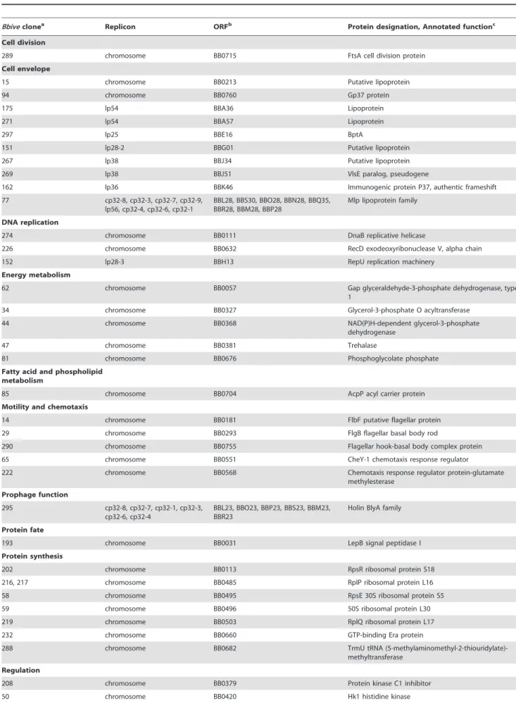
Table 3.B. burgdorferi in vivo-expressed candidate genes organized by functional category.
Bbiveclonea Replicon ORFb Protein designation, Annotated functionc
Cell division
289 chromosome BB0715 FtsA cell division protein
Cell envelope
15 chromosome BB0213 Putative lipoprotein
94 chromosome BB0760 Gp37 protein
175 lp54 BBA36 Lipoprotein
271 lp54 BBA57 Lipoprotein
297 lp25 BBE16 BptA
151 lp28-2 BBG01 Putative lipoprotein
267 lp38 BBJ34 Putative lipoprotein
269 lp38 BBJ51 VlsE paralog, pseudogene
162 lp36 BBK46 Immunogenic protein P37, authentic frameshift
77 cp32-8, cp32-3, cp32-7, cp32-9,
lp56, cp32-4, cp32-6, cp32-1
BBL28, BBS30, BBO28, BBN28, BBQ35, BBR28, BBM28, BBP28
Mlp lipoprotein family
DNA replication
274 chromosome BB0111 DnaB replicative helicase
226 chromosome BB0632 RecD exodeoxyribonuclease V, alpha chain
152 lp28-3 BBH13 RepU replication machinery
Energy metabolism
62 chromosome BB0057 Gap glyceraldehyde-3-phosphate dehydrogenase, type
1
34 chromosome BB0327 Glycerol-3-phosphate O acyltransferase
44 chromosome BB0368 NAD(P)H-dependent glycerol-3-phosphate
dehydrogenase
47 chromosome BB0381 Trehalase
81 chromosome BB0676 Phosphoglycolate phosphate
Fatty acid and phospholipid metabolism
85 chromosome BB0704 AcpP acyl carrier protein
Motility and chemotaxis
14 chromosome BB0181 FlbF putative flagellar protein
29 chromosome BB0293 FlgB flagellar basal body rod
290 chromosome BB0755 Flagellar hook-basal body complex protein
65 chromosome BB0551 CheY-1 chemotaxis response regulator
222 chromosome BB0568 Chemotaxis response regulator protein-glutamate
methylesterase
Prophage function
295 cp32-8, cp32-7, cp32-1, cp32-3,
cp32-6, cp32-4
BBL23, BBO23, BBP23, BBS23, BBM23, BBR23
Holin BlyA family
Protein fate
193 chromosome BB0031 LepB signal peptidase I
Protein synthesis
202 chromosome BB0113 RpsR ribosomal protein S18
216, 217 chromosome BB0485 RplP ribosomal protein L16
58 chromosome BB0495 RpsE 30S ribosomal protein S5
59 chromosome BB0496 50S ribosomal protein L30
219 chromosome BB0503 RplQ ribosomal protein L17
232 chromosome BB0660 GTP-binding Era protein
288 chromosome BB0682 TrmU tRNA
(5-methylaminomethyl-2-thiouridylate)-methyltransferase
Regulation
208 chromosome BB0379 Protein kinase C1 inhibitor
amount of flaB expression detected in the stationary phase temperature-shifted spirochetes (Figure 3B) was dramatically decreased compared to the amount of flaB expression detected in log phase andin vivogrown spirochetes (Figure 3A), suggesting
that flaB is not expressed at the same level under all growth conditions. Together these data demonstrated that bbk46 was highly induced during murine infection andbbk46expression was not controlled by RpoS duringin vitrogrowth.
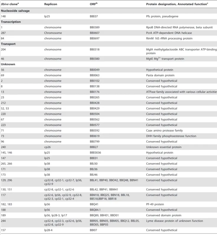
Table 3.Cont.
Bbiveclonea Replicon ORFb Protein designation, Annotated functionc
Nucleoside salvage
148 lp25 BBE07 Pfs protein, pseudogene
Transcription
1 chromosome BB0389 RpoB DNA-directed RNA polymerase, beta subunit
287 Chromosome BB0607 PcrA ATP-dependent DNA helicase
84 chromosome BB0697 RimM 16S rRNA processing protein
Transport
204 chromosome BB0318 MglA methylgalactoside ABC transporter ATP-binding
protein
46 chromosome BB0380 MgtE Mg2+transport protein
Unknown
56 chromosome BB0049 Hypothetical protein
69 chromosome BB0063 Pasta domain protein
2 chromosome BB0102 Conserved hypothetical
8 chromosome BB0138 Conserved hypothetical
13 chromosome BB0176 ATPase family associated with various cellular activities
23 chromosome BB0265 Conserved hypothetical
212 chromosome BB0428 Conserved hypothetical
52, 53 chromosome BB0429 Conserved hypothetical
220 chromosome BB0504 Conserved hypothetical
67 chromosome BB0562 Conserved hypothetical
223 chromosome BB0577 Conserved hypothetical
71 chromosome BB0592 Caax amino protease family
73 chromosome BB0619 DHH family phosphoesterase function
96 chromosome BB0799 Conserved hypothetical
240 cp26 BBB27 Unknown essential protein
145, 146 lp25 BBE0036 Hypothetical protein
147 lp25 BBE01 Conserved hypothetical
265, 266 lp38 BBJ30 Conserved hypothetical
171 lp38 BBJ36 Conserved hypothetical
173 lp38 BBJ46 Conserved hypothetical
129, 296 cp32-8, cp32-1, cp32-7, lp56,
cp32-9
BBL41, BBP40, BBO42, BBQ48, BBN41 Conserved hypothetical
130, 151 cp32-8, cp32-1, cp32-6 BBL42, BBP41, BBM41 Conserved hypothetical
117 cp32-6, lp56, cp32-9, cp32-8,
cp32-3, cp32-1, cp32-4
BBM18, BBQ25, BBN18, BBL18, BBS18,BBP18, BBR18
Conserved hypothetical
182, 183 lp56 BBQ41 PF-49 protein
188 lp56 BBQ84.1 Conserved hypothetical
189 lp56, lp28-3, lp17 BBQ89, BBH01, BBD01 Conserved domain protein
244 cp32-4, cp32-3, cp32-6, lp56,
cp32-8, cp32-9
BBR05, BBN05, BBM05, BBQ12, BBL05, BBO05, BBP05
Lyme disease protein of unknown function
157 lp28-4 BBI07 Conserved hypothetical
aIn some cases twoBbiveclones shared overlapping, non-identical sequence, as indicated by twoBbiveclone numbers. bORF, open reading frame that maps just downstream and in the same orientation to theBbivesequence.
Thebbk46open reading frame fails to produce detectable amounts of protein duringin vitrogrowth
The bbk46 gene is a member of paralogous gene family 75, which also includes lp36-encoded genesbbk45,bbk48andbbk50, all of which are annotated to encode putative P37 immunogenic lipoproteins [43–45] (Figure 4). These genes are located on the right arm of lp36 in B. burgdorferi clone B31, which is a highly variable region among distinct B. burgdorferi isolates [44]. The
members of paralogous gene family 75 are conserved withinB. burgdorferi isolates but are not present in the relapsing fever spirochetes. The B. burgdorferi clone B31 BBK45, BBK48 and BBK50 proteins are predicted to be 301, 288 and 332 amino acids, respectively. However,B. burgdorfericlone B31bbk46is annotated as a pseudogene as a result of an authentic frame shift resulting in a TAA stop codon at nucleotide 625 [43–45], thereby producing a putative 209 amino acid protein. In contrast, the BBK46 homolog
Figure 2. The nucleotide and putative amino acid sequence of the BBK46 open reading frame.The reverse complement of nucleotides 28391 to 29474 on lp36, encompassingbbk46(Genbank GeneID: 1194234) and its putative promoter sequence. The nucleotide sequence ofBbive162 is underlined. The putative ribosome binding site is shown in bold italics. The putative BBK46 amino acid sequence is shown in bold. The stop codons at nucleotides 625 and 820 are highlighted in gray. The position of the inserted FLAG-epitope tag sequence is indicted with a star (*). The position of the inserted cMyc-epitope tag sequence is indicated with a number sign (#).
in clone N40, BD04, harbors a CAA codon at nucleotide 625, resulting in a glutamic acid residue at amino acid 209 and producing a putative 273 amino acid protein [46]. Sequence analysis of the cloned bbk46 open reading frame confirmed the presence of the TAA stop codon at nucleotide 625 (Figure 2). To experimentally determine the size of the BBK46 protein produced inB. burgdorferiB31 thebbk46ORF along with a FLAG epitope tag sequence prior to the stop codon at nucleotide 625 and a cMyc epitope tag sequence prior to the stop codon at nucleotide 820 (Figure 2) was cloned into theB. burgdorferishuttle vector pBSV2G under the control of either the constitutive flaBpromoter or the putative endogenousbbk46promoter. A mutant clone lacking the entire BBK46 open reading frame was constructed by allelic exchange and verified by PCR analysis (Figure 5A and 5B). The bbk46 mutant clone was transformed with the shuttle vectors carrying the epitope tagged bbk46 constructs. All transformants were verified to contain the plasmid content of the parent clone.
BBK46 protein production was assessed in both E. coli and B. burgdorferi. Immunoblot analyses using aFLAG and acMyc monoclonal antibodies resulted in detection of a FLAG-epitope tagged protein of an approximate molecular mass of 23 kDa, which is the predicted size of the 209 amino acid BBK46 protein, in theE. coliclones carrying both theflaBp-driven and thebbk46p -driven constructs (Figure 6). Surprisingly, no FLAG epitope tagged protein was detected in either B. burgdorferi clone (Figure 6), althoughbbk46gene expression was observed in these clones (data not shown), indicating that the lack of detectable BBK46 protein was not likely the result of a transcription defect. Furthermore, no cMyc epitope tagged protein was detected in eitherE. coliorB. burgdorferi. Together these data suggested that although thebbk46 ORF is competent to produce a 23 kDa protein inE. coliand the transcript is expressed inB. burgdorferiduringin vitro growth, the protein is either not produced or is rapidly turned over in log phasein vitrogrownB. burgdorferi.
As a putative member of the P37 immunogenic lipoprotein family, BBK46 is predicted to localize to the spirochete outer surface and to be immunogenic during mammalian infection. Therefore, recombinant BBK46, lacking the first 32 amino acids that are predicted to comprise the signal sequence for the lipoprotein, was produced in E. coli as an N-terminal fusion to glutathioneS-transferase (GST). To assess the immunogenicity of the BBK46 protein, immunoblot analysis was performed using purified rGST-BBK46 probed with mouse immune serum collected 21 days post inoculation with 16104 wild-type B. burgdorferi. The rGST-BBK46 protein was found to be non-immunoreactive with mouse immune serum, in contrast to the control antigen BmpA (Figure S2). These data suggest that, if produced in B. burgdorferi, BBK46 is not an immunoreactive antigen. However, these data do not rule out the possibility that the immunogenic epitope is not present or available in the recombinant protein produced inE. coli.
Thebbk46gene is required forB. burgdorferipersistence in immunocompetent mice
In vitrogrowth analysis demonstrated that thebbk46mutant and complemented clones had no detectable in vitro phenotypes (Figure 5C). Therefore, to examine the role of bbk46 in mouse infectivity, groups of five C3H/HeN female mice were needle inoculated intradermally under the skin of the upper back with 16104 wild-type, Dbbk46/vector or Dbbk46/bbk46+
spirochetes. Three weeks post inoculation, mice were assessed forB. burgdorferi infection by serology and reisolation of spirochetes from the inoculation site, ear, bladder and joint tissues. All five mice from each infection group were seropositive for anti-B. burgdorferi antibodies (Figure 7, Table 4). Surprisingly however, no spirochetes were reisolated from all tissues examined from the five mice inoculated with the Dbbk46/vector clone (Table 4), whereas, all five mice inoculated with the wild-type or theDbbk46/ bbk46+
clone resulted in reisolation of spirochetes from all tissues analyzed (Table 4). Together these data demonstrated that spirochetes lacking thebbk46gene transiently infected and elicited a humoral response in mice, but were unable to maintain a persistent infection in mouse tissues. To further define the contribution of the host immune response to the inability of spirochetes lacking thebbk46genes to cause a persistent infection, groups of five severe combined immunodeficiency (scid) mice were inoculated with 16104 wild-type, Dbbk46/vector or Dbbk46/ bbk46+
spirochetes. Three weeks post inoculation the animals were assessed for infection by reisolation of spirochetes from the ear, bladder and joint tissues. Consistent with the hypothesized role of
Figure 3. Expression of thebbk46gene is upregulated during murine infection and is RpoS-independent. Total RNA was isolated from bladder tissue collected from (A) mice infected with 16105wild-type B. burgdorferithree weeks post-inoculation (in vivo,
gray bars) and from log phasein vitrogrown spirochetes (in vitro, white bars) or (B) stationary phase temperature-shifted stationary phase in vitrogrown wild-type (white bars) orDrpoS(gray bars)B. burgdorferi. RNA was reverse transcribed to cDNA using random hexamer primers. The expression of bbk46, flaB and ospC were quantified using quantitative reverse transcription polymerase chain reaction (qRT-PCR) and a standard curve analysis method. The mRNA levels of thebbk46, flaB and ospC gene transcripts were normalized to that of the constitutiverecAgene. The data are expressed as the gene transcript/ recAtranscript. The data represent the average of triplicate qRT-PCR analyses of 3 biological replicates. Error bars represent the standard deviation from the mean.
bbk46 in immune evasion and persistence, four out of five immunodeficient mice inoculated with theDbbk46 mutant were positive for spirochete reisolation from all tissues examined (Table 4). These data demonstrated that a functional host immune response is required for the clearance of spirochetes lackingbbk46 from mouse tissues 3 weeks post infection, indicating thatbbk46is essential for the ability ofB. burgdorferito avoid killing by the host immune system in order to establish a persistent infection.
Discussion
In this study we have successfully adapted and applied for the first time an IVET-based genetic screen for use inB. burgdorferifor the purpose of identifying spirochete genes that are expressed
during mammalian infection. Historically, genetic manipulation of low passage, infectiousB. burgdorferihas been challenged by the low transformation frequencies of these spirochetes, preventing appli-cation of classicin vivogenetic screening techniques such asin vivo expression technology (IVET) and signature-tagged mutagenesis (STM) [47] to identifyB. burgdorferigenetic elements important for pathogenicity. However, advances in the understanding of theB. burgdorferirestriction modification systems that inhibit transforma-tion [34,48–51] have recently allowed constructransforma-tion and charac-terization of a comprehensive STM mutant library in infectiousB. burgdorferi[52]. The foundation for our strategy for development of IVET inB. burgdorferiwas based upon the spirochete’s requirement of lp25 for both restriction modification and virulence functions. Spirochetes lacking lp25 are highly transformable but
non-Figure 4. Amino acid alignment of the putative members of the immunogenic protein P37 family encoded on lp36.Shown is an amino acid alignment of the P37 protein family members BBK45 (GenBank accession no. NP_045617.2), BBK46 (translatedbbk46, Genbank GeneID: 1194234), BBK48 (GenBank accession no. NP_045619.1) and BBK50 (GenBank accession no. NP_045621.1). Amino acids identical to the consensus sequence are shaded. The predicted SpLip lipobox sequence [81] is indicted with five stars. Dashes represent spaces introduced for optimal sequence alignment. The positions of the two stop codons in thebbk46translation are indicated with arrows. Amino acid sequences were aligned using the CLUSTAL W algorithm in the MEGALIGN program from the DNASTAR Lasergene suite.
infectious in mice [27–29,34]. Restoration of the lp25-encoded pncA gene to lp252spirochetes restores wild-type infectivity [24] but maintains high transformation frequency. At the time of the development of the pBbIVET system the true start codon of the pncAgene was not defined; therefore, the promoter-lesspncAgene construct in the pBbIVET plasmid used an engineered AUG start codon and was missing the first 24 nucleotides of the now defined pncA ORF [23]. Furthermore, this construct was purposefully
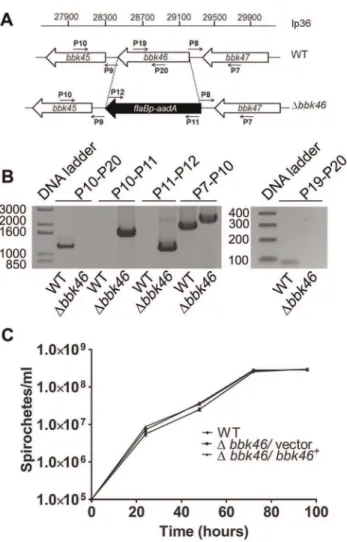
Figure 5. Generation of the Dbbk46 mutant and genetic complemented clones inB. burgdorferi.(A) Schematic representa-tion of the wild-type (WT) andDbbk46loci on lp36. The sequence of the entire bbk46 open reading frame was replaced with a flaBp-aadA
antibiotic resistance cassette [37,82]. Locations of primers for analysis of the mutant clones are indicated with small arrows and labels P7–P12, P19 and P20. Primer sequences are listed in Table 5. (B) PCR analysis of theDbbk46mutant clone. Genomic DNA isolated from WT andDbbk46/ vector spirochetes served as the template DNA for PCR analyses. DNA templates are indicated across the bottom of the gel image. The primer pairs used to amplify specific DNA sequences are indicated at the top of the gel image and correspond to target sequences as shown in A. Migration of the DNA ladder in base pairs is shown to the left of each image. (C)In vitrogrowth analysis of mutant clones. A3-68DBBE02 (WT), bbk46::flaBp-aadA/pBSV2G (Dbbk46/vector) and bbk46::flaBp-aadA/
pBSV2G-bbk46(Dbbk46/bbk46+
) spirochetes were inoculated in tripli-cate at a density of 16105spirochetes/ml in 5 ml of BSKII medium.
Spirochete densities were determined every 24 hours under dark field microscopy using a Petroff-Hausser chamber over the course of 96 hours. The data are represented as the number of spirochetes per ml over time (hours) and is expressed as the average of 3 biological replicates. Error bars indicate the standard deviation from the mean. doi:10.1371/journal.ppat.1003567.g005
Figure 6. BBK46 protein production is detectable inE. colibut not in B. burgdorferi. Immunoblot analysis of total protein lysate prepared from 1.56108B. burgdorferiDbbk46(Bb) orE. coliharboring either pBSV2GflaBp-bbk46-FLAG-cMyc(flaBp) or pBSV2Gbbk46p-bbk46
-FLAG-cMyc(bbk46p). Protein lysates were separated by SDS-PAGE and
immunoblots performed using anti-FLAG monoclonal antibodies (a FLAG) and anti-cMyc monoclonal antibodies (a cMyc). 300 ng of purified PncA-FLAG [23] and GST-BmpA-cMyc [77] proteins served as positive controls (+) for each antibody. The positions of markers to the
left of the panel depict protein standard molecular masses in kilodaltons.
doi:10.1371/journal.ppat.1003567.g006
Figure 7. Spirochetes lacking bbk46 retain seroreactivity in mice. Immunoblot analysis of sera collected three weeks post inoculation from groups of five C3H/HeN mice inoculated with clone A3-68DBBE02 (WT), bbk46::flaBp-aadA/pBSV2G (Dbbk46/vector) and
bbk46::flaBp-aadA/pBSV2G-bbk46(Dbbk46/bbk46+) at a dose of 16104
spirochetes per mouse. (A) Total protein lysate fromB. burgdorfericlone B31 A3 was probed with the serum from each individual mouse (1–5). (B) Purified recombinant GST-OspC protein was probed with pooled sera from the five mice in each infection group oraOspC polyclonal antibodies. The positions of markers to the left of the panel depict protein standard molecular masses in kilodaltons.
designed without a ribosome binding site (RBS) and was dependent upon the cloned B. burgdorferi DNA fragments to contain both a promoter and a functional RBS. Although we acknowledge that this requirement may have limited the number of clones identified in our screen, during development of the BbIVET system we found that inclusion of an RBS sequence in the promoterless pncA construct resulted in vector-driven PncA production in the absence of a promoter. Thus, in order to reduce the possibility of recovering false positive clones, the pBbIVET system was designed without an RBS. The enzyme Tsp509I was selected to generate the DNA fragments for the pBbIVET library because the AATT restriction site of this enzyme is present approximately every 58 bp in the B. burgdorferi B31 genome. However, it is possible that DNA fragments generated with this enzyme will not result in sequences that contain a 39 RBS appropriately distanced from the start codon of the pncA ORF, thereby limiting the number of clones identified in the screen.
Screening of a 15,000 clone B. burgdorferi genomic library in mice identified 289 DNA sequences from across all 22B. burgdorferi replicons capable of promoting pncA expression resulting in an infectious phenotype. It is likely that the BbIVET screen did not achieve saturation because the number of clones analyzed was only estimated to cover theB. burgdorferigenome one time, under the assumption that each cloned DNA fragment in the library was unique. Analysis of the pBbIVET library inB. burgdorferisuggested that the library was composed of 15,000 unique clones. However, because only a small fraction of the library was examined for the sequences of the DNA fragment inserts, our findings do not rule out the potential that the library was composed of less than 15,000 non-identical clones and therefore, may represent less than 16 coverage of the genome. Of the 175 mice infected with the pBbIVET library, 10% resulted in reisolation of a single clone, 62% resulted in reisolation of two to five unique clones, and 28% resulted in reisolation of six to eleven unique clones. Furthermore, 57% of the 289 Bbive sequences were only recovered once; whereas, 39% of the sequences were recovered two to five times and 4% of the sequences were recovered six to twelve times. These data are indicative of the amount of redundancy in the screen and suggest that although the screen may not have been representative of the entire B. burgdorferi genome, a large percentage of mice became infected with multiple clones and many of the Bbive sequences were recovered more than once.
We found that 71 of theBbivesequences mapped to canonical promoter positions upstream of annotated open reading frames in
theB. burgdorferigenome. Unexpectedly, the well characterizedin vivo-expressed ospC promoter was not among these sequences. However, theospCp was successfully recovered in our functional validation of the BbIVET system, suggesting that the BbIVET screen had not reached complete saturation of the genome and with further screening of the BbIVET library theospCpsequence may be recovered. Alternatively, given that ospC expression is known to be down-regulated after the initial stages of infection [11,53–56] it is possible that in the context of a mixed infection individual pBbIVET clones carrying the ospCp lack a fitness advantage due to decreased expression three weeks post inocula-tion and may not be recovered in our screen. This explanainocula-tion may appear to conflict with the findings reported herein thatospC expression is high at three weeks post inoculation and theospCp served as a robust positive control promoter for the BbIVET system. However, down-regulation ofospCexpression at this time point in infection is a stochastic process that occurs at the level of the individual spirochete and does not occur simultaneously across the entire population [55]. Although at the population level the ospCpis expressed at this time point in our studies, in the context of the BbIVET screen individual clones carrying the ospCp may express reduced amount of pncA and may be out competed by other BbIVET clones carrying stronger promoters.
A subset of the genes identified in the BbIVET screen included knownin vivo-expressed genes, which provided validation that our genetic system was working as expected and was sufficiently powerful. The screen recovered the promoter for genes bba36 (Bbive175), bba57 (Bbive271), bbb27 (Bbive240), bbj34 (Bbive267), bbj36 (Bbive171), bbj51 (Bbive269), bb0213 (Bbive15) and bb0760 (Bbive94), all of which have been shown previously to be expressed during mammalian infection [11]. Furthermore, bba57 was recently reported to be up-regulatedin vivoand to contribute to pathogenesis in the mouse [57]. ThebptAgene encodes a function that has been shown to be required forB. burgdorferisurvival in the tick and to contribute to mouse infectivity [30,31]. In addition, Bbive14,58,232, 84, 269, 295and 77are associated with genes that have been shown to be up-regulated inin vivo-like conditions and/or gene products that are immunogenic in humans and mice [5,8,58,59]. Notably, few in vivo-expressed candidate genes identified using BbIVET were previously observed to be up-regulated in mammalian host-adapted spirochetes derived from growth within rat dialysis membrane chambers (DMCs). Genes identified in our analyses that have also been detected by microarray analysis of DMC grown spirochetes include bba36 Table 4.Thebbk46gene is required for persistent infection of immunocompetent mice.
Clone Serologya Positive reisolation of spirochetes from mouse tissuesb
Inoculation site Ear Bladder Joint
Immunocompetent mice
wild-type 5/5 5/5 5/5 5/5 5/5
Dbbk46/vector 5/5 0/5 0/5 0/5 0/5
Dbbk46/bbk46+ 5/5 5/5 5/5 5/5 5/5
Immunodeficient mice
wild-type NA NA 5/5 5/5 5/5
Dbbk46/vector NA NA 4/5 4/5 4/5
Dbbk46/bbk46+ NA NA 5/5 5/5 5/5
aDetermined 3 weeks post inoculation by serological response to
B. burgdorferitotal protein lysate and recombinant OspC protein. NA, not applicable. bNumber of mice positive for spirochete reisolation/number of mice analyzed. NA, not applicable.
[8,10],bbj51[7,8], bb0551,bbm28[8],bb0495, and bb0660[7]. The results of the DMC microarray studies are reported as genes that are significantly up-regulated in DMC-derived spirochetes relative to spirochetes grown in vitro; whereas, the BbIVET screen does not distinguish between genes that are specifically inducedin vivoand genes that are expressed both in vitroandin vivo. Furthermore, the environmental cues within the DMCs may not fully reflect those experienced by B. burgdorferi during an active infection. Finally, the BbIVET system specifically selects for promoters that are capable of driving expression ofpncAallowing the spirochetes to survive through-out a three week mouse infection. Together, these technical and biological differences between the DMC microarray and BbIVET screen likely contributed to the distinct results obtained from the two methods of gene expression analysis. In addition, few genes that have been previously established to be RpoS-regulated in vitroand/or within DMCs [10,60] were identified by the BbIVET screen. RpoS-regulated genes bba36, bba57, bb0265andbbh01[10,60] were among thein vivo-expressedBbive candidate genes. Similarly, only one putative BosR-regulated gene, bb0592 [61], was identified in the BbIVET screen. Although it is unclear why only a small number of know RpoS-regulated promoters were recovered, the recently identi-fied AT-rich BosR binding site [61] contains the restriction site for the Tsp509I restriction enzyme used to generate the BbIVET library. Therefore, it is possible that the BosR binding sites were subject to cleavage by Tsp509I, perhaps resulting in a limited number of DNA fragments that contained BosR-dependent promoters.
The BbIVET screen was carried out in such a way that both DNA fragments that are expressedin vitroandin vivo, as well as those fragments that are specifically induced in vivo, could be recovered. Therefore, it was not surprising that genes encoding cell division, DNA replication, energy metabolism, protein synthesis and transcription functions were identified, all of which are likely functions essential for spirochete growth under all condition. These findings were consistent with those categories of genes not recovered by genome-wide transposon mutagenesis, suggesting that these genes encode essential functions [52]. The BbIVET screen identified genes that encode proteins in functional categories that may contribute to B. burgdorferi infectivity and pathogenesis including, putative lipoproteins, motility and chemotaxis proteins, transport pro-teins and propro-teins of unknown function. Similarly, transposon mutagenesis analysis indicated that motility and chemotaxis genes as well as transport genes are important forB. burgdorferi survival in the mouse [52].
Linear plasmid 36 is known to be critical for B. burgdorferi survival in the mouse; however, the genes on lp36 that contribute to this requirement have not been fully characterized [37]. The recently published comprehensive STM study suggests that many of the genes encoded on lp36 participate inB. burgdorferiinfectivity [37,52]. BbIVET identified gene bbk46on lp36. We found that bbk46 was expressed both in vitro and in vivo. However, bbk46 expression was dramatically induced in spirochetes isolated from infected mouse tissues as compared to spirochetes grownin vitro, suggesting a possible role for this gene inB. burgdorferiinfectivity. Moreover, consistent with lack of identification of bbk46 as an RpoS-regulated genes in previous studies of the RpoS regulon [10,62],control of bbk46 expression was found to be RpoS-independent underin vitrogrowth conditions that typically induce expression of rpoS regulated genes [4,8,42,60]. These findings highlight the power and uniqueness of the IVET-based approach for identification of B. burgdorferi in vivo-expressed genes, which
might not be discovered using other genome-wide gene expression methods. Surprisingly, BBK46 protein was not detected in spirochetes expressing FLAG epitope tagged bbk46 under the control of the putative native promoter or the constitutive flaB promoter. Moreover, sera fromB. burgdorferi infected mice were non-immunoreactive against recombinant BBK46 protein. In support of these data, no peptide corresponding to BBK46 has been detected in genome-wide proteome analysis ofB. burgdorferi under different environmental conditions [63]. Our findings suggest that despite high gene expression, the encoded BBK46 protein is produced at low levels in the spirochete and/or BBK46 is rapidly turned over in the cell. Alternatively,bbk46may function as an RNA. The molecular nature of the functional product of bbk46is currently under investigation.
Deletion of bbk46 from low-passage, infectious B. burgdorferi resulted in no observablein vitrogrowth defect. Immunocompetent mice needle inoculated with spirochetes lackingbbk46were found to be seropositive for B. burgdorferi antibodies three weeks post-infection, although the serological responses appeared to be slightly diminished relative to those of mice infected with the wild-type and complemented clones. Surprisingly, however, no live spirochetes were reisolated from all tissues examined from the mutant infected mice at this same time point. Conversely, all mice infected with the wild-type or complemented clone were both seropositive and reisolation positive. Furthermore,bbk46was not required for spirochete survival in immunocompromised mice. These data indicate thatbbk46is dispensable for the initial stages of B. burgdorferimurine infection but this gene is essential forB. burgdorferi persistence in mouse tissues and may contribute to a mechanism of spirochete evasion of host-acquired immune defenses.
B. burgdorferi survival in the mammalian host requires diverse mechanisms that allow the spirochete to resist and evade the host’s immune responses. However, the genetic components of these important properties of the pathogen have yet to be well defined. Here we demonstrate that spirochetes lacking bbk46 establish an initial infection and are seroreactive but are unable to persist in murine tissues following host antibody production. To our knowledge a similar phenotype has been documented for only two other B. burgdorferi genes, the lp28-1 encoded vls antigenic variation locus [27,29,64–66] and the chromosomally encoded lmp-1(bb0210) gene [67]. Moreover, analogous to the bbk46mutant, the phenotypes of spirochetes lacking a functional vlslocus as well as spirochetes lackinglmp-1have been shown to be dependent on the host immune response as these mutants demonstrate wild-type survival under immune privileged growth conditions and in immunocompromised mice [27,29,65–67]. Although it is clear that the antigenic switching mechanism conferred by thevlslocus is essential forB. burgdorferipersistence in the host [65,66], the precise mechanism of vls-dependent immune evasion remains unknown. Similarly the mechanism of lmp-1-dependent protection of B. burgdorferi against the host’s humoral immune response is unknown [67]. VlsE and Lmp-1 are highly antigenic proteins present on the outer surface of the spirochete [67–69]. The BBK46 open reading frame appears to encode a lipoprotein with a predicted signal sequence for outer surface localization; however, recombinant BBK46 protein produced in E. coli was not found to be seroreactive when analyzed by immunoblot using immune sera collected from mice infected with wild-typeB. burgdorferi. Future studies are focused on elucidation of the role of bbk46 in the pathogenesis of B. burgdorferi.
during mammalian infection. This represents the first use of this system inB. burgdorferi. The power of this system was validated by identification of a subset of genes that have been demonstrated previously to be upregulatedin vivo. Furthermore, IVET identified bbk46, a novel, uncharacterized gene located on essential virulence plasmid lp36. We have presented evidence that bbk46 is highly upregulated duringB. burgdorferimurine infection and is critical for the spirochete’s ability to persistently infect immunocompetent mouse tissues. Further analysis of the molecular mechanism of bbk46-promoted survival, as well as identification and character-ization of other putative virulence factors identified by BbIVET, will contribute to advancing understanding ofin vivo persistence and pathogenicity ofB. burgdorferi.
Materials and Methods
Ethics statement
The University of Central Florida is accredited by the International Association for Assessment and Accreditation of
Laboratory Animal Care. Protocols for all animal experiments were prepared according to the guidelines of the National Institutes of Health and were reviewed and approved by the University of Central Florida Institutional Animal Care and Use Committee (Protocol numbers 09-38 and 12-42).
Bacteria clones and growth conditions
AllB. burgdorfericlones used were derived from clone B31 A3. Clone A3 68-1, which lacks lp25 and lp56 [35] was used for the pBbIVET library. The B31 A3 wild-type andrpoS::kan B. burgdorferi clones [70] were used for gene expression experiments. All low-passageB. burgdorferimutant and complemented clones generated herein were derived from infectious clone A3-68DBBE02, which lacks cp9, lp56 and gene bbe02 on lp25 [51]. B. burgdorferi was grown in liquid Barbour-Stoenner-Kelly (BSK) II medium supplemented with gelatin and 6% rabbit serum [71] and plated in solid BSK medium as previously described [72,73]. All spirochete cultures were grown at 35uC supplemented with 2.5% CO2. Kanamycin was used at 200mg/ml, streptomycin Table 5.List of primers used in this study.
Primer number Designation Sequence (59– 39)a
1 pncA 59EcoRI A cggaattcatgGCACTTATTTTAATAGATATAC
2 pncA 39XbaI gctctagaTTATATATTAAGCTTACTTTGGCTG
3 ospC prom 59EcoRI cggaattcTTCTTTTTCATTAATTTGTGCCTCC
4 ospC prom 39EcoRI cggaattcTTAATTTTAGCATATTTGGCTTTGCTTATGTCG
5 pUC18R BSV2 AGCGGATAACAATTTCACACAG
6 pncA prom 39seq ACTGTTAGATACTGGCAAAGTGCC
7 bbk46Fup500 GTTCTTTTATGGAGCAAGCAACTAA
8 bbk46Rup500 CGGAAGCCACAAGAGGCGACAGACACTATCTTAGTACCTCTTCTTAGAATCTG
9 bbk46Fdown500 GGCGAGATCACCAAGGTAGTCGGCAAATAAATAATACTAATCTTAGATAGCTCAGCTTT
10 bbk46Rdown500 CTAGCTTCACTAGTTTCCCTAGA
11 flaBpaadA F TGTCTGTCGCCTCTTGTG
12 flaBpaadA R TTATTTGCCGACTACCTTGGTG
13 K4659kpn1fwd cggggtaccCTTCCAGTGTAGGCTTTAGTTT
14 K4639FLAGrev TTAtttatcatcatcatctttataatcTGCCTCAACTGCCTTTCTC
15 K4659FLAGfwd gattataaagatgatgatgataaaTAAAATGCTTCAAAGGAAAATTATGAATGG
16 K4639C-mycSalIrev acgcgtcgacTTAcagatcttcttcagaaataagtttttgttcATAAGCAGCTTCATATGCTTTATTT
17 K4659PCR3fwd CGGGGTACCCTTCCAGTGTAG
18 K4639PCR3rev ACGCGTCGACTTACAGATCTTCTTCAGAAATA
19 Lp3629018F AGCATTATTTGTACTTCTAGGC
20 Lp3629013R ACATACTAGACAACAACAAGTC
21 flaBF3 GCATTAACGCTGCTAATCTTAG
22 flaBR3 GCATTAATCTTACCAGAAACTCC
23 recA F AATAAGGATGAGGATTGGTG
24 recA R GAACCTCAAGTCTAAGAGATG
25 ospC1 F ACGGATTCTAATGCGGTTTTACCT
26 ospC1 R CAATAGCTTTAGCAGCAATTTCATCT
27 flaBp 59KpnI gggggtaccTGTCTGTCGCCTCTTGTGGCT
28 flaBp 39BamHI gggggatccGATTGATAATCATATATCATTCCT
29 bbk46+S 59BamHIF cgggatccATGAATTTAATTGCTAAATTATTTATTTTATCCAC
30 bbk46-S 59BamHIF cgggatcc ATGTGTAACCTATATGATAATCTTGCAGAC
31 bbk46 39XhoIR ccgctcgag TTAATAAGCAGCTTCATATGCTTTATTTAG
a
was used at 50mg/ml and gentamicin was used at 40mg/ml, when appropriate. All cloning steps were carried out using DH5aE. coli, which were grown in LB broth or on LB agar plates containing 50mg/ml kanamycin, 300mg/ml spectinomycin or 10mg/ml gentamicin.
Generation of the pBbIVET plasmid
The promoterless pncA gene was amplified from B. burgdorferi B31 genomic DNA using primers 1 and 2 (Table 5) and Taq DNA polymerase (New England Biolabs). The EcoRI/XbaI-digested pncA fragment was cloned into EcoRI/XbaI-linearized plasmid pBSV2*TT [23], creating plasmid pBbIVET. The in vivo-expressedospCpromoter with EcoRI ends was amplified fromB. burgdorferiB31 genomic DNA using primers 3 and 4 (Table 5) and cloned into the EcoRI-cut, Antarctic phosphatase-treated (New England Biolabs) pBbIVET plasmid in front of the promoterless pncAgene, resulting in plasmid pBbIVETospCp. All plasmids were analyzed and verified by restriction digest and sequence analysis. The pBbIVET and pBbIVET ospCp plasmids were each trans-formed by electroporation into A3 68-1 [35] as described [37] and transformants selected in solid BSK medium containing kanamy-cin and confirmed by PCR using primers 1 and 2 (Table 5). Total genomic DNA was prepared from PCR-positive clones and screened for the presence of theB. burgdorferiplasmid content [70]. The clones that retained the plasmid content of the parent clone were used in further experiments.
Generation of the BbIVET library
Total genomic DNA was isolated from a 250 ml culture ofB. burgdorferiB31 clone A3 grown to a density 16108spirochetes/ml using the Qiagen genomic DNA buffer set and Genomic-tip 500/ G, according to the manufacturer’s protocol (Qiagen). A3 genomic DNA was partially digested with Tsp509I (New England Biolabs). The partial digests were electrophoretically separated on a 0.8% agarose gel and the 300 to 500 bp range of DNA fragments extracted and ligated in a 1:1 molar ratio with EcoRI-digested and Antarctic phosphatase-treated pBbIVET. Library ligations were electroporated into E. coli Top10 cells (Life Technologies) and transformants selected on LB agar containing 50mg/ml kanamy-cin, resulting in approximately 30,000 independent clones. Plasmid DNA was isolated from these cells and 20mg aliquots of the plasmid library were transformed by electroporation into B. burgdorferiA3 68-1, as previously described [73]. One fifth of each transformation was plated on solid BSK medium containing kanamycin. B. burgdorferi pBbIVET colonies were verified to containB. burgdorferiDNA fragments by PCR using primers 5 and 6 (Table 5) and the number of transformants recovered quantitated. The approximately 15,000 B. burgdorferi clones recovered over 40 transformations were stored in aliquots of pools of approximately 100 BbIVET clones each in 25% glycerol at 280uC.
Selection ofB. burgdorfericlones havingin vivo -expressed DNA fragments
Each BbIVET pool (,100 clones) was grown in 10 ml of fresh BSKII medium to a density of 16108spirochetes/ml. In groups of approximately 20 animals, 144 6–8 week old C3H/HeN female mice were each inoculated (80% intraperitoneal and 20% subcutaneous) with a dose 16106spirochetes of a unique pool of ,100 BbIVET clones, under the assumption that each clone was present at dose 16104 spirochetes. A fraction of each inoculum was plated on solid BSK medium and colonies screened for the presence of virulence plasmid lp28-1. Three weeks post
inocula-tion, spirochetes were reisolated from ear, heart, bladder and joint tissues in 10 ml BSKII medium containing 20mg/ml phospho-mycin (Sigma), 50mg/ml rifampicin (Sigma) and 2.5 mg/ml amphotericin B (Sigma) in 0.2% dimethyl sulfoxide (Sigma). Total genomic DNA was isolated from each spirochete cultures using the Wizard genomic DNA purification kit (Promega) and transformed into chemically competentE. coliDH5acells and colonies selected on LB agar containing kanamycin to recover the pBbIVET plasmids. Twenty four transformants were chosen at random from each plasmid rescue and colony PCR performed using primers 5 and 6 (Table 5) to amplify thein vivo-expressed DNA fragment. PCR products were subsequently digested with a cocktail of restriction enzymes (DraI, SspI and AseI) and visualized on a 1% agarose gel. Approximately 14,000E. coliclones were analyzed in this manner. All unique BbIVET fragments, as determined by the restriction digest pattern (Figure S1), were analyzed by direct sequencing of the PCR product using primer 5. Each individual sequence was identified by blastn analysis and mapped to its location in theB. burgdorferiB31 genome.
Deletion ofbbk46
We used a PCR-based overlap extension strategy to delete the bbk46 gene. A spectinomycin/streptomycin resistance cassette, flaBp-aadA[74] with blunt ends, was amplified from genomic DNA isolated from cloneDguaAB[35] using Phusion High-fidelity DNA polymerase (Thermo Scientific) and primers 11 and 12 (Table 5). The 500 bp flanking region upstream of the bbk46 ORF was amplified from theB. burgdorferiB31 clone A3 genomic DNA using the Phusion High-fidelity DNA polymerase and primers 7 and 8 (Table 5). This introduced a 25 bp sequence at the 39end of this fragment that was complementary to the 59end of theflaBp-aadA cassette. Similarly, the 500 bp flanking region downstream of the bbk46ORF was amplified using the primers 9 and 10 (Table 5), which introduced a 59sequence of 30 bp that was complementary to the 39end of the resistance cassette. The PCR products from the above 3 reactions were mixed in equal volumes and used as a template for a fourth amplification reaction using Phusion High-fidelity DNA polymerase and primers 7 and 10 (Table 5) in order to generate a product containing the resistance cassette flanked by the 500 bp sequences upstream and downstream of the bbk46 ORF. This product was ligated with linear pCR-Blunt using a Zero Blunt PCR cloning Kit (Life technologies), yielding the allelic exchange plasmid pCR-Blunt-Dbbk46-flaBp-aadA.B. burgdorferi A3-68DBBE02 was transformed with 20mg of pCR-Blunt-D bbk46-flaBp-aadA purified from E. coli as previously described [37]. Streptomycin-resistant colonies were confirmed to be true transformants by PCR using primer pairs 7 and 10 and 11 and 12 (Table 5). PositiveDbbk46-flaBp-aadAclones were screened with a panel of primers [70] for the presence of all of theB. burgdorferi plasmids of the parent A3-68DBBE02 clone [51], and a single clone was selected for further experiments.
Complementation of theDbbk46mutant
pBSV2G [75] and cloned inE. coli. The pBSV2Gbbk46p -bbk46-FLAG-cMyc plasmid structure and sequence were confirmed by restriction digest and DNA sequence analysis. In addition, a 400 bp DNA fragment encompassing theflaBpromoter with KpnI and BamHI ends was amplified from B31 A3 genomic DNA using primers 27 and 28 (Table 5). The KpnI+BamHI-digested PCR product was ligated into KpnI+ BamHI-digested B. burgdorferi shuttle vector pBSV2G [75]. Thebbk46-FLAG-cMycgene without the putativebk46 promoter sequence and with BamHI and SalI ends was amplified from pBSV2G bbk46p-bbk46-FLAG-cMyc plasmid DNA using Phusion High-fidelity DNA polymerase (New England Biolabs) and primers 29 and 18 (Table 5). The BamHI+SalI-digested PCR product was ligated into BamHI+ SalI-digested pBSV2GflaBpand cloned inE. coli. The pBSV2GflaBp -bbk46-FLAG-cMycplasmid structure and sequence were confirmed by restriction digest and DNA sequence analysis. The Dbbk46 mutant was transformed with 20mg of pBSV2G bbk46p -bbk46-FLAG-cMyc, pBSV2G flaBp-bbk46-FLAG-cMyc or pBSV2G alone isolated from E. coli and positive transformants selected as previously described [37,76]. The clones that retained the B. burgdorferiplasmid content of the parent clone were selected for use in further experiments.
Immunoblot analysis of BBK46-FLAG-cMyc
Production of the BBK46-FLAG-cMyc protein was examined in bothE. coli andB. burgdorferi carrying pBSV2Gbbk46p -bbk46-FLAG-cMyc or pBSV2G flaBp-bbk46-FLAG-cMyc. Total E. coli protein lysates were prepared from 26109cells harvested following overnight growth in LB medium at 37uC with aeration.E. colicells were resuspended and lysed in 200ml B-PER protein extraction reagent (Pierce), followed by the addition of 200ml 26Laemmli sample buffer plus 2-mercaptoethanol (Bio-rad). TotalB. burgdorferi protein lysates were prepared from 26109spirochetes harvested at mid-log phase. The spirochetes were washed twice in 1 ml cold HN buffer (50 mM Hepes, 50 mM NaCl, pH 7.4) and lysed in 200ml B-PER protein extraction reagent (Thermo Scientific), followed by the addition of 200ml 26Laemmli sample buffer plus 2-mercaptoethanol (Bio-rad). 30 ml of each protein lysate (,1.56108cells) were separated by SDS-PAGE and transferred to a nitrocellulose membrane. 300 ng of PncA-FLAG [23] and GST-BmpA-cMyc [77] proteins served as positive controls. Immunoblot analysis was performed using anti-FLAG monoclonal primary antibody (Genscript) diluted 1:500 in Tris-buffered saline, pH 7.4 and 0.5% Tween20 (TBST) and goat anti-mouse IgG+IgM-HRP secondary antibody (EMD Millipore) diluted 1:10,000 in TBST and the signal detected using SuperSignal West Pico chemluminescent substrate kit (Thermo Scientific). The membrane was then stripped using 0.2 M NaOH, reblocked using 5% skim milk in TBST and probed with anti-cMyc primary antibody (Genscript) diluted 1:500 in TBST and goat anti-mouse IgG+IgM-HRP (EMD Millipore) and visualized as described above.
Cloning, purification and seroreactivity analysis of rGST-BBK46
An in-frame glutathinone S-transferase (GST)-BBK46 fusion protein lacking the putative BBK46 signal sequence was generated using primers 30 and 31 (Table 5) and purified, as previously described [77]. Approximately 1mg of GST-BBK46 was separated by SDS-PAGE, transferred to a nitrocellulose membrane and analyzed by immunoblot for seroreactivity using immune serum collected 3 weeks post inoculation from mice infected with wild-typeB. burgdorferias previously described [77]. Controls included 1mg of GST alone and total protein lysates generated from BL21
E. coli,B. burgdorferiB31 A3 andE. coliexpressingB. burgdorferi bmpA [37] prepared as described above. The membrane was stripped as described above and reprobed with anti-GST primary monoclonal antibody (EMD Millipore) diluted 1:1000 in TBST and goat anti-mouse IgG+IgM-HRP (EMD Millipore) and visualized as described above.
In vitrogrowth analysis
Wild-type (A3-68DBBE02), Dbbk46/vector and Dbbk46/ bbk46+
spirochetes were inoculated in triplicate at a density of 16105 spirochetes/ml in 5 ml of BSK II medium. Spirochete densities were determined every 24 hours under dark field microscopy using a Petroff-Hausser chamber over the course of 96 hours.
RNA isolation fromin vitrogrown spirochetes
To obtainin vitrogrown log phase spirochetes, wild-type (B31 A3) spirochetes were grown in triplicate in 5 ml of BSKII medium pH 7.5 at 35uC to a density of 36107spirochetes/ml. To obtain stationary phase, temperature-shifted spirochetes, wild-type (B31 A3) spirochetes were grown in triplicate in 5 ml of BSKII medium pH 7.5 at 35uC to a density of 36107spirochetes/ml, transferred to 25uC for 48 hours and then returned to 35uC for an additional 24–36 hours to a density of 26108 spirochetes/ml. A total of 16107 spirochetes were harvested from each culture and total RNA was isolated using TRIzol reagent (Life Technologies) according to the manufacturer’s instructions. RNA was resus-pended in 100ml DEPC-treated dH2O. RNA was treated with TURBO DNA-free (Life Technologies) to remove any contami-nating genomic DNA. 1ml of Riboguard (40 U/ml) RNAse inhibitor (Epicentre) was added to all samples and RNA stored at280uC.
RNA isolation from infected mouse tissue
B. burgdorferi-infected mouse bladders (see mouse infection experiments below) were manually macerated on ice using sterile scalpels and transferred to a 2 ml sterile tube containing lysing Matrix D (MP Biomedicals). 1 ml of RNA pro solution (FastRNA Pro Green kit, MP Biomedicals) was added to each sample on ice. Tissues were homogenized using a PowerGen High-Throughput Homogenizer (Fisher Scientific) following six cycles of beating for 45 sec and 2 minute incubations on ice. Samples were centri-fuged at 13,000 rpm for 5 minutes at 4uC. The upper aqueous phase was transferred to new tubes and incubated for 5 minutes at room temperature. 500ml of 1-bromo-3-chloropropane (Sigma Aldrich) and 45ml of 5 M sodium acetate were added to each sample and samples were incubated for an additional 5 minutes at room temperature. Samples were centrifuged at 13,000 rpm for 5 minutes at 4uC. The upper aqueous phase was transferred to new tubes and RNA precipitated with the addition of 500ml of absolute ethanol and 1ml GlycoBlue (Life technologies). RNA was pelleted by centrifugation at 13,000 rpm for 10 minutes at 4uC. RNA was washed with 70% ethanol in DEPC-treated dH20 and resuspended in 100ml DEPC-treated dH20. RNA was treated with TURBO DNA-free (Life Technologies) to remove any contaminating genomic DNA. 1ml Riboguard (40 U/ml) RNAse inhibitor (Epicentre) was added to all samples and RNA stored at280uC.
Gene expression analysis