195
INTRODUCTION
Acute coronary syndrome (ACS) represents a global epidemic, and is emerging as a public health problem, especially in the Indian sub-co ntinent. Acsub-cording t o the National Commission on Macro-economics and Health, there would be an estimated 62 million patients with coronary artery disease (CAD) by 2015 in India.1 Of these, 23 million would be younger
than 40 years of age.1 Indians also show higher
incidence, morbidity and mortality due to CAD than other ethnic groups. Many of these deaths are attributed to the development of arrhythmias during periods of myocardial infarction.2,3
The purpose of this study was to evaluate the burden and study the profile of cardiac arrhyt hmias in the first o ne week of hospitalisation in patients wit h acute myocardial infarction.
MATERIAL AND METHODS
This observational, descriptive clinical study
was conducted in PES Hospital, Kuppam, from December 2013 to November 2014. Fifty patients aged over 18 years admitted to the intensive coronary care unit (ICCU) with acute myocardial infarction were studied. Patients
aged less than 18 years of age, and patients who had sustained a myocardial infarction 48 hours or earlier were excluded from the study. The diagnosis of acute myocardial infarction was based on the following criteria.4 If there was a
typical rise and gradual fall in troponin or more rapid rise and fall of biochemical marker of myocardial necrosis [creatinine kinase - muscle brain fraction (CK-MB)] with at least one of the following: (i) ischaemic symptoms; (ii) development of pathologic Q waves on the electrocardiogram (ECG); (iii) ECG changes indicative of ischaemia (ST-segment elevation or depression); (iv) coronary artery intervention (e.g., coronary angioplasty).
RESULTS
Most patients with acute myocardial infarction were in the age group 50-59 years followed by older than 70 years. Only 2/50 cases were aged below 40 years. Overall, men (n=34, 68%) out numbered women (n=16, 32%) (Table 1). In subject s aged o ver 70 years, wo men outnumbered men. Both patients under age of 40 years were men. The site(s) of occurrence of myocardial infarction is shown in Table 2.
Special Feature:
“Short Communication”A study of arrhythmias in the first week of acute myocardial infarction-an
experience of a rural medical college hospital
Chinta Rajkumar, E. Kiran Kumar, Avin Subhash, Nilam Kumari Singh, M .V. Nagabushana, Y.J. Visweswara Reddy
Department of General Medicine, PES Institute of Medical Sciences and Research, Kuppam
Rajkumar C, Kiran Kumar E, Subhash A, Singh NK, Nagabushana MV, Reddy YJV. A study of arrhythmias in the first week of acute myocardial infarction-an experience of a rural medical college hospital. J Clin Sci Res 2016;5:195-7. DOI: http:// dx.doi.org/10.15380/2277-5706.JCSR.15.042.
Online access
http://svimstpt.ap.nic.in/jcsr/jul-sep16_files/sf.15.042.pdf DOI: http://dx.doi.org/10.15380/2277-5706.JCSR.15.042
Corresponding author: Dr Y.J. Visweswara Reddy, Professor and Head, Department of General Medicine, PES Institute of Medical Sciences and Research, Kuppam, India.
e-mail: yjvreddy@gmail.com
Received: July 11, 2015; Revised manuscript received: May 11, 2016; Accepted: May 12, 2016
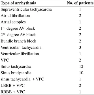
196 In the present study 45.2% of arrhythmias occurred during the first hour and 35.7% during 1-12 hours of hospitalization. According to the site, anterior wall (i.e., anterior + anterolateral + anteroseptal) was most commonly affected (52%) followed by inferior wall (i.e., inferior + infero-lateral) and lateral wall (24 % each) (Table 3). Sinus tachycardia was observed most frequently in anterior (50%) and antero-lateral (16.6%) followed by inferior (8.3%) myocardial infarction. Arrhythmias were noted in 80% of patients with left ventricular dysfunction. Arrhythmia were noted in 75% patients who were thrombolysed compared to 36/42 patients who were not thrombolysed. Sinus bradycardia (24%) and sinus tachycardia (28.5%) were the most frequently seen arrhythmia (Table 3).
DISCUSSION
Myocardial ischaemia is characterised by ionic and biochemical alterations, creating an unstable elect rical substrate capable of initiating and sustaining arrhythmia. Acute myocardial infarction creates areas of electrical
inactivity and blocks conduction, which also promotes arrhythmias. At least 75% of patients with acute myocardial infarction develop arrhythmias in the peri-infarct period, and majorit y of deaths occur secondary to development of arrhythmias. The age and gender distribution in the present study was similar to that observed in the report by Lincoff et al5 and the Framingham study.6
A substantial number of patients with acute myocardial infarction have some cardiac rhythm abnormality, and approximately 25% have cardiac conduction disturbances within 24 hours following of infarct onset. Almost any rhythm disturbance can be associated with acute myocardial infarction, including bradyarrhythmias, supraventricular tachyarr-hythmias, ventricular arrtachyarr-hythmias, and atrio-ventricular block, among others. With the advent of thrombolytic therapy, it was found that some rhythm disturbances in patients with acute myocardial infarction may be related to coronary artery reperfusion.3
Our observations suggest that arrhythmias are an important cause of morbidity in patients with
Table 2: Site of involvement in 50 patients with acute myocardial infarction
Site No. (%)
Anteroseptal 12 (24)
Lateral 12 (24)
Anterolateral 9 (18)
Inferior 7 (14)
Inferolateral 5 (10)
Anterior 5 (10)5
Total 50 (100)
Table 1: Age and gender incidence in 50 patients with acute myocardial infarction
Age No. of Males Females
(years) patients
30-39 2 2 0
40-49 9 8 1
50-59 19 12 7
60-69 7 6 1
> 70 13 6 7
Total 50 34 16
Table 3: Various types of arrhythmias seen in 50 patients with acute myocardial infarction
Type of arrhythmia No. of patients
Supraventricular tachyacardia 1
Atrial fibrillation 2
Atrial ectopics 1
1st degree AV block 2
2nd degree AV block 2
Bundle branch block 2
Ventricular tachycardia 3
Ventricular fibrillation 1
VPC 2
Sinus tachycardia 12
Sinus bradycardia 10
sinus tachycardia + VPC 1
LBBB + VPC 2
RBBB + VPC 1
VPC = ventricular premature complex; LBBB = left bundle branch block; RBBB = right bundle branch block AV = atrioventricular
197 acute myocardial infarction. Diligent monitoring for arrhythmias and instituting appropriate treatment can be life-saving.
REFERENCES
1. Rissam HS, Kishore S, Trehan N. Coronary artery
disease in young Indians. The missing link. J Indian Acad Clin Med 2001;2:128-32.
2. Enas EA, Dhawan J, Petkar S. Coronary artery diseases in Asian Indians: lessons learnt and the role of lipoprotein(a). Indian Heart J 1997;49:25-34.
3. Aufderheide TP. Arrhythmias associated with acute myocar dial in far ction an d th r ombolysis. EmergMed Clin North Am 1998;16:583-600.
4. Alpert JS, Thygesen K, Antman E, Bassand JP.Myocardial infarction redefined – A consensus document of the Joint European Society of Cardiology/ American College of Cardiology Committee for the redefinition of myocardial infarction. J Am Coll Cardiol 2000;36:959-69.
5. Lincoff AM, Califf RM, Ellis SG, SigmonKN, Lee KL, Leimberger JD, et al. Thrombolytic therapy for women with myocardial infarction: is there a gender gap? Thrombolysis and Angioplasty in Myocardial Infarction Study Group. J Am Coll Cardiol 1993:22;1780-7.
6. Lerner DJ, Kannel WB. Pattern of coronary heart disease morbidity and mortality in sexes: a 26-year follow of the Framingham population. Am Heart J 1986;111:383-90.