CENTRO DE CIÊNCIAS EXATAS E DA NATUREZA
PROGRAMA DE PÓS-GRADUAÇÃO EM BIOLOGIA CELULAR E MOLECULAR
THAÍS BEZERRA MANGEON HONORATO
EFEITO DA SALINIDADE EM CÉLULAS DO SISTEMA IMUNE DO OURIÇO-DO- MAR Echinometra lucunter
THAÍS BEZERRA MANGEON HONORATO
EFEITO DA SALINIDADE EM CÉLULAS DO SISTEMA IMUNE DO OURIÇO-DO- MAR Echinometra luncunter
Dissertação apresentada ao Programa de Pós-Graduação em Biologia Celular e Molecular do Centro de Ciências Exatas e da Natureza, da Universidade Federal da Paraíba, como parte dos requisitos para obtenção do título de MESTRE EM BIOLOGIA CELULAR E MOLECULAR
Orientador: Luis Fernando Marques-Santos Co-orientadora: Patrícia Mirella da Silva Scardua
Honorato, Thaís Bezerra Mangeon.
Efeito da salinidade em células do sistema imune do ouriço-do-mar Echinometra lucunter / Thaís Bezerra Mangeon
Honorato.- João Pessoa, 2016. 88f. : il.
Orientador: Luis Fernando Marques-Santos Coorientadora: Patrícia Mirella da Silva Scardua Dissertação (Mestrado) - UFPB/CCEN
1. Biologia celular e molecular. 2. Salinidade. 3. Ouriço-do- mar. 4. Sistema imune. 5. Celomócitos.
EFEITO DA SALINIDADE EM CÉLULAS DO SISTEMA IMUNE DO OURIÇO-DO- MAR Echinometra luncunter
Dissertação de Mestrado avaliada em / /
BANCA EXAMINADORA
Prof. Dr. Patricia Mirella da Silva Scardua
Programa de Pós-Graduação em Biologia Celular e Molecular Universidade Federal da Paraíba
Co-orientadora
Prof. Dr. Jose Antonio Novaes da Silva Universidade Federal da Paraíba
Examinador Externo
Profa. Dra. Naila Francis Paulo de Oliveira Universidade Federal da Paraíba
Examinadora Interna
Profa. Dra. Regina Célia Bressan Queiroz de Figueiredo Fundação Oswaldo Cruz
Suplente Externo
Prof.. Dr. Savio Torres de Farias Universidade Federal da Paraíba
‘’Você vai ser uma revolucionária porque qualquer mulher que está sendo autêntica em seu trabalho trará ideias e formas de trabalho que vão contra o status quo de sua empresa, indústria, comunidade - um status quo definido por valores masculinos e modos masculinos de trabalho’’
incentivadora durante toda minha vida e por acreditar em mim mesmo quando nem eu acreditava.
Ao meu pai, Ricardo Honorato, pelo apoio em todas as minhas escolhas e presença em cada pequeno passo dado, por me guiar e mostrar o valor do estudo e dedicação e por me apresentar à paixão pela ciência desde a minha infância;
Ao meu orientador Prof. Dr. Luis Fernando Marques dos Santos pelas cobranças, incentivo e total atenção e orientação durante esses 4 anos. Agradeço pela presença constante, por acreditar em mim e pela amizade que construímos.
À Profa. Drª. Patrícia Mirella da Silva Scardua, pela excelente co-orientação e confiança durante o desenvolvimento desse trabalho, por todas as sugestões, dúvidas, críticas e debates essenciais para a conclusão dessa dissertação.
Aos membros da Banca Examinadora por aceitarem contribuir com o nosso trabalho.
Aos meus colegas de trabalho e amigos do LABID: Jocelmo, Elis, Leo, Raianna, Thyago, Tainá, Dalliane, Leanderson e Luiz por serem um exemplo de trabalho em equipe e por cada momento de apoio, ajuda, companheirismo, diversão e sorrisos diários. Tem muito dessa família neste trabalho;
A todo o pessoal do LABIPI: Natan, Cairé, Lucas, Fernando, Jaíse e Sâmia pela companhia diária e prontidão para ajudar sempre;
Ao técnico do laboratório, Bosco Carlos, pelo trabalho, carinho, preocupação, sorrisos e atenção dia após dia.
A secretária do PPGBCM Ludmilla Maul por exercer sua profissão de forma exemplar e nos ajudar da melhor forma possível em todas as dúvidas, prazos e burocracias necessárias.
de desespero e um sorriso cheio de orgulho nos momentos de sucesso.
Aos meus avós, Remilson, Josefina e Hermelinda, por serem minha fonte de amor diária, por me acolherem, amarem e sempre estarem presentes durante toda a minha vida.
Ao meu amigo e namorado Victor Lisboa, pela enorme paciência, por abraçar o mestrado junto comigo, pelas abdicações, pelo carinho, amor e confiança que no fim valeria a pena.
A todos da minha família que de alguma forma me incentivaram e acreditaram em mim: minha madrasta Fernanda, todos os tios e tias, primos, meus padrinhos Fernando e Teresa e às minhas irmãs, Mariana e Julia que conseguem alegrar qualquer momento com um simples sorriso.
oceanos. A salinidade é um dos fatores que limitam a distribuição e sobrevivência de organismos marinhos. Celomócitos são as células do sistema imune dos equinodermos e têm sido estudados como biomarcadores em situações de estresse. O objetivo do presente estudo foi investigar o efeito da salinidade em celomáticos do ouriço-do-mar tropical Echinometra lucunter. Os animais foram coletados na costa de João Pessoa (Nordeste do Brasil). Os animais ou os celomócitos foram expostos a diferentes salinidades (25‰ e 45‰) e parâmetros fagocíticos, produção de espécies reativas de oxigênio (ROS), atividade mitocondrial e atividade dos transportadores ABC analisados. Os parâmetros fagocíticos não alteraram quando os animais ou as células foram expostos a 25‰ ou 45‰ nos intervalos de tempo monitorados. Porém, foi observado um aumento na concentração de celomócitos quando os animais foram expostos a 25‰. Os níveis de ROS foram maiores quando as células foram incubadas a 25‰, e menores quando as células foram cultivadas a 45‰. Foi observada uma perda do potencial de membrana mitocondrial interna quando os celomócitos foram incubados a 45‰. A atividade dos transportadores ABC diminuiu quando as células foram incubadas a 25‰ e aumentou quando as células foram incubadas a 45‰. O presente trabalho demonstra que o sistema imune do ouriço-do-mar E. lucunter tolera mudanças de salinidade (25‰ até 45‰), e sugere dois parâmetros celulares (níveis de ROS e atividade de transportadores ABC) como potenciais biomarcadores no monitoramento de mudanças na salinidade ambiental.
Human activities have caused climate changes and altered the salinity of the oceans. Salinity is one of the factors that limit the distribution and the survival of marine organisms. Coelomocytes are the immune system cells of the echinoderms and have been studied as biomarkers in stress situations. The aim of the present study was to investigate the effect of the salinity in the immune system cells of the tropical sea urchin Echinometra lucunter. Animals were collected in João Pessoa coast (Brazilian Northeast). Animals or coelomocytes were exposed to different salinity (25‰ to 45‰) and phagocytic parameters, production of reactive oxygen species (ROS), mitochondrial activity and ABC transporter activity analyzed. The phagocytic parameters did not change when animals or cells were exposed to low or high salinity in any time intervals monitored. However, our data showed an increase in the coelomocytes concentration when animals were exposed to 25‰. ROS levels were higher when cells were incubated at 25‰ and lower when cells were cultured at 45‰. We noted a loss of the mitochondrial inner membrane potential when coelomocytes were incubated at 45‰. The activity of ABC transporters decreased when cells were incubated at low salinity and increased when cells were incubated at high salinity. Our work shows that the immune system of the tropical sea urchins E. lucunter tolerates salinity changes from 25‰ to 45‰ and suggests two cellular parameters (ROS levels and ABC transporters activity) as potential biomarkers on the monitoring of the impact of environmental salinity changes.
TLR Receptores do tipo Toll (do inglês, Toll-like receptors)
PAMPS Padrões moleculares associados a patógenos (do inglês, Pathogen-associated molecular patterns)
SRCR Receptores scavenger com domínio rico em cisteína (do inglês, Scavenger receptor cysteine-rich protein domain)
PRR Receptores de reconhecimento de padrão (do inglês, Pattern recognition receptors)
ROS Espécies reativas de oxigênio (do inglês, reactive oxygen species) ABC (do inglês, ATP-binding cassette transporter)
MDR Resistência a múltiplas drogas (do inglês, Multiple drug resistance)
MXR Resistência a múltiplos xenobióticos (do inglês Multiple xenobiotic resistance) ΔΨm Potencial de membrana mitocondrial
FSW Água-do-mar filtrada (filtered seawater) ASW Água-do-mar artificial (artificial seawater)
DiOC6(3) (3,3' iodeto diexiloxacarbocianina)
C-A M Calceína-AM
CCCP Carbonilcianeto m-clorofenil-hidrazona
H2DCFDA 2′,7′-dihidro-diclorofluoresceína H2O2 Peróxido de Hidrogênio
MFI Média da intensidade de fluorescência (mean of fluorescence intensity) UV Radiação ultravioleta
Figura 1: Tipos celulares de celomócitos do ouriço-do-mar Echinometra lucunter ... 22
Figura 2: Echinometra lucunter em seu habitat ... 24
Figure 1: Effect of salinity on the phagocytic capacity and phagocytic capacity of sea urchin coelomocytes at different time spans - in vivo assay ... 66
Figure 2: Effect of salinity on the phagocytic capacity and phagocytic capacity of sea urchin coelomocytes at different time spans - in vitro assay ... 67
Figure 3: In vitro effect of salinity on coelomocytes ROS production ... 68
Figure 4: In vitro effect of salinity on the ΔΨm of coelomocytes ... 68
1. Introdução ... 14
1.1 Histórico ... 14
1.2 Imunidade Inata ... 15
1.2.1 Receptores de Reconhecimento de Padrão (PRR) ... 16
1.2.2 Fagocitose e a imunidade inata...17
1.2.3 Transportadores ABC como mecanismo de defesa ...18
1.3 Celomócitos ... 20
1.4 Equinodermos e a salinidade...22
2 Objetivos ... 25
2.1 Objetivo Geral ...25
2.2 Objetivos específicos ... 25
3 Resultados ... 26
4 Artigo ... 27
4.1 Introduction ... 30
4.2 Material and Methods ... 33
2.1 Drugs ... 33
2.2 Animals Capture and Maintenance ... 33
2.3 n vivo exposure to different salinities ... 34
2.4 Coelomocytes sampling and preparation ... 34
2.5 Coelomic fluid preparation ... 34
2.6 In vitro exposure of coelomocytes to different salinities ... 35
2.7 Total and differential coelomocytes concentration ... 35
2.8 Flow cytometry analyses ... 35
2.9 Phagocytic capacity ... 35
2.10 Investigation of ROS production ... 36
2.11 Mitochondrial inner membrane potential ... 36
2.12 ATP-binding cassette (ABC) transporters activity ... 37
2.13 Statistical analysis ... 37
4.3 Results ... 37
3.1 Effect of salinity on the phagocytosis capacity at different time spans - in vivo assays ... 37
3.2 Effect of salinity on the cell concentration of sea urchin coelomocytes at different time spans - in vivo assays ... 38
3.3 Effects of salinity on phagocytosis of sea urchin coelomocytes at different time spans - in vitro assays... 41
1. Introdução
1.1 Histórico
1.2 Imunidade inata
Atualmente tem-se o conhecimento de que a imunidade consiste em uma reação contra elementos não próprios, incluindo células, macromoléculas - tais como proteínas e polissacarídeos - e substâncias químicas que podem ser reconhecidas como elementos estranhos ao organismo. O sistema imune adaptativo apresenta uma resposta imune mais rápida e mais específica em invasões recorrentes, o que ocorre graças a uma vasta variedade de tipos celulares e de receptores de antígenos, além da produção de anticorpos com especificidade de ligação ao antígeno. Porém, antes da evolução para um sistema imune adaptativo, mecanismos inatos de autodefesa foram adquiridos. A estratégia básica da imunidade inata é que o organismo produza, constitutivamente, receptores genéricos que reconheçam padrões conservados em diferentes patógenos, sendo assim capazes de desencadear uma resposta que limita a invasão do patógeno (JANEWAY; MEDZHITOV, 2002). Essas moléculas alvo são componentes indispensáveis dos microrganismos e, por isso, não são facilmente alteradas por mutação e seleção. O reconhecimento e a resposta imune ocorrem para que a interdição da infecção seja possível antes que os microrganismos possam proliferar, disseminar e debelar o hospedeiro (BEUTLER, 2004). Assim, a base da imunidade inata é a diferenciação entre o próprio e o não próprio a partir do reconhecimento de padrões conservados em patógenos.
1.2.1 Receptores de Reconhecimento de Padrão (PRR)
O ouriço-do-mar tem uma alta diversidade de receptores envolvidos na imunidade inata em relação a outras espécies de animais já caracterizados (SODERGREN et al., 2006). O sequenciamento genômico do ouriço-do-mar Strongylocentrotus purpuratus demonstrou que estes animais possuem um sistema imune bastante diversificado e mediado por um repertório surpreendente de genes que codificam proteínas de reconhecimento de patógenos (SODERGREN, 2006). Uma das características mais marcantes do genoma de S. purpuratus, no que diz respeito à imunidade, é uma enorme expansão de três classes de genes que codificam proteínas de reconhecimento inato do sistema imunológico que compõem os receptores Toll-like (TLRs, do inglês “Toll-like receptors”), NACHT e genes que codificam proteínas contendo repetições ricas em leucina (NLR) e receptor scavenger com domínios ricos em cisteína (SRCR). Cada um desses receptores de reconhecimento padrão (do inglês PRR) participa do reconhecimento de potenciais agentes patogênicos através de ligação direta ou indireta a padrões moleculares associados a patógenos (PAMPs) (HIBINO et al., 2006b). Foram identificados, ainda, genes homólogos dos reguladores imunológicos e hematopoiéticos, muitos dos quais foram previamente identificados somente em cordados, bem como genes que são fundamentais na imunidade adaptativa de vertebrados com mandíbula (RAST et al., 2006). Acredita-se que cerca de 4 a 5% dos genes identificados no genoma do S. purpuratus estejam envolvidos diretamente com o sistema imune (HIBINO et al., 2006a), o que aponta estes organismos como excelentes modelos para o estudo das funções imunológicas.
Os receptores do tipo Toll são elementos cruciais da imunidade inata que interagem com PAMPs para iniciar uma resposta antimicrobiana que culmina na fagocitose do organismo. Os TLRs são evolutivamente conservados, sendo encontrados desde o nematódeo Caenorhabditis elegans até os mamíferos (AKIRA; TAKEDA, 2004; BEUTLER, 2004; HOFFMANN, 2003; JANEWAY; MEDZHITOV, 2002). Os PAMPs reconhecidos pelos TLRs incluem lipídeos, lipoproteínas, proteínas e ácidos nucleicos derivados de uma vasta gama de microrganismos como bactérias, vírus, parasitas e fungos. O reconhecimento de PAMPs por TLRs ocorre em diversas estruturas celulares, incluindo a membrana plasmática, os endossomos e os lisossomos (KAWAI; AKIRA, 2010).
microrganismos são os principais ligantes (ARESCHOUG; WALDEMARSSON; GORDON, 2008; JEANNIN et al., 2005; PEISER et al., 2006; PLÜDDEMANN et al., 2009). A função mais conhecida dos receptores scavengers é sua atuação no reconhecimento e fagocitose, além de atuar como co-receptores para os receptores de tipo Toll modulando respostas inflamatórias (ARESCHOUG; GORDON, 2009).
1.2.2 Fagocitose e a imunidade inata
A fagocitose é um mecanismo em que as células migram, reconhecem e englobam partículas (partículas inertes ou vivas). Organismos unicelulares usam a fagocitose para a captação de nutrientes, porém, a fagocitose em metazoários ocorre principalmente nas células fagocíticas do sistema imune e se tornou um mecanismo de eliminação de partículas não-próprias ao organismo (ADEREM; UNDERHILL, 1999). Receptores, como os TLRs e scavengers, citados anteriormente, são responsáveis pelo reconhecimento e iniciação da fagocitose, causando aumento na taxa fagocítica dessas células especializadas (ADEREM; UNDERHILL, 1999).
Inúmeros estudos demostram que células fagocíticas respondem a estresses fisiológicos e ambientais (BACCAN; OLIVEIRA; MANTOVANI, 2004; RIBAS et al., 2011; SESTI-COSTA et al., 2010; TSUKAMOTO; MACHIDA, 2014). Alterações na taxa de fagocitose em animais marinhos vem sendo observadas mediante agentes estressores (BRANCO et al., 2013; MALAGOLI et al., 2007; MATOZZO; MARIN, 2011). Assim, estudos sobre os fagócitos têm sido utilizados como uma ferramenta para avaliação do sistema imune de invertebrados.
ROCHER; DURAND, 2005). Espécies reativas de oxigênio são continuamente gerados como subprodutos de uma série de processos celulares e também são produzidos quando as células são expostas a vários estímulos externos, tais como: citocinas, radiação ultravioleta (UV), agentes quimioterápicos, hipertermia e até mesmo fatores de crescimento (CHEN, 2014; MENG; LV; FANG, 2008; WANG et al., 2013). Além disso, são necessárias para a função celular adequada devido ao seu papel na sinalização celular e a resposta imunitária, no entanto, quando ROS excede um nível de homeostase ocorre estresse oxidativo (KOHCHI et al., 2009; SCHIEBER; CHANDEL, 2014; THANNICKAL; FANBURG, 2000). O estresse oxidativo é o resultado de um desequilíbrio entre a produção de ROS e a capacidade da célula para mitigar os danos através de vias antioxidantes, mecanismos de reparo ou eliminação das moléculas danificadas (SCHIEBER; CHANDEL, 2014). A produção de ROS foi documentada em invertebrados marinhos como ouriços-do-mar (DU et al., 2013), pepino-do-mar (SUN et al., 2008), e ostras (DONAGHY et al., 2012). Estudos em hemócitos da ostras Crassostrea gigas, mostraram que a produção de ROS em hemócitos não estimulados possui origem mitocondrial. Nesse caso, espécies reativas de oxigênio são geradas através da redução do oxigênio que ocorre nos Complexos I, II e III da cadeia transportadora de elétrons (LAMBERT et al., 2012). A avaliação de ROS tem sido utilizada no monitoramento do efeito de fatores exógenos sobre o sistema imune de ouriços-do-mar, tais como: choque térmico agudo, redução do pH ou exposição à compostos tóxicos (COTEUR; DANIS; DUBOIS, 2005; MATRANGA; BONAVENTURA; DI BELLA, 2002; MATRANGA et al., 2000). Esse conjunto de dados ressalta a importância da investigação da produção de ROS em situações de estresse em células somáticas de ouriços-do-mar e a sua correlação com a atividade mitocondrial.
1.2.3 Transportadores ABC como mecanismo de defesa
do ATP. Em eucariotos, os transportadores ABC são encontrados na membrana plasmática e nas membranas que constituem o sistema de endomembranas (TER BEEK; GUSKOV; SLOTBOOM, 2014), como, por exemplo, na membrana do retículo endoplasmático, nas membranas de peroxissomos, na membrana mitocondrial e no envelope nuclear. (BABAKHANIAN; BENDAYAN; BENDAYAN, 2007; BURKE; ARDEHALI, 2007; GIBBONS et al., 2003; KIM et al., 2013; MORITA; IMANAKA, 2012; ZUTZ et al., 2009) Todos os transportadores ABC apresentam um núcleo com a mesma arquitetura modular: dois domínios transmembrana (DTM) e dois domínios de ligação de nucleotídeos (NBDs) (TER BEEK; GUSKOV; SLOTBOOM, 2014). Estas proteínas estão envolvidas no transporte de peptídeos, aminoácidos, polissacarídeos, drogas, antibióticos e toxinas (DEAN; RZHETSKY; ALLIKMETS, 2001). Em ouriços-do-mar a família de transportadores ABC divide-se em oito subfamílias (ABCA a ABCH), comparado com as sete subfamílias presentes em mamíferos (ABCA a ABCG) (SHIPP; HAMDOUN, 2012).
A diversidade na especificidade de substratos reflete a heterogeneidade funcional dos transportadores ABC na fisiologia celular (BURKE; ARDEHALI, 2007; GADSBY; VERGANI; CSANÁDY, 2006; HARTZ, 2010; HO; PIQUETTE-MILLER, 2006; JONKER et al., 2000; LIPTROTT et al., 2009; MARQUES-SANTOS et al., 1999; RUSSEL, 2010; SARKADI; HOMOLYA, 2006; SCHINKEL et al., 1995; ZHOU; SCHUETZ; BUNTING, 2001). Recentemente, os transportadores ABC foram associados a desintoxicação celular, estando relacionados com a proteção de organismos marinhos contra xenobióticos (MXR; do inglês multi-xenobiotic resistance) (FERREIRA; COSTA; REIS-HENRIQUES, 2014; KURELEC; PIVČEVIĆ, 1989, 1991; KURELEC, 1992).
Dois processos celulares inerentes aos celomócitos, a fagocitose e a migração celular, têm sido correlacionados com proteínas da superfamília ABC. Dados da literatura demonstram o envolvimento de transportadores ABC na fagocitose mediada por macrófagos (IWAMOTO et al., 2006; JEHLE et al., 2006; LUCIANI; CHIMINI, 1996) e na fagocitose de células tumorais por células dendríticas (KOPECKA et al., 2011). Proteínas da superfamília ABC também têm sido descritas em processos de migração celular em diversos organismos e tipos celulares, tais como: células germinativas de Drosophila melanogaster (RICARDO; LEHMANN, 2009); células dendríticas humanas (RANDOLPH et al., 1998); linfócitos T (HONIG et al., 2003); e mastócitos (MITRA et al., 2006).
superfamília ABC (ABCB1 e ABCC1) em gametas e células embrionárias do ouriço-do-mar Echinometra lucunter (DE SOUZA et al., 2010). Dados mais recentes do nosso grupo demonstram que estas proteínas se encontram funcionais nos estágios mais tardios do desenvolvimento, apresentando um claro padrão de expressão diferencial em larva plúteos (TORREZAN; FIGUEIREDO; MARQUES-SANTOS, 2012), esses mesmos transportadores são capazes de proteger gametas e embriões de ouriços-do-mar contra os efeitos nocivos da radiação ultravioleta (LEITE et al., 2014). Estes trabalhos sugerem que a expressão dos transportadores ABC em invertebrados marinhos seja crucial na proteção contra estressores físicos e químicos. Além disso, nosso grupo identificou a presença de transportadores ABC em celomócitos de ouriços-do-mar E. lucunter (dados não publicados).
Este conjunto de dados encoraja o desenvolvimento de estudos sobre a atividade de transportadores ABC em celomócitos de equinodermos. O fato destas células estarem em contato direto com estressores ambientais, e proteínas da superfamília ABC estarem diretamente envolvidas na resistência a estressores químicos e físicos torna ainda mais interessante a investigação da correlação entre os transportadores ABC e os processo fisiológicos mediados pelos celomócitos.
1.3 Celomócitos
O sistema imune de ouriços-do-mar atua a partir de resposta celular e/ou humoral (BRANCO; FIGUEIREDO; SILVA, 2014). As células responsáveis pelo reconhecimento de patógenos e pela resposta do sistema imune em ouriços-do-mar são denominadas celomócitos. Os celomócitos são comumente encontrados nas espaços celômicos de equinodermos, principalmente a cavidade celômica perivisceral, sistema hemal e sistema vascular de água e também são encontrados no tecido conjuntivo, porém, frequentemente, infiltram outros tecidos e órgãos (GLIŃSKI; JAROSZ, 2000; MUNOZ-CHAPULI et al., 2005; SMITH et al., 2006). Os celomócitos são divididos em quatro subpopulações com diferentes funções, são estas: fagócitos, células vibráteis, esferulócitos vermelhos e esferulócitos incolores (BOOLOOTIAN; GIESE, 1958; GEDDES, 1880; SMITH et al., 2006).
também podem estar envolvidos em outros processos celulares como: quimiotaxia, aglutinação e reações de coagulação (HILLIER; VACQUIER, 2003; SMITH et al., 2006), migração celular (METCHNIKOFF, 1968) , produção de ROS (DOLMATOVA et al., 2013; ITO et al., 1992; JOHNSON, 1969b), encapsulamento (COFFARO; HINEGARDNER, 1977). Além de participar também dos processos humorais: citotoxicidade (BERTHEUSSEN, 1979), opsonização e produção de substâncias antibacterianas (GERARDI; LASSEGUES; CANICATTI, 1990). Assim, estudos sobre os fagócitos têm sido utilizados como uma ferramenta para avaliação do sistema imune de invertebrados.
As células vibráteis são células flageladas que se deslocam em movimento rotacional (BORGES et al., 2005; SMITH et al., 2006). Essas células compõe o segundo tipo celular mais populoso de celomócitos (BERTHEUSSEN; SELJELID, 1978; JOHNSON, 1969b; SMITH; BRITTEN; DAVIDSON, 1992). Foram inicialmente descritas como responsáveis por movimentar o fluido celômico, porém Johnson (1969) ao observar essas células em uma gota em suspensão não detectou nenhuma movimentação do fluido. (JOHNSON, 1969b). Adicionalmente as células vibráteis também foram associadas à reações de coagulação (BERTHEUSSEN; SELJELID, 1978).
Figura 1. Tipos celulares de celomócitos do ouriço-do-mar Echinometra lucunter. (A) Fagócito; (B) Célula vibrátil; (C) Esferulócito incolor; (D) Esferulócito vermelho. Fonte: Lima-Santos, dados não publicados. Aumento 1000x.
1.4 Equinodermos e a salinidade
vetorial de sal (BINYON, 1966; HYMAN, 1955; SANTOS-GOUVEA; FREIRE, 2007; WARNAU et al., 1998). Equinodermos não possuem, aparentemente, qualquer órgão excretor específico (BOOLOOTIAN, 1966; DIEHL, 1986; HYMAN, 1955), e a amônia facilmente atravessa a parede do corpo do animal, sendo o método essencial de excreção de nitrogênio, como é típico da maior parte dos animais aquáticos (BOOLOOTIAN, 1966). No entanto, apesar de serem considerados osmoconformes estenohalinos, os equinodermos frequentemente suportam variações de salinidade, quer em seus ambientes (especialmente intertidal), ou experimentalmente em laboratório (BINYON, 1966).
A redução da salinidade é um dos principais fatores que limita a distribuição e a sobrevivência de espécies marinhas (LI et al., 2013). Curry e colegas (2003) relataram que o aquecimento global e as mudanças no ciclo hidrológico têm alterado a distribuição de água nos oceanos de todo o mundo (CURRY; DICKSON; YASHAYAEV, 2003). Estas mudanças têm efeitos sobre a sobrevivência e fisiologia de muitos organismos (ALLEN; PECHENIK, 2010; CARBALLEIRA; MARTÍN-DÍAZ; DELVALLS, 2011; CHOI et al., 2013; KUMAR et al., 2010; LUO; LIU, 2011). Alguns relatos têm descrito que a taxa de fertilização, de clivagem embrionária e da ocorrência de polispermia - em equinodermos - são sensíveis às variações de salinidade. No entanto, alguns estudos têm mostrado que as espécies de Echinoidea são capazes de tolerar mudanças de salinidade moderadas (DROUIN; HIMMELMAN; BÉLAND, 1985; STICKLE; DENOUX, 1976; WOLFF, 1968).
2. Objetivos
2.1 Objetivo Geral
Investigar o efeito da salinidade nas células do sistema imune do ouriço-do-mar E. lucunter.
2.2 Objetivos Específicos
Os objetivos específicos foram caracterizar o efeito da salinidade na:
Fagocitose de celomócitos de ouriços-do-mar in vivo e in vitro;
3. Resultados
JOURNAL OF EXPERIMENTAL MARINE BIOLOGY AND ECOLOGY
EFFECT OF SALINITY ON IMMUNE SYSTEM CELLS OF SEA URCHIN Echinometra lucunter
Thaís Mangeon Honoratoa, Raianna Bonia, Patricia Mirella da Silvaa and Luis Fernando
Marques-Santosa
a Laboratório de Biologia Celular e do Desenvolvimento (LABID), Departamento
de Biologia Molecular, Universidade Federal da Paraíba, João Pessoa, Paraíba, Brazil.
4. Artigo
EFFECTS OF SALINITY ON THE IMMUNE SYSTEM CELLS OF THE TROPICAL SEA URCHIN Echinometra lucunter
Thaís Mangeon Honoratoa, Raianna Bonia, Patricia Mirella da Silvab and Luis Fernando
Marques-Santosa
a Laboratório de Biologia Celular e do Desenvolvimento (LABID), Departamento de Biologia Molecular, Universidade Federal da Paraíba, João Pessoa, Paraíba, Brazil.
b Laboratório de Imunologia e Patologia de Invertebrados (LABIPI), Departamento de Biologia Molecular, Universidade Federal da Paraíba, João Pessoa, Paraíba, Brazil.
Abbreviations: ROS, reactive oxygen species; FSW, filtered seawater; ASW, artificial seawater; M.F.I., mean of fluorescence intensity; LB, latex beads; MK, MK-571; Rev, Reversin 205; ΔΨm, mitochondrial inner membrane potential.
ABSTRACT
Human activities have caused climate changes and altered the salinity of the oceans. The reduction of the salinity is one of the factors that limit the distribution and the survival of marine organisms. Coelomocytes are the immune system cells of the echinoderms and have been studied as stress biomarkers. The aim of the present study was to investigate the effect of the salinity on the immune system cells of the tropical sea urchin Echinometra lucunter. Animals were collected in João Pessoa coast (Brazilian Northeast). Animals or coelomocytes were exposed to different salinity (25‰, 35‰, 45‰) and times. Phagocytic parameters, production
of reactive oxygen species (ROS), mitochondrial activity and ABC transporter activity were analyzed. The phagocytic parameters did not change when animals or cells were exposed to 25‰ or 45‰ in any time intervals monitored. However, our data showed an increase in the
coelomocytes concentration when animals were exposed to 25‰. Reactive oxygen species levels were higher when cells were incubated at 25‰ and lower when cells were kept at 45‰. We noted a loss of the mitochondrial inner membrane potential when coelomocytes were incubated at 45‰. The activity of ABC transporters decreased when cells were incubated at the lowest salinity and increased when cells were incubated at the highest salinity tested. Our work shows that the salinity does not compromise the immune system of sea urchin E. lucunter and suggests two cellular parameters (ROS levels and ABC transporters activity) as potential biomarkers for the monitoring of the impact of environmental salinity changes.
Highlights
. Effect of the salinity on sea urchin coelomocytes
. Low salinity increased coelomocytes concentration in the coelomic fluid
. Salinity altered ROS levels, ΔΨm and ABC transporters activity in coelomocytes
1. Introduction
Deuterostome invertebrates have developed a variety of immune responses against foreign pathogens and molecules (Gross et al., 1999). The genomic sequencing of the sea urchin Strongylocentrotus purpuratus revealed that these animals have a robust immune system which is comprised by a vast repertoire of genes involved in the innate recognition of pathogen molecules (Sodergren et al., 2006). The main mechanisms of immune defense against infectious agents used by most of the invertebrates are: the synthesis and secretion of recognition, neutralizing and lytic proteins/molecules, which participate in nodule formation; encapsulation, cell lysis, and phagocytosis of foreign particles (Cervello et al., 1996; Gerardi et al., 1990; Li et al., 2014; Loker et al., 2004; Majeske et al., 2013; Stabili et al., 1996; Tahseen, 2009).
In echinoderms, the immune response is divided into humoral (mediated by molecules present in the body fluids) and cellular (cell mediated). The cells responsible for the innate immunity are known as coelomocytes. The coelomocytes are found in the coelomic perivisceral cavity, vascular water system, circulatory system, connective tissue and the tissues of various organs (Tahseen, 2009), and consist of four subpopulations: phagocytes; vibratile cells; red spherule cells and colorless spherule cells (Johnson, 1969). The phagocytes are involved in cell migration and phagocytosis, and represent the major subpopulation of the coelomocytes. In addition, phagocytosis is the main immune defense mechanism of the marine invertebrates (Ellis et al., 2011). Phagocytosis is a mechanism in which immune cells migrate to the infected site, recognize, ingest and destroy the foreign particle (inert or alive). A variety of phagocytic receptors, such as Toll-like receptors and scavengers receptors are responsible for the phagocytic process in the specialized cells (Aderem e Underhill, 1999). It is estimated that about 4 to 5% of the genes in the genome of S. purpuratus are directly involved with the immune system (Hibino et al., 2006).
2
destroy the internalized particles (Forman e Torres, 2002). The process starts on the cell or phagosome membranes by activating NADPH oxidase, with a strong oxygen consumption, a mechanism called oxidative burst, causing the reduction of molecular oxygen to superoxide
anion (O -); which can be spontaneously or enzymatically (superoxide dismutase) converted into hydrogen peroxide (H2O2). Others reactive radicals such as hydroxyl radical (·OH), or singlet oxygen (1O
2) are also produced (Dupré-crochet et al., 2013). In addition, ROS are also produced by the mitochondrial electron transport chain under physiological or stress conditions (Banh et al., 2016; Bhat et al., 2015; Figueira et al., 2013; Kandola et al., 2015; Orrenius et al., 2007). When an imbalance between ROS production and ROS scavenger occurs, cells enter a condition named oxidative stress (Costantini e Verhulst, 2009). Several works have been using ROS levels of the sea urchins immune system cells as indicative of stress from different sources, such as: UV radiation, acute heat shock, pH reduction or exposure to heavy metals (Coteur et al., 2005; Lu e Wu, 2005; Matranga et al., 2000).
Another important defense mechanism present in deuterostome invertebrates which acts against physical and chemical stressors is the activity of the ABC transporters (Bonaventura et al., 2011, 2005; Dean, 2001; Miller, 2010; Russo et al., 2010). The ABC transporters constitute a large group of integral membrane proteins that promotes the active transport of a substrate across the membrane coupled to ATP hydrolysis. In eukaryotes, ABC transporters are found in the plasma membrane, and membranes that constitute the endomembrane system (Babakhanian et al., 2007; Burke e Ardehali, 2007; Gibbons et al., 2003; Higgins e Gottesman, 1992; ter Beek et al., 2014; Zutz et al., 2009). ABC proteins are widely distributed - from microorganisms to human - and their structures are highly conserved (Dean et al., 2001). ABC transporters were firstly associated with the multidrug resistance phenomenon in cancer cells (Gottesman et al., 2002; Rumjanek et al., 2001). Recently, ABC transporters have been linked to cellular detoxification and associated with the protection of marine organisms against xenobiotics (multixenobiotic resistance, also known as MXR) (Ferreira et al., 2014; Kurelec e Pivčević, 1991, 1989; Branko Kurelec, 1992). The sequenced
genes (65 genes) (Sodergren et al., 2006). ABC transporters expression is modulated in response to xenobiotics, stress and diseases (Bonaventura et al., 2005; de Araujo Leite et al., 2014; Felix e Barrand, 2002; Miller, 2010). Recently, it has been reported that ABC transporters can also play an important role in the immune system processes, such as phagocytosis and cell migration (Hinz e Tampé, 2012; Seyffer e Tampé, 2014; van de Ven et al., 2009).
Marine ecosystems play a key role for the ecology of the planet. Human activities have caused climate changes that have altered the hydrological cycle of the planet, including: increase of ultraviolet radiation incidence, increase in the sea surface temperature, acceleration in the global rainfall, changes in the rate of evaporation and changes in the salinity of the oceans (Haerter et al., 2010; Semenov et al., 2012; Talley et al., 2002; Trenberth, 1998; Williamson et al., 2014). The reduction of the salinity is one of the main factors that limits the distribution and the survival of marine species (Kaiser, 2011; Li et al., 2013; Russell, 2013; Tomanek et al., 2012). Curry and colleagues (2003) reported that global warming and changes in the hydrological cycle have altered the distribution of water in the oceans all over the world (Curry et al., 2003). These changes have effects on the survival and physiology of many organisms (Allen e Pechenik, 2010; Carballeira et al., 2011; Choi et al., 2013; Kumar et al., 2010; Luo e Liu, 2011). Some reports have described that fertilization rate, embryo cleavage and polyspermy occurrence - in echinoderms - are sensitive to salinity variations (Allen e Pechenik, 2010; Allen et al., 2015; Carballeira et al., 2011). In spite of being an osmoconformer, varying their internal medium according to the external environmental, echinoderms are stenohaline animals and do not tolerate large variations in the salinity of the environment (Freire et al., 2011). For a sea urchin, fluctuations in environmental salinity may be reflected in the coelomic fluid and impact the physiology of the coelomocytes. However, some studies have shown that Echinoidea species are able to tolerate moderate salinity changes (Drouin et al., 1985; Stickle e Denoux, 1976; Wolff, 1968).
Pinsino et al., 2008), UV radiation (Matranga et al., 2006) and pollutants (Pinsino et al., 2008). It has been also described that salinity can affect the immune system of marine invertebrate , reducing the immune response to foreign agents (Fisher et al., 1987). The tropical sea urchin
Echinometra lucunter inhabits intertidal areas and is subject to environmental changes such as temperature and salinity (Lima et al., 2009). The investigation of the status of sea urchin immune system cells under different salinity conditions may helps in the understanding the effects of environmental changes in the marine ecosystem. Adding to this, sea urchin reproduction play an important role in the marine ecosystem, contributing to the marine food chain. So, the aim of the present study was to investigate the effect of salinity on immune system cells of the tropical sea urchin E. lucunter, contributing to the knowledge about the effects of climate change on marine invertebrates.
2. Material and Methods
2.1 Drugs
Calcein-AM (C/AM), carbonyl cyanide m-chlorophenyl hydrazone (CCCP), 2',7'- dichlorodihydrofluorescein diacetate (H2DCFDA), MK571, reversin 205, hydrogen peroxide (H2O2) and DiOC6(3) (3,3′-Dihexyloxacarbocyanine,iodide) were purchased from Sigma- Aldrich (St. Louis. USA). Fluorescent latex beads were purchased from Polysciences, Inc. (Pensylvania, USA). NaCl, KCl, CaCl22H2O, MgCl26H2O, MgSO47H2O and NaHCO3 were purchased from VETEC Química Fina (Rio de Janeiro, Brazil).
2.2 Animals Capture and Maintenance
Adult sea urchins E. lucunter (Linnaeus, 1758) were sampled at Ponta do Seixas, João Pessoa, Paraíba, Brazil (7°08'54.1"S; 34°47'43.2"W). Animals were transported to the laboratory in plastic containers filled with local seawater. They were extensively washed with
filtered local seawater (FSW, 80 m), 35‰ to remove gross biological organisms and disposed
was authorized by ICMBio (Instituto Chico Mendes de Conservação da Biodiversidade/Authorization code number: 32105-1).
2.3 In vivo exposure to different salinities
Before the assay, sea urchins were acclimatized in a glass tank containing FSW (35‰, ambient salinity) at 25oC and under constant aeration.
For the assay, animals were distributed into plastic tanks (20 L; 4 L per animal; N = 2 sea urchin per tank), containing FSW with different salinities (2 replicates per condition): 25‰ (low salinity), 35‰ (ambient salinity; control group), and 45‰ (high salinity). To adjust the salinities, synthetic sea salt (Tetra Marine Salt Pro) was added to FSW. To prepare the ambient salinity and low salinity, FSW was firstly diluted with distilled water. All tanks received constant aeration and the water temperature was 25 oC. The experiment was repeated twice.
The analyses of coelomocyte phagocytosis activity and total and differential coelomocytes concentration were performed 6h and 24h after salinity challenge.
2.4 Coelomocytes sampling and preparation
First, the coelomic fluid was withdrawn through a puncture in the peristomial membrane by inserting a needle (21 gage) coupled to a sterile syringe (3 mL) containing anticoagulant solution ISO-EDTA (20 mM Tris, 0.5 M NaCl, 70 mM EDTA, pH 7.5) at a ratio of 1:1 (ISO- EDTA : coelomic fluid).
Coelomocytes were obtained by centrifugation of the coelomic fluid, at 600 x g for 5 minutes, at 4° C. The pelleted cells were resuspended in coelomic fluid previously prepared (see item 2.5), at each salinity, and the concentration of cell suspension adjusted to 5 x 10⁵ cells/ml.
2.5 Coelomic fluid preparation
at 1067 x g for 20 minutes, at 4° C. The supernatant was filtered (0.22 μm) and stored at -20oC until use. Before the assay, the salinity of the coelomic fluid was adjusted to 25‰, 35‰ and 45‰ by addition of salts according to ASW formula (artificial seawater; Vogel et al., 1999) or
by dilution with distilled water.
2.6 In vitro exposure of coelomocytes to different salinities
Coelomocytes (N = 5) were separately resuspended in the coelomic fluid adjusted to the three salinities (25‰, 35‰, and 45‰) for all in vitro assays. Cells were then incubated in
flow cytometry tubes (500 μL/tube, 3 replicates) at 25 oC.
Analyses of four cell parameters were performed at time 0h, 4h, 8h or 24h after salinity challenge. The following cell parameters were analysed by flow cytometry: phagocytic activity, reactive oxygen species production, mitochondrial inner membrane potential, and ABC transporters activity.
2.7 Total and differential coelomocytes concentration
Total and differential coelomocytes concentrations were analyzed under phase- contrast microscopy by using a Neubauer chamber.
2.8 Flow cytometry analyses
Flow cytometry analyses were performed using a FACSCalibur flow cytometer (BD Biosciences, San Jose, California, USA). All cell parameters were analyzed using FL1 detector (green fluorescence, 530 nm). Fluorescence intensity was acquired in a total of 10,000 events per sample. The results were represented by the mean of fluorescence intensity (M.F.I.) in arbitrary units (a.u.).
2.9 Phagocytic capacity
Fluoresbrite® Yellow Green, final concentration 2% in MilliQ water) in the ratio of 1:10 (cells:beads; 3 technical replicates). The samples were immediately fixed in 4% formaldehyde (1:1) and preserved at 4 ºC for a maximum of 2 days.
Two phagocytic activity parameters were calculated: the phagocytic capacity and the phagocytic index. The phagocytic capacity expresses the percentage of cells from the whole cell population, which internalized at least one fluorescent microsphere. The phagocytic index indicates the capacity of the cells to internalize more than one fluorescent microspheres (see below).
Phagocytic capacity (%) = number of phagocytes containing engulfed beads 100 coelomocytes
Phagocytic index = number of microspheres internalized number of phagocytes containing engulfed beads
2.10 Investigation of ROS production
After in vitro salinity exposure (4h) coelomocyte suspensions were harvested from wells and incubated with dichlorofluorescein diacetate (DCFH-DA, final concentration 10 µM) for 30 min, at 25 ºC, in flow cytometry tubes protected from light (2 technical replicates). Hydrogen peroxide (H2O2, final concentration 1 mM) was used as a positive control.
2.11 Mitochondrial inner membrane potential
After in vitro salinity exposure (4h), coelomocyte suspensions were harvested from wells and incubated with 3,3′-dihexyloxacarbocyanine iodide (DiOC6(3), final concentration 20 nM)
for 30 min, at 25 ºC, in flow cytometry tubes protected from light (2 technical replicates). The protonophore carbonyl cyanide m-chlorophenyl hydrazone (CCCP, final concentration 200
2.12 ATP-binding cassette (ABC) transporters activity
After in vitro salinity exposure (4h), coelomocyte suspensions were harvested from wells and incubated with calcein-AM (final concentration 100 nM) for 30 min, at 25 ºC, in flow cytometry tubes protected from light (2 technical replicates). Two ABC transporters blockers, reversin 205 (ABCB1 blocker; final concentration 10 µM) and MK-571 (ABCC1 blocker; final concentration 10 µM) were used as positive controls and separately added 30 minutes before calcein-AM incubation.
Calcein-AM is a non-fluorescent ABC transporters substrate. When entering the cell, the acetomethoxy (AM) group is cleaved by esterases, making calcein a fluorescent molecule. As calcein-AM is pumped out of cells by ABC transporters, the level of fluorescence is inversely proportional to the activity of the transporters.
2.13 Statistical analysis
All results are expressed as mean ± standard error of the mean (SEM). Percentage data were transformed to arcsin before analysis. All data were analyzed by one-way ANOVA followed by Tukey’s post-test. A statistically significant difference was determined when
P<0.05.
3. Results
3.1 Effect of salinity on the phagocytosis capacity at different time spans - in vivo
assays
maintained at 35‰ - ambient salinity - the phagocytic capacity ranged from 63.27 ± 3.10% (6h)
to 52.52 ± 4.61% (24h; Fig. 1). At low salinity (25‰), the phagocytic capacity ranged from 57.58 ± 4.22% (6h) to 62.46 ± 3.15% (24h; Fig. 1). The phagocytic capacity of coelomocytes from animals kept at high salinity (45‰) ranged from 62.91 ± 4.91% (6h) to 56.67 ± 5.83%
(24h; Fig. 1).
The phagocytic index also did not change when animals were maintained at different salinities (25‰ to 45‰; Fig. 2). The basal phagocytic index (time 0h) ranged from 2.18 ± 0.15
to 2.78 ± 0.53 according to the treatment group (Fig 2). The phagocytic index ranged from 5.96 ± 0.83 to 3.55 ± 1.10 when animals were kept at ambient salinity (6 and 24h, respectively; Fig 2). When animals were exposed to the lowest salinity (25‰), the phagocytic index ranged from
4.25 ± 1.42 to 4.35 ± 1.35 (6 and 24h, respectively; Fig 2); and phagocytic index ranged from 6.56 ± 0.25 to 6.87 ± 1.73 when animals were exposed to the highest salinity (45‰) (6 and 24h, respectively; Fig 2).
3.2 Effect of salinity on the cell concentration of sea urchin coelomocytes at different
time spans - in vivo assays
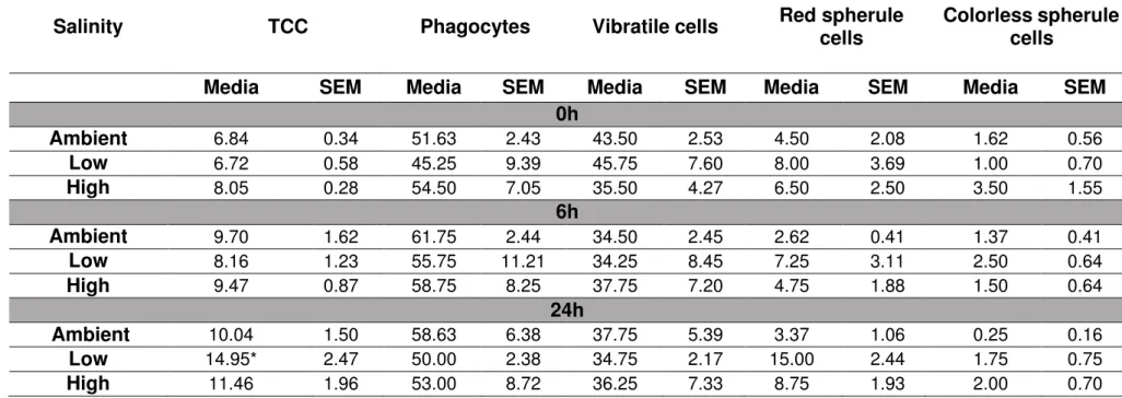
Coelomocytes concentration may alter under stress conditions due to cell migration to the peristomial membrane (de Faria e Machado; da Silva, 2008). Therefore, we evaluated the total (TCC) and differential (DCC) coelomocyte count when animals were exposed to different salinities. The analysis was performed immediately before (time 0h) and 6h and 24h after the exposure to different salinities. The total cellular concentration from sea urchins kept at ambient salinity was 9.70 ± 1.62 and 10.04 ± 1.50 (6h and 24h, respectively) (Tab. 1). However, TCC increased when animals were exposed to low salinity for 24h (6.72 ± 0.58 to 14.95 ± 2.47, 0h and 24h, respectively; Tab. 1). No difference was observed in TCC when animals were exposed to high salinity in the monitored time intervals.
40
Table 1 - In vivo effect of salinity on the total and differential coelomocytes concentration at different time spans. Ambient: Ambient salinity: 35‰; Low:
Low salinity: 25‰; High: High salinity: 45‰. TCC: total coelomocyte count. Data are expressed as the mean (cells/ml x 106, total coelomocytes count; %,
coelomocytes subpopulations) and standard error of the mean (SEM) of four independent experiments performed in duplicate (N = 4). *p<0.05 when compared
to low salinity (basal cellular concentration and 6h) (One-way ANOVA followed by Tukey’s post-test).
Salinity
TCC
Phagocytes
Vibratile cells
Red spherule
cells
Colorless spherule
cells
Media
SEM
Media
SEM
Media
SEM
Media
SEM
Media
SEM
0h
Ambient
6.84 0.34 51.63 2.43 43.50 2.53 4.50 2.08 1.62 0.56Low
6.72 0.58 45.25 9.39 45.75 7.60 8.00 3.69 1.00 0.70High
8.05 0.28 54.50 7.05 35.50 4.27 6.50 2.50 3.50 1.556h
Ambient
9.70 1.62 61.75 2.44 34.50 2.45 2.62 0.41 1.37 0.41Low
8.16 1.23 55.75 11.21 34.25 8.45 7.25 3.11 2.50 0.64High
9.47 0.87 58.75 8.25 37.75 7.20 4.75 1.88 1.50 0.6424h
Ambient
10.04 1.50 58.63 6.38 37.75 5.39 3.37 1.06 0.25 0.16Low
14.95* 2.47 50.00 2.38 34.75 2.17 15.00 2.44 1.75 0.753.3 Effects of salinity on the phagocytosis of sea urchin coelomocytes at different time spans - in vitro assays
We then evaluate the effect of salinity on cell parameters when the assay was carried out in vitro (coelomocytes incubated in coelomic fluid with different salinities). We firstly investigated the effect of different salinities on the phagocytosis. The phagocytic capacity decreased according to the increasing in the incubation time (from 0h to 24h) to all salinities tested. However, there was no difference between the treatments (Fig. 2A). After 24h of cell exposure to different salinities, the decrease in the phagocytic capacity ranged from 40.41% to 56.13% (25‰ and 45‰, respectively; Fig. 2A).
The phagocytic index also decreased throughout the time of cell culture to all salinities. After 24h of cell exposure to different salinities, the phagocytic index was reduced in 17.18%, 16.15% and 26.35% when compared to time 0h (25‰, 35‰, 45‰, respectively; one way ANOVA followed by Tukey’s post-test; Fig. 2B).
3.4 In vitro effect of salinity on coelomocytes ROS production
Since our results showed a similar phagocytosis behavior under different salinities - both in vivo and in vitro assays - corroborating the osmoconformer behavior of the sea urchin
E. lucunter (Freire et al., 2011), we decided to investigate the other cellular parameters directly exposing the coelomocytes to coelomic fluid with different salinities (in vitro assays).
3.5 In vitro effect of salinity on the ΔΨm of coelomocytes
We then investigated if changes in ROS production were followed by alterations in the mitochondrial activity. The basal ΔΨm (time 0h) was similar when the assay was performed at
ambient or low salinity (276.10 ± 16.01 and 249.70 ± 20.03, respectively, M.F.I.). However, we observed a slight decrease in the M.F.I. of cells stained with DiOC6(3) when the assay was performed at high salinity (231.10 ± 10.02).
The M.I.F. of coelomocytes exposed at ambient salinity for 4h was 265.40 ± 5.80 (Fig. 4). The incubation of cells for 4h - at low salinity - did not alter the ΔΨm (M.F.I. = 248.70 ±
14.88, Fig 4). However, the M.F.I. also showed a slight decreased when cells were incubated at high salinity for 4h (225.60 ± 11.35, Fig. 4). The depolarizing agent CCCP (200 µM) was used as positive control, and the M.F.I. were 55.24 ± 3.12 (0h) and 54.71 ± 2.10 (4h) (data not shown).
3.6 In vitro effect of salinity on ABC transporters activity.
Subsequently, we evaluated the effect of salinity on ABC transporters activity with the calcein intracellular accumulation assay. The basal calcein level (time 0h) was higher when the assay was performed at the lowest salinity (677.30 ± 43.89 against 407.20 ± 12.75, ambient salinity; M.F.I.; Fig. 5A) and lower when the assay was performed at the highest salinity (265.30
± 7.46 against 407.20 ± 12.75, ambient salinity; M.F.I.; Fig. 5A). Similar results were obtained when cells were incubated for 4h (Fig. 5B).
Two ABC transporters blockers (reversin 205, ABCB1 blocker; and MK-571, ABCC1 blocker) were used as positive control. The increase in calcein fluorescence was higher when cells were incubated at low salinity in the presence of MK-571 (1,177.00 ± 54.34 against 830.90
4. Discussion
In the present study, we investigated the effect of salinity on sea urchin immune system cells. The sea urchins - whole animals - or coelomocytes were exposed to ambient (35‰), low (25‰) and high salinity (45‰) in order to assess their sensitivity to the abiotic factor. Our
results showed that changes in the salinity did not affect the phagocytic capacity, but ROS production and ABC transporters activity were sensitive to different salinities.
Firstly, we investigated the effect of animal exposures to different salinities (25‰, 35‰ and 45‰) on the immune system response to foreign particles. In sea urchins, the phagocytes
are the only cells that perform phagocytosis (Borges et al., 2005; Metchnikoff, 1968). In E.
lucunter, these cells represent around 50% of the coelomocytes (Table 1). The phagocytosis of inert particles is a non-specific process mediated by the extension of specialized areas of the cell membrane, named filopodia (Aderem e Underhill, 1999). We did not observe differences in the phagocytic parameters of coelomocytes when animals (in vivo assay) were exposed up to 24h to low or high salinity (25‰ and 45‰, respectively; Fig. 1A and B). It was not possible to increase the exposure time to low or high salinity since animals exhibited signs of health impairments, such as loss of spines, loss of attachment to the substrate and gamete spawning, after 48 hours of exposure to these salinities (data not shown). We also addressed the phagocytic parameters of coelomocytes that was directly exposed to different salinities (in
vitro assays). No alteration in the phagocytic parameters was observed to any salinity conditions (Fig. 2A and B).
Other authors have also investigated the phagocytosis in marine organisms exposed to different salinities. Studies with the sea cucumber Apostichopus japonicus demonstrated that hemocyte’s phagocytic capacity was higher after 0.5 and 1h at low (25‰) or high salinity (35‰)
(F. Wang et al., 2008). Gagnaire et al. (2006) reported that the exposure of the oyster
phagocytosis exhibited a salinity-dependent behavior, ranging from 20%, at 18‰, to 46.5%, at 38‰ (Jauzein et al., 2013). Nevertheless, our data showed that the phagocytic parameters of
E. lucunter coelomocytes were not affected when animals or coelomocytes were exposed to lower or higher salinity up to 24h. Therefore, this data set corroborates the osmoconformer behavior to the sea urchin E. lucunter.
In echinoderms under stress conditions, changes in coelomocytes concentration can occur due to cell migration from the perivisceral coelom to the peristomial membrane (de Faria e Machado; da Silva, 2008). We then investigated if salinity affects the total and differential concentration of sea urchin coelomocytes. Other studies have addressed this question in marine organisms exposed to different salinities. A study with the shrimp Litopenaeus
vannamei showed that the total hemocyte count of shrimps kept at 2.5‰ and 5‰ salinities was lower than those kept at higher salinities (15‰,25‰, and 35‰) (Lin et al., 2012). Jauzein et al. (2013) observed that the sunray venus clam M. nimbosa kept at 18‰ for 7 days exhibited a lower concentration of circulating hemocytes. The authors suggest that the reduction in the concentration of circulating hemocytes may be due to a massive infiltration of hemocytes in tissues that are in direct contact with the seawater. In contrast, our data showed an increase in coelomocytes concentration (122.47%) when animals were kept at low salinity up to 24h (Tab. 1), suggesting a recruitment of cells from peripheral tissues to the coelomic fluid or incorporation of new cells from the axial organ. Further studies must be conducted to address this point.
The oxidative stress is an important component of the response to environmental changes in marine organisms (Coteur et al., 2005; Lesser, 2006). The increase in ROS levels is a common feature observed in animals exposed to abiotic stress (Apel e Hirt, 2004); and many studies have identified high levels of ROS in organisms under salt stress (Borsani et al., 2001; Pérez-López et al., 2009; Schwarzländer et al., 2009; Skopelitis et al., 2006). In addition, ROS generation is an essential mechanism during the phagocytosis (Buggé et al., 2007; Forman e Torres, 2002). So, we investigated the effect of different salinities on coelomocytes’s ROS levels. Our results showed that ROS levels increased 31.28% at low salinity (25‰) and decrease 30.22% at high salinity (45‰) after 4h of exposure (Fig. 3). Our data suggest that
alterations in ROS levels are not associated to the phagocytosis process since we did not observed any changes in the phagocytic parameters when coelomocytes were exposed to different salinities.
The effect of salinity on ROS production has been investigated in vivo in other marine organisms. Study with M. nimbosa observed that the level of ROS from hemocytes was twice higher when animals were kept at low salinity (18‰) for 7 days (Jauzein et al., 2013). In
contrast, Perrigault et al. (2011) observed a lower generation of ROS in hemocytes from the clam Mercenaria mercenaria kept at low salinity (17‰) for 2 weeks and a higher production of ROS in hemocytes from clams kept at high salinity (30‰). To prevent oxidative damage caused by ROS, cells possess antioxidants systems, including enzymes (i.e., catalase and superoxide dismutase) and non-enzymatic antioxidants (glutathione) (Pamplona e Costantini, 2011). It has been demonstrated that antioxidant enzymes play an important role in reducing the oxidative stress during salinity changes in marine organisms as the ark shell Scapharca broughtonii (An e Choi, 2010) and the olive flounder fish Paralichthys olivaceus (Choi et al., 2008). At the same time, it is known that salinity changes can affect the antioxidant system. In coelomocytes from the sea cucumber A. japonicus, the antioxidant enzyme superoxide dismutase exhibited an increased activity at low (20‰ and 25‰) and high salinity (35‰) when animals were kept up to 72h; but it was observed a decrease in the activity of catalase at low salinity (20‰) just after
coelomocytes exposed to 25‰ or 45 ‰ may be due to changes in the status of the antioxidant
systems.
Since mitochondria are an important ROS source under stress conditions (Orrenius et al., 2007), we decided to investigate if salinity alters coelomocytes’s mitochondrial activity. Previous studies have reported that mitochondria can be affected by salinity alterations (Gao et al., 2011; Jacoby et al., 2011). Paital and Chainy (2014), studying salinity effects on gills mitochondria of the mud crab Scylla serrata, showed alterations on the mitochondria respiration according to the salinity conditions. Authors suggested that high salinity (35‰)
causes a hypoxic state in mitochondria and, consequently, the generation of ROS. Hypoxia can leads to the disruption of mitochondrial inner membrane potential (Δψm) (Solaini et al., 2010). Our results showed a slight decrease (16.29%) in mitochondrial activity (loss of Δψm)
when E. lucunter coelomocytes were exposed to high salinity (45‰) (Fig. 4). However, under the same salinity, we observed a decrease in coelomocytes ROS generation (Fig. 3). Adding to this, we also showed that the increase in coelomocytes ROS levels - when cells were exposed to 25 ‰ - is not related to mitochondrial activity since no change in Δψm was observed.
Therefore, we discard an association between mitochondrial activity and ROS generation in E.
lucunter coelomocytes. So, further studies must be conducted to address the antioxidant mechanism under salt-stress in sea urchin coelomocytes.
that the blockade of ABCC1 leads to a decrease in ROS levels due to the accumulation of glutathione into the cells (Muanprasat et al., 2013; Mueller et al., 2010). However, our results showed that the activity of ABC transporters was reduced (increased calcein accumulation) around 40% when coelomocytes were exposed, for 4h, to 25‰ (Fig. 5B), but at the same
condition, we observed an increase in ROS production (Fig. 4). Similarly, we observed an increase in ABC transporters activity (decreased calcein accumulation) around 50% when coelomocytes were incubated at 45‰ for 4h (Fig. 5B), but a decrease in ROS level when cells
were exposed to the same salinity (Fig. 4). So, our data suggest that the activity of ABC transporters - in sea urchin coelomocytes - is not related to ROS levels as observed by other authors. Additionally, the results obtained with MK-571 treatment (increased calcein accumulation) confirmed that calcein fluorescence levels reflect ABC transporters activity. Previous works from our group have shown that reversin 205 - or even verapamil, another ABCB1 blocker - do not increase calcein accumulation in E. lucunter coelomocytes (unpublished data).
Most studies of ABC transporter activity through salinity changes were achieved in plants. These studies have been focused in ABC transporters gene expression under salt- stress and the reestablishment of ionic and osmotic homeostasis (Peng et al., 2014). Gu et al. (2004) reported the down-regulated of an ABC transporter gene (clone 253) in the plant
affect the activity of ABC transporters in coelomocytes, since it was observed even at time 0h in which cells were exposed to low or high salinity for a short time interval (30 min - calcein staining). Moreover, we also observed an increase in the cell size (FSC parameter - flow cytometry analyses - data not shown) and a decrease in the ABC transporter activity when coelomocytes were incubated for 4h at low salinity (hypoosmotic condition). Nevertheless, cell exposure to 45‰, for the same time interval, did not alter cell size. So, additional studies must
be performed to elucidate the mechanisms that control the activity of ABC transporters under different salinities and their effects on MXR phenotype in sea urchin coelomocytes.
Acknowledgements
This work was supported by Conselho Nacional de Desenvolvimento Científico e
Tecnológico (CNPq) (Project number: 479206/2012-0). Thaís Bezerra Mangeon Honorato is
Author Contribution
5. References
Aderem, A., Underhill, D.M., 1999. Mechanisms of phagocytosis in macrophages. Annu. Rev. Immunol. 17, 593–623. doi:10.1146/annurev.immunol.17.1.593
Allen, J.D., Armstrong, A.F., Ziegler, S.L., 2015. Environmental induction of polyembryony in echinoid echinoderms. Biol. Bull. 221–231. doi:10.1371/journal.pone.0142994
Allen, J.D., Pechenik, J. a, 2010. Understanding the effects of low salinity on fertilization success and early development in the sand dollar Echinarachnius parma. Biol. Bull. 218, 189–199.
An, M.I., Choi, C.Y., 2010. Activity of antioxidant enzymes and physiological responses in ark shell, Scapharca broughtonii, exposed to thermal and osmotic stress: Effects on hemolymph and biochemical parameters. Comp. Biochem. Physiol. Part B 155, 34–42.
doi:10.1016/j.cbpb.2009.09.008
Apel, K., Hirt, H., 2004. Reactive oxygen species: metabolism, oxidative stress, and signal transduction. Annu. Rev. Plant Biol. 55, 373–399. doi:10.1146/annurev.arplant.55.031903.141701
Arizza, V., Giaramita, F.T., Parrinello, D., Cammarata, M., Parrinello, N., 2007. Cell cooperation in coelomocyte cytotoxic activity of Paracentrotus lividus coelomocytes. Comprative Biochem. Physiol. 147, 389–394. doi:10.1016/j.cbpa.2007.01.022
Babakhanian, K., Bendayan, M., Bendayan, R., 2007. Localization of P-glycoprotein at the nuclear envelope of rat brain cells. Biochem. Biophys. Res. Commun. 361, 301–6.
doi:10.1016/j.bbrc.2007.06.176
Banh, S., Wiens, L., Sotiri, E., Treberg, J.R., 2016. Mitochondrial reactive oxygen species production by fish muscle mitochondria: Potential role in acute heat-induced oxidative stress. Comp. Biochem. Physiol. B. Biochem. Mol. Biol. 191, 99–107.
doi:10.1016/j.cbpb.2015.10.001
10. doi:10.1016/j.biopha.2015.07.025
Bonaventura, R., Poma, V., Costa, C., Matranga, V., 2005. UVB radiation prevents skeleton growth and stimulates the expression of stress markers in sea urchin embryos. Biochem. Biophys. Res. Commun. 328, 150–157. doi:10.1016/j.bbrc.2004.12.161
Bonaventura, R., Zito, F., Costa, C., Giarrusso, S., Celi, F., Matranga, V., 2011. Stress response gene activation protects sea urchin embryos exposed to X-rays. Cell Stress Chaperones 16, 681–7. doi:10.1007/s12192-011-0277-3
Borges, J.C., Porto-Neto, L., Mangiaterra, M.B., Jensch, B., Silva, J.R., 2002. Phagocytosis in vitro and in vivo in the Antarctic sea urchin Sterechinus neumayeri at 0 ° C. Polar Biol. 1–
15. doi:10.1007/s00300-002-0431-6
Borges, J.C.S., Jensch-Junior, B.E., Garrido, P.A.G., Mangiaterra, M.B.B.C.D., Silva, J.R.M.C., 2005. Phagocytic amoebocyte sub populations in the perivisceral coelom of the sea urchin Lytechinus variegatus (Lamarck, 1816). J. Exp. Zool. Part A 303, 241–8.
doi:10.1002/jez.a.151
Borsani, O., Valpuesta, V., Botella, M., 2001. Evidence for a role of salicylic acid in the oxidative damage generated by NaCl and osmotic stress in Arabidopsis seedlings. Plant Physiol. 126, 1024–1030. doi:10.1104/pp.126.3.1024
Buggé, D.M., Hégaret, H., Wikfors, G.H., Allam, B., 2007. Oxidative burst in hard clam (Mercenaria mercenaria) haemocytes. Fish Shellfish Immunol. 23, 188–96.
doi:10.1016/j.fsi.2006.10.006
Burke, M. a, Ardehali, H., 2007. Mitochondrial ATP-binding cassette proteins. Transl. Res. 150, 73–80. doi:10.1016/j.trsl.2007.03.002
Carballeira, C., Martín-Díaz, L., DelValls, T. a., 2011. Influence of salinity on fertilization and larval development toxicity tests with two species of sea urchin. Mar. Environ. Res. 72, 196–203. doi:10.1016/j.marenvres.2011.08.008
Cervello, M., Arizza, V., Cammarata, M., Matranga, V., Parrinello, N., 1996. Properties of sea urchin coelomocyte agglutinins. Ital. J. Zool. 63, 353–356.
Choi, C.Y., An, K.W., An, M.I., 2008. Molecular characterization and mRNA expression of glutathione peroxidase and glutathione S-transferase during osmotic stress in olive flounder (Paralichthys olivaceus). Comp. Biochem. Physiol. A. Mol. Integr. Physiol. 149, 330–337. doi:10.1016/j.cbpa.2008.01.013
Choi, K., Cope, W.G., Harms, C. a., Law, J.M., 2013. Rapid decreases in salinity, but not increases, lead to immune dysregulation in Nile tilapia, Oreochromis niloticus (L.). J. Fish Dis. 36, 389–399. doi:10.1111/j.1365-2761.2012.01417.x
Cole, B.J., Hamdoun, A., Epel, D., 2013. Cost, effectiveness and environmental relevance of multidrug transporters in sea urchin embryos. J Exp Biol 216, 3896–3905.
doi:10.1242/jeb.090522
Cole, S.P.C., Deeley, R.G., 2006. Transport of glutathione and glutathione conjugates by MRP1. Trends Pharmacol. Sci. 27, 438–446. doi:10.1016/j.tips.2006.06.008
Costantini, D., Verhulst, S., 2009. Does high antioxidant capacity indicate low oxidative stress? Funct. Ecol. 23, 506–509. doi:10.1111/j.1365-2435.2009.01546.x
Coteur, G., 2004. Environmental factors influencing the immune responses of the common European starfish (Asterias rubens). Fish Shellfish Immunol. 16, 51–63. doi:10.1016/S1050-4648(03)00030-5
Coteur, G., Danis, B., Dubois, P., 2005. Echinoderm reactive oxygen species (ROS) production measured by peroxidase, luminol-enhanced chemiluminescence (PLCL) as an immunotoxicological tool. Prog. Mol. Subcell. Biol. 39, 71–83. doi:10.1007/3-540-
27683-1_4
Curry, R., Dickson, B., Yashayaev, I., 2003. A change in the freshwater balance of the Atlantic Ocean over the past four decades. Nature 426, 826–829. doi:10.1038/nature02206
de Araujo Leite, J.C., de Vasconcelos, R.B., da Silva, S.G., de Siqueira-Junior, J.P., Marques- Santos, L.F., 2014. ATP-binding cassette transporters protect sea urchin gametes and embryonic cells against the harmful effects of ultraviolet light. Mol. Reprod. Dev. 81, 66–
83.