Oxidative stress: molecular perception
and transduction of signals triggering
antioxidant gene defenses
Department of Genetics, North Carolina State University, Raleigh, NC, USA J.G. Scandalios
Abstract
Molecular oxygen (O2) is the premier biological electron acceptor that
serves vital roles in fundamental cellular functions. However, with the beneficial properties of O2 comes the inadvertent formation of reactive
oxygen species (ROS) such as superoxide (O2•-), hydrogen peroxide,
and hydroxyl radical (OH•). If unabated, ROS pose a serious threat to or cause the death of aerobic cells. To minimize the damaging effects of ROS, aerobic organisms evolved non-enzymatic and enzymatic antioxidant defenses. The latter include catalases, peroxidases, super-oxide dismutases, and glutathione S-transferases (GST). Cellular ROS-sensing mechanisms are not well understood, but a number of transcription factors that regulate the expression of antioxidant genes are well characterized in prokaryotes and in yeast. In higher eukary-otes, oxidative stress responses are more complex and modulated by several regulators. In mammalian systems, two classes of transcrip-tion factors, nuclear factor κB and activator protein-1, are involved in the oxidative stress response. Antioxidant-specific gene induction, involved in xenobiotic metabolism, is mediated by the “antioxidant responsive element” (ARE) commonly found in the promoter region of such genes. ARE is present in mammalian GST, metallothioneine-I and MnSod genes, but has not been found in plant Gst genes. However, ARE is present in the promoter region of the three maize catalase (Cat) genes. In plants, ROS have been implicated in the damaging effects of various environmental stress conditions. Many plant defense genes are activated in response to these conditions, including the three maize Cat and some of the superoxide dismutase (Sod) genes.
Correspondence
J.G. Scandalios Department of Genetics North Carolina State University Raleigh, NC 27695 USA
E-mail: [email protected]
Based on a Plenary Lecture presented at the XXXIII Annual Meeting of the Brazilian Society of Biochemistry and Molecular Biology, Caxambu, MG, Brazil, May 15-18, 2004.
Received January 31, 2005 Accepted April 18, 2005
Key words
•Catalase •Aging •Telomeres •Gene regulation •Superoxide dismutase •Genomics
Introduction
The environment in which they exist af-fects all living organisms. Whether internal or external to the organism, the environment is continually changing, and the organism must adapt if it is to survive. An organism apparently well adapted to its environment at any one time may be poorly adapted only
and to which its genome must respond in a programmed manner for the organism to survive. How the genome perceives and transduces environmental signals to effect the expression and/or repression of pertinent genes in a selective manner remains a key question. Examples are responses to light, oxidative stress, pathogenicity, wounding, anaerobiosis, thermal shock, and the “SOS” response in microorganisms (1). Some sens-ing mechanism(s) must be present to alert the cell to imminent danger, and to trigger the orderly sequence of events that will miti-gate this danger. In addition, there are ge-nomic responses to unanticipated, unpro-grammed challenges for which the genome is unprepared, but to which it responds in discernible though initially unforeseen and unpredictable ways (2). Many, though not all, signals are perceived at the cell surface by plasma membrane receptors. Activation of such receptors by mechanisms such as ligand binding may lead to alterations in other cellular components, ultimately result-ing in alterations in cell shape, ion conduc-tivity, gene acconduc-tivity, and other cellular func-tions (3). Identification and isolation of mu-tants that are unable to respond, or that re-spond abnormally to a particular signal, may provide ways to decipher the mechanisms by which a particular signal is transduced into a given response. Long before humans began manipulating and altering their envi-ronment, organisms from the simplest to the most complex began evolving methods to cope with stressful stimuli. Consequently, most living cells possess an amazing capac-ity to cope with a wide diverscapac-ity of environ-mental challenges, including natural and syn-thetic toxins, pathogens, extreme tempera-tures, high metal levels, and radiation. Many studies in the past have demonstrated clear “cause-effect” relationships upon exposure of a given organism or cell to a particular environmental factor or stressor. But only recently have certain environmental insults been shown to elicit specific genomic
re-sponses (4). At present, relatively little is known of the underlying molecular mechan-isms by which the genome perceives envi-ronmental signals and mobilizes the organ-ism to respond. Such information is not only interesting in and of itself, but is also essen-tial in any future attempts to engineer organ-isms for increased tolerance to environmen-tal adversity. Recent dramatic advances in molecular biology and genomics have made it possible to investigate the underlying mechanisms utilized by organisms to cope with environmental stresses. Investigations of genomic responses to challenge are shed-ding light on unique DNA sequences ca-pable of perceiving stress signals, thus al-lowing the cell or organism to mobilize its defenses (5). The general picture emerging from recent studies involves the sensing of a signal and the transduction of the signal to the transcription apparatus to catalyze trans-cription initiation. A signal transduction path-way contains elements that enable a signal to be transmitted within and between cells and to be translated into an appropriate response. Cells can respond to a variety of environ-mental, physical, and chemical stimuli using a diverse range of transduction and response mechanisms. The essential features of a sig-nal transduction pathway comprise a recep-tor (recognition element) capable of detect-ing a stimulus, second messengers (trans-mission elements) such as calcium or phos-phorylation cascades, and response elements (e.g., gene transcription). Such signaling networks are now amenable to study and dissection by biochemical and genetic ap-proaches that may elucidate the underlying mechanisms and lead to the identification of the molecules responsible. A thorough under-standing of how organisms perceive, respond, and adapt to changing environmental and de-velopmental stimuli is now attainable.
Gene responses
an array of genes required for their stable functioning and precise metabolic roles. A genome can respond in a rapid and specific manner by selectively decreasing or increas-ing the expression of specific genes. Genes whose expression is increased during times of stress presumably are critical to the organism’s survival under adverse condi-tions. Examination and study of such “stress-responsive” genes have implications for hu-man health and well being, for agricultural productivity, and for furthering basic bio-logical knowledge. In addition to aiding the organism under stress, genomes that are modified by stress can be utilized to study the molecular events that occur during peri-ods of increased or decreased gene expres-sion. The mechanisms by which an organ-ism recognizes a signal to alter gene expres-sion and responds to fill that need are impor-tant physiologically and render possible the examination of gene regulation under vari-ous environmental regimens. The mechan-isms of induction of stress response genes are similar among various organisms exam-ined. Similarities in stress-induced changes in gene expression have been observed for a variety of stressors (3,6-8). Some that have been studied in detail include radiation, ther-mal shock, pathogenic infections, anaero-biosis, trauma, photostress, physical wound-ing, oxidative stress, water stress, and heavy metals. In all cases, specific changes in tran-script and/or protein expression have been observed in various organisms subjected to such challenges.
Recent studies by several laboratories, using a variety of organisms, indicate that oxidative stress is a common denominator underlying many diseases and environmen-tal insults which can lead to cell death in virtually all aerobes (9). It is also becoming clear that a variety of different biotic and abiotic stresses cause their deleterious ef-fects, directly or indirectly, via reactive oxy-gen species (ROS) oxy-generation.
For example, numerous toxic
environ-mental chemicals such as xenobiotics, pesti-cides, herbipesti-cides, fungipesti-cides, ozone, ciga-rette smoke, and radiation cause their harm-ful environmental effects via generation of free radicals and other ROS.
Herein, I will focus on oxidative stress, its causes and consequences, and mechan-isms employed by organmechan-isms to cope with it.
Oxygenation of the Earth
Earth is the only planet in our solar sys-tem known to contain molecular oxygen (O2) in its atmosphere and to support aerobic
life. However, when Earth was formed about 4.5 billion years ago, its atmosphere was unlike the present, being primarily reducing and essentially free of oxygen. Most likely, the earliest living organisms were anaerobic heterotrophs living in the primitive ocean depths, shielded from the damaging effects of solar ionizing radiation. The earliest rela-tively low levels of oxygen were probably the result of photolytic dissociation of water by the sun’s ionizing radiation. The bulk of Earth’s present oxygen concentration (21% O2) is derived from the photosynthetic
ac-tivities of cyanobacteria and plants. It has been estimated that Earth contains about 410 x 103 Erda moles (Emol = 108 mol) of
oxy-gen, and of this, 38.4 x 10³ Emol is in the
hydrosphere as water. Molecular oxygen is present in the atmosphere (37 Emol) and in the hydrosphere (0.4 Emol) and undergoes continuous turnover, with the total oxygen exchange estimated at ~15 x 103 Emol/106
years. Aerobic life is responsible for the major portion of oxygen turnover, with pho-tosynthesis being the main input into the oxygen reservoir, and respiration the main output. The two processes are in approxi-mate equilibrium, and fossil fuel combus-tion is the major source of oxygen loss from the reservoir (10,11).
The accumulation of dioxygen (O2) in
organ-isms that use O2 as the electron acceptor,
thus providing a higher yield of energy com-pared with fermentation and anaerobic res-piration.
O2, ROS, and oxygen toxicity
In its ground state (its normal configura-tion, O2) molecular oxygen is relatively
unreactive. However, during normal meta-bolic activity, and as a consequence of vari-ous environmental perturbations (e.g., ex-treme temperatures, radiation, xenobiotics, toxins, air pollutants, various biotic and abi-otic stresses, and diseases) O2 is capable of
giving rise to frightfully reactive excited states such as free radicals and derivatives (9,12).
The oxidation powers of O2 are restricted
because electrons can only be absorbed from another species whose electron spin is anti-parallel to the two unpaired, anti-parallel-spin electrons in diatomic oxygen. This spin re-striction renders ground state molecular oxy-gen sufficiently unreactive, so that it cannot abstract electrons from other species. How-ever, removal of the spin restriction by add-ing a sadd-ingle e
-, or upon transfer of energy to oxygen from a photosensitizer (e.g., chloro-phyll, flavcontaining compounds), in-creases the reactivity of oxygen. Photosensi-tizers can harvest light and energize O2 to
form singlet oxygen (1∆
gO2), which can
in-teract directly with another molecule, trans-ferring the additional energy to the target
molecule.
The complete reduction of O2 to water
requires four electrons. O2 has a preference
for a stepwise univalent pathway of reduc-tion resulting in partially reduced intermedi-ates (Figure 1). The reactive species of re-duced dioxygen include the superoxide radi-cal (O2•−), hydrogen peroxide (H2O2), and
the hydroxyl radical (OH•
). The latter can also be generated by the interaction of O2
•−
and H2O2 in the presence of metal ions. Both
O2
•−
and OH•
are extremely reactive and can cause molecular damage, leading to cell death. The hydroxyl radical reacts with vir-tually anything, inflicting indiscriminate and extensive intracellular damage. The O2•− is
the conjugate base of a weak acid, the perhydroxyl radical (HO2), whose pKa is
4.69 ± 0.08. Thus, under acidic conditions, the very reactive perhydroxyl radical may predominate following a one electron reduc-tion of dioxygen, while at higher pH values the O2•− is predominant. These and the
physi-cally energized form of dioxygen, singlet oxygen (1O
2), are the biologically most
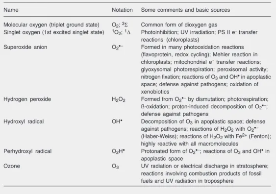
im-portant ROS (Table 1). An activation energy of ~22 kcal/mol is required to raise molecu-lar oxygen (O2) from its ground state to its
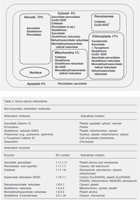
first singlet state. In higher plants, this en-ergy is readily obtained from light quanta via such transfer molecules as chlorophyll. Unless abated, all of these intermediate oxy-gen species are extremely reactive and cyto-toxic in all organisms (9,13). ROS can inter-act with proteins, lipids, and nucleic acids to cause severe molecular damage (Table 2). Thus, oxygen provides a paradox, in that it is essential for aerobic life, yet in its reduced forms is one of the most toxic substances with which life on Earth must cope. ROS are found in virtually all intracellular organelles or compartments as a consequence of nor-mal metabolic activity. Each organelle or compartment has potential targets for oxida-tive damage, as well as mechanisms for the elimination of excess ROS accumulation (Figure 2).
Table 1. Reactive oxygen species of interest in oxidative stress.
Name Notation Some comments and basic sources
Molecular oxygen (triplet ground state) O2; 3Σ Common form of dioxygen gas
Singlet oxygen (1st excited singlet state) 1O2; 1∆ Photoinhibition; UV irradiation; PS II e- transfer
reactions (chloroplasts)
Superoxide anion O2•− Formed in many photooxidation reactions
(flavoprotein, redox cycling); Mehler reaction in chloroplasts; mitochondrial e- transfer reactions;
glyoxysomal photorespiration; peroxisomal activity; nitrogen fixation; reactions of O3 and OH• in apoplastic
space; defense against pathogens; oxidation of xenobiotics
Hydrogen peroxide H2O2 Formed from O2•− by dismutation; photorespiration;
ß-oxidation; proton-induced decomposition of O2•−;
defense against pathogens Hydroxyl radical OH•
Decomposition of O3 in apoplastic space; defense
against pathogens; reactions of H2O2 with O2•−
(Haber-Weiss); reactions of H2O2 with Fe2+ (Fenton);
highly reactive with all macromolecules
Perhydroxyl radical O2H• Protonated form of O2•−; reactions of O3 and OH• in
apoplastic space
Ozone O3 UV radiation or electrical discharge in stratosphere;
reactions involving combustion products of fossil fuels and UV radiation in troposphere
Table 2. Examples of reactive oxygen species (ROS) damage to lipids, proteins and DNA.
Oxidative damage to lipids
• Occurs via several mechanisms of ROS reacting with fatty acids in the membrane lipid bilayer, leading
to membrane leakage and cell death.
• In foods, lipid peroxidation causes rancidity and development of undesirable odors and flavors.
Oxidative damage to proteins
• Site-specific amino acid modifications (specific amino acids differ in their susceptibility to ROS attack) • Fragmentation of the peptide chain
• Aggregation of cross-linked reaction products • Altered electrical charge
• Increased susceptibility to proteolysis
• Oxidation of Fe-S centers by O2•− destroys enzymatic function
• Oxidation of specific amino acids “marks” proteins for degradation by specific proteases • Oxidation of specific amino acids (e.g., Try) leads to cross-linking
Oxidative damage to DNA
• DNA deletions, mutations, translocations • Base degradation, single-strand breakage • Cross-linking of DNA to proteins Defenses against reactive oxygen
To minimize the damaging effects of ROS, aerobic organisms evolved both non-enzymatic and non-enzymatic antioxidant
Table 3. Some natural antioxidants.
Non-enzymatic antioxidant molecules
Antioxidant molecule Subcellular location
Ascorbate (vitamin C) Plastid; apoplast; cytosol; vacuole
ß-Carotene Plastid
Glutathione, reduced (GSH) Plastid; mitochondrion; cytosol Polyamines (e.g., putrescine, spermine) Nucleus; plastid; mitochondrion; cytosol α-Tocopherol (vitamin E) Cell and plastid membranes
Zeaxanthin Chloroplast
Antioxidant enzymes
Enzyme EC number Subcellular location
Ascorbate peroxidase 1.11.1.11 Plastid stroma and membranes Peroxidases (non-specific) 1.11.1.7 Cytosol; cell wall-bound
Catalase 1.11.1.6 Glyoxysome; peroxisome; cytosol; mitochondria
Superoxide dismutase (SOD) 1.15.1.1 Cytosol (Cu/ZnSOD); plastid (Cu/ZnSOD; FeSOD); mitochondrion (MnSOD); peroxisome Dehydroascorbate reductase 1.8.5.1 Cytosol; plastid
Glutathione reductase 1.6.4.2 Mitochondrion; cytosol; plastid Monodehydroascorbate reductase 1.6.5.4 Plastid stroma
Glutathione S-transferases 2.5.1.18 Cytosol; microsomal Figure 2. Intracellular antioxidant
resources in plant cells. SOD = superoxide dismutase.
(SOD), catalases (CAT) and peroxidases, protect by directly scavenging superoxide radicals and hydrogen peroxide, converting them to less reactive species. SODs catalyze
the dismutation of O2•− to H2O2, and CAT
and peroxidases reduce H2O2 to 2H2O. The
dria; FeSODs are generally found in prokary-otes, in algae and in some higher plant chlo-roplasts; NiSODs have been found in Strep-tomyces. Unlike most other organisms that have only one of each type of SOD in the various cellular compartments, plants have multiple forms of each type encoded by more than one gene (Figure 3), indicative that plants have far more complex antioxi-dant defenses (14,15). Plants also produce a large variety of small non-enzymatic anti-oxidant compounds as second tier defenses, such as glutathione, ascorbate, tocopherols, flavonoids, alkaloids, and carotenoids in high concentrations that are capable of quench-ing ROS. The dismutation of O2•− to O2 +
H2O2 by SOD is hardly a bargain, as the
resulting H2O2 can react with metal ions,
giving rise to the highly toxic OH•.
Fortu-nately, CAT come to the rescue by degrad-ing H2O2 to O2 and H2O. Most aerobes,
including mammals, possess at least one form of homotetrameric CAT with ferriheme at the active sites.
Catalase
CAT is largely, but not exclusively, lo-calized in peroxisomes, wherein many H2O2
-producing enzymes reside. Thus CAT, which
Figure 3. Zymograms showing multiplicity of superoxide dismutase (SOD, left) and catalase (CAT, right) in maize. The cytosolic CuZnSOD-4 and SOD-4A co-migrate to the same position, as do all members of the Sod3 multigene family (14). Intracellular location is indicated in parentheses. Catalases are tissue-specific in their expression: LE = milky endosperm; COL = coleoptile; SC = scutellum; ALEU = aleurone; PER = pericarp; LF = green leaf. When CAT-1 and CAT-2 are co-expressed (ALEU) their subunits interact to generate intergenic heterotetramers. CAT-3 does not form heterotetramers in vivo when co-expressed with either CAT-1 or CAT-2 (16). Arrow shows direction of migration in gel.
which the substrate, O2•− for SOD and H2O2
for CAT, is both reductant and oxidant, whereas different reductants are required for the peroxidases, depending upon their speci-ficities. Under some conditions CAT can act as an efficient peroxidase. SODs deal with the first product of the univalent reduction of O2, converting it to H2O2, which must then
be destroyed by CAT and/or peroxidases. Thus, the SOD and CAT serve, in tandem, as front-line antioxidant defenses:
O2•− + O2•− + 2H+ SOD O2 + H2O2 (K2 = 2.4 x 10⁹ M-1 s-1)
H2O2 + H2O2
CAT
2 H2O + O2 (K1 = 1.7 x 10⁷ M-1 s-1)
H2O2 + R(OH)2
Px 2H2O + R(O)2 (K4 = 0.2-1 x 10³ M-1 s-1)
Superoxide dismutase
SODs have been isolated and character-ized from a wide variety of organisms. One class consists of SODs with Cu(II) plus Zn(II) at the active site (Cu/ZnSOD), another with Mn(III) (MnSOD), a third with Fe(III) (FeSOD), and a fourth with Ni(II/III) (NiSOD). Cu/ZnSODs are generally found in the cytosol of eukaryotic cells, in chloro-plasts, and in some prokaryotes; MnSODs are found in prokaryotes and in
mitochon-CAT
CuZnSOD-1 (Chl)
CuZnSOD-2 (Cyt)
MnSOD-3 (Mit)
CuZnSOD-4/4A (Cyt)
CuZnSOD-5 (Cyt)
CAT 3
CAT 1 CAT 2
SOD
CAT 1 CAT 2
CAT 3
CAT 1 CAT 2
LE COL SC10 ALEU PER LF
exhibits a high Km for H2O2, can act upon the
H2O2 produced before it diffuses to other
parts of the cell. CAT is a tetrameric heme-containing enzyme that is found in all aero-bic organisms. Because of its wide distribu-tion, evolutionary conservadistribu-tion, and capac-ity to rapidly degrade hydrogen peroxide, it has been proposed that CAT plays an impor-tant role in systems which have evolved to allow organisms to live in aerobic environ-ments.
CAT is one of the most active catalysts produced by nature. It decomposes hydro-gen peroxide at an extremely rapid rate, corresponding to a catalytic center activity of about 107 min-1. Depending upon the
con-centration of H2O2, it exerts a dual function.
At low concentrations (<1µM) of H2O2, it
acts “peroxidatically”, i.e., a variety of hy-drogen donors (e.g., ethanol, ascorbic acid) can be oxidized in the following manner: RH2 + H2O2→ R + 2H2O.
At high concentrations of substrate, CAT decomposes toxic hydrogen peroxide at an extremely rapid rate using the “catalatic” reaction in which H2O2 acts as both an
ac-ceptor and donor of hydrogen molecules: 2H2O2→ 2H2O + O2.
CAT is unique among H2O2 degrading
enzymes in that it degrades H2O2 without
consuming cellular reducing equivalents. Hence, CAT provides the cell with a very energy efficient mechanism to remove hy-drogen peroxide. Therefore, when cells are stressed for energy and are rapidly generat-ing H2O2 through “emergency” catabolic
pro-cesses, H2O2 is degraded by CAT in an
energy-efficient manner. This results in a net gain of reducing equivalents and, therefore, cellular energy. It has been proposed that CAT may be uniquely suited to regulate the homeostasis of H2O2 in the cell. In the
cata-latic mode, CAT has a very high apparent Michaelis constant and, therefore, is not eas-ily saturated with substrate. Thus, the en-zyme activity increases linearly over a wide range of H2O2 concentrations, thereby
main-taining a controlled intracellular H2O2
con-centration. In mammalian systems, organs with high concentrations of CAT (i.e., liver and kidney) have low levels of endogenous H2O2, and organs with low concentrations of
CAT (i.e., lung and heart) have high endog-enous levels of H2O2. Further, if CAT
activ-ity is inhibited, H2O2 concentrations rise in
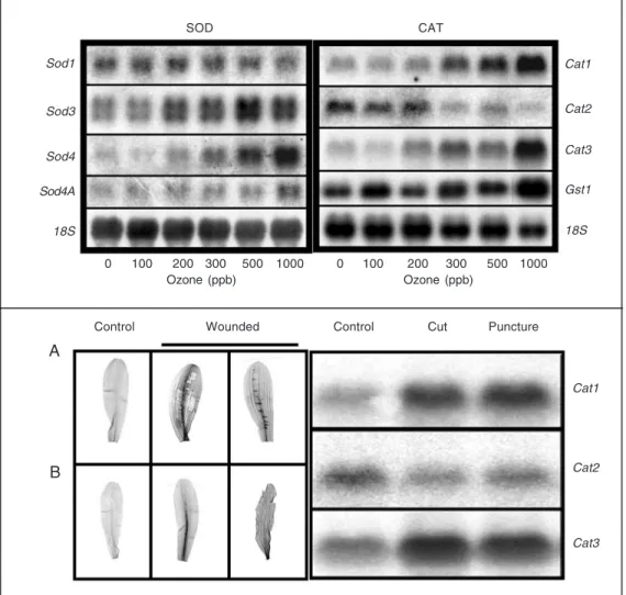
the liver. As in the case of SOD, multiple CATs (isozymes; Figure 3) encoded by spe-cific genes are found in plants, whereas ani-mals exhibit one form of CAT (16,17). Both Cat and Sod genes respond differentially to various stresses known to generate ROS (Fig-ure 4).
ROS, telomeres, and aging
Many hypotheses have been proposed to explain the root cause of aging. One broad-based hypothesis is that generalized homeo-static failure leads to age-related decline. Another is the “free-radical theory of aging” suggesting that endogenous ROS continu-ally damage cellular macromolecules, in-cluding DNA (18). Incomplete repair of such damage would lead to its accumulation over time resulting in age-related deterioration.
tox-icity plays an important role in longevity (20,21).
Much attention has also recently been given to the role of telomere shortening as a causative factor in aging/senescence (22,23). Telomeres are stretches of repetitive DNA of tandem short sequence repeats (TTAGGG) that “cap” the ends of eukaryotic chromo-somes to protect against degradation. Te-lomeres in most human cells shorten with each round of DNA replication, because they lack the enzyme telomerase. Telomer-ase is a specialized reverse transcriptTelomer-ase, which helps to replicate the telomere ends of chromosomes. However, telomerase is nor-mally expressed only in germ-line cells and is derepressed in tumor cells in which telom-eres are stabilized. It has recently been
dem-onstrated that oxidative stress is the main reason for telomere shortening (24). It has been observed that H2O2 plus Cu(II) induced
8-oxo-7,8-dihydro-2'-deoxyguanosine for-mation in the telomere sequences more effi-ciently than in non-telomere sequences. Oxi-dative damage is not repaired as well in telomeric DNA as elsewhere in the chromo-some. Oxidative stress accelerates telomere loss, while antioxidants decelerate it. Thus, oxidative stress is a critical modulator of telomere loss and telomere-driven replica-tive senescence is primarily a stress response. In a recent study, individuals under general-ized stress were found to have shorter telom-eres and less telomerase activity, and more oxidative stress (25). Given the available information, aging is likely to be a
multifac-Figure 4. Response to acute ozone exposure of the Sod (top-left) and Cat (top-right) genes in maize leaves after 6 h of O3
fu-migation. As noted, some of the transcripts are upregulated (e.g., Sod3, Sod4, Cat1, Cat3), while others are downregulated (e.g., Sod1, Cat2). The 18S rRNA is a loading control. The production of H2O2 in maize leaves
(bot-tom-left) in response to wound-ing. Seven-day-old plants were excised at the base of the stems and supplied with diaminobenzi-dine (DAB) for 6 h. The plants then were wounded and continu-ously supplied with DAB for 4 h. The production of H2O2 can be
visualized by the deposition of the brown-red (dark areas) color products in the leaves. A, Con-trol and wounded leaves (wounding was conducted near the main vein). B, Control and wounded leaves (wounding was conducted at the main vein and cut at the leaf edges). Cat tran-script accumulation in response to wounding (bottom-right). RNA was isolated from similarly treated leaves, indicative that differential transcript accumula-tion is effected by inducaccumula-tion of H2O2.
Sod1
Sod3
Sod4
Sod4A
18S
Cat1
Cat2
Cat3
Gst1
18S
0 100 200 300 500 1000
Ozone (ppb) Ozone (ppb)
0 100 200 300 500 1000
Wounded Puncture
Control Control Cut
A
B
Cat1
Cat2
Cat3
torial process; whether ROS are peripheral targets that correlate with longevity or cen-tral regulators of human aging remains to be resolved.
Oxidative stress
ROS such as O2
•−
, H2O2, and OH
•
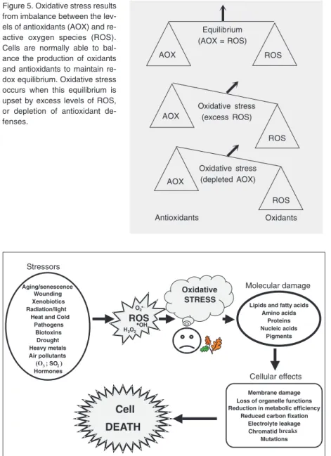
are produced in all aerobic organisms and nor-mally exist in the cell in balance with
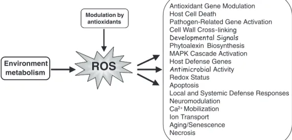
anti-oxidant molecules. Oxidative stress occurs when this critical balance is disrupted due to depletion of antioxidants or excess accumu-lation of ROS, or both (Figure 5). That is, when antioxidants are depleted and/or if the formation of ROS increases beyond the abil-ity of the defenses to cope, then oxidative stress and its detrimental consequences en-sue. Such stress occurs when severely ad-verse environments or physiologic condi-tions overwhelm biological systems. One rapid and clear indicator of oxidative stress is the induction of antioxidant defenses and/ or increases in endogenous ROS levels. The formation of ROS can be accelerated as a consequence of various environmental stress conditions, including UV-radiation, high light intensities, exposure to herbicides, ex-treme temperatures, toxins such as cerco-sporin and aflatoxin, air pollutants, metals, wounding, and xenobiotics. Many inducers of oxidative stress are known carcinogens, mutagens, and toxins. ROS production and accumulation is a common denominator in many diseases and environmental insults and can lead to severe cellular damage leading to physiological dysfunction and cell death in virtually all aerobes (Figure 6).
When oxidative stress occurs, cells func-tion to counteract the oxidant effects and to restore redox balance by resetting critical homeostatic parameters. Such cellular activ-ity leads to activation or silencing of genes encoding defensive enzymes, transcription factors, and structural proteins (26,27).
ROS perform essential cellular functions
It has recently become apparent that ROS are not always harmful metabolic byprod-ucts as generally believed (27). When tightly regulated, ROS perform critical functions in the cell. In fact, a significant body of evi-dence indicates that ROS, particularly O2
•−
and H2O2, act as intracellular signaling
mol-ecules. In bacteria, the transcription factor
Figure 5. Oxidative stress results from imbalance between the lev-els of antioxidants (AOX) and re-active oxygen species (ROS). Cells are normally able to bal-ance the production of oxidants and antioxidants to maintain re-dox equilibrium. Oxidative stress occurs when this equilibrium is upset by excess levels of ROS, or depletion of antioxidant de-fenses.
Figure 6. Scheme showing some of the initiators (stressors) of reactive oxygen species (ROS) and the biological consequences leading to a variety of physiological dysfunctions that can lead to cell death.
Equilibrium (AOX = ROS)
Oxidative stress (excess ROS) Oxidative stress (depleted AOX) Antioxidants Oxidants
#
#
#
AOX AOX AOX ROS ROS ROS Aging/senescence Wounding Xenobiotics Radiation/light Heat and ColdPathogens Biotoxins Drought Heavy metals Air pollutants
(O3; SO2)
Hormones
ROS
Lipids and fatty acids Amino acids Proteins Nucleic acids Pigments Cellular effects Molecular damage Cell DEATH Stressors
H2O2
O2•−
•OH
Oxidative STRESS
Membrane damage Loss of organelle functions Reduction in metabolic efficiency
Reduced carbon fixation Electrolyte leakage
Chromatidbreaks
OxyR activates a number of genes inducible by H2O2, while the transcription factors SoxR/
SoxS mediate responses to O2
•−
(28,29). Yeast also has two distinct adaptive stress re-sponses, one directed towards H2O2 and one
towards O2
•−
(30-32). In higher eukaryotes, both animals and plants, oxidative stress responses are more complex and are modu-lated by several regulators (9). ROS-depend-ent redox cycling of cysteinyl thiols is criti-cal for establishing protein-protein and pro-tein-DNA interactions that determine many aspects of signal transduction pathways by regulating the activity of many transcription factors (26,32). For example, activation of nuclear factor κB (NF-κB) and activator protein-1 (AP-1), known to have critical roles in proliferation, differentiation and morpho-genesis, can result from stimulation by di-verse agents proceeding through a common pathway involving ROS generation. The pathway leading to H2O2 production and
sub-sequent redox activation of NF-κB has been shown to involve the Rho family of small GTP-binding proteins (33).
Both intracellular and extracellular sources of ROS are capable of modulating gene expression. Low doses of H2O2 (<20
µM) can elicit changes in phosphorylation of specific regulatory proteins including pro-tein kinase B. Direct signaling action of
H2O2 in the differential regulation of
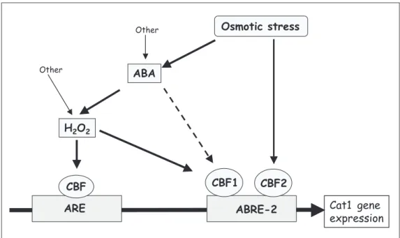
anti-oxidant genes in plants likely occurs via protein-DNA interactions in the region of the antioxidant-responsive element (ARE; TGACTCA), NF-κB, and abscisic acid-re-sponsive element 2 (ACGT) in the promot-ers of these genes. In addition to induction of defense gene expression, other roles of ROS in plants include direct killing of pathogens, involvement in cell wall structure, and pro-motion of programmed cell death (9). In yeast and animal cells, ROS have been shown to arrest cell division, and cell cycle progres-sion is under negative ROS control (34). A clear and powerful example of how ROS are put to constructive uses was the observation that O2•− plays an important role against
invading microbes, in effect serving as a broad-spectrum antibiotic (35). In response to invasion by pathogens, plants also mount a broad range of defense responses, includ-ing a rapid and transient production of large amounts of ROS (“oxidative burst”) (36).
Thus, ROS play different, or even oppos-ing roles, duroppos-ing different cellular processes (Figure 7). For example, under physiologic conditions, H2O2 may play an important role
in signal transduction pathways and in acti-vation of the transcription factor NF-κB, while under pathologic (stress) conditions, H2O2 can lead to apoptosis or necrosis.
sequently, the steady-state level of ROS within cells is critical and is determined by the interplay between ROS-generating mechanisms and the subtle modulating roles played by cellular antioxidants. In plants, H2O2 is generated under a diverse range of
biotic and abiotic conditions, and its accu-mulation in specific tissues at specific devel-opmental times, in the appropriate quanti-ties, benefits plants and can mediate cross-tolerance to other stresses (37). H2O2 affects
gene expression and activates MAP kinases (MAPK), which in turn function as regula-tors of transcription (3).
ROS and gene expression
Numerous studies indicate that cells have the means to sense ROS and to induce spe-cific responses, but the underlying mechan-isms are not fully understood (9,26). The transcriptional network that responds to ROS in eukaryotes is currently being deciphered, whereas the prokaryotic system is better un-derstood.
Nearly three decades ago, it was shown that the expression of ~30 proteins was in-duced by H2O2 in bacteria (38). Of these 30
proteins, 12 were maximally induced within 10 min and 18 between 10-30 min. The OxyR regulatory protein was subsequently shown to regulate expression of 9 of the 12 rapidly induced proteins. The tetrameric OxyR protein is a member of the LysR fam-ily of transcription activators and exists in two forms, reduced and oxidized; only the oxidized form is able to activate transcrip-tion. Further studies led to the identification of a number of OxyR-activated genes (39). Similarly, the SoxRS regulatory proteins were found to regulate expression of O2•−
-responsive proteins in bacteria (40). Regula-tion of the SoxRS regulon occurs by conver-sion of SoxR to an active form that enhances soxS transcription. The enhanced levels of SoxS in turn activate expression of the regulon (40). In addition to SoxR and OxyR,
several other transcriptional regulators modu-late the expression of antioxidant genes in bacteria, indicative of the complexity and connectivity of overlapping regulatory net-works. No apparent homologs of OxyR, SoxR, or SoxS have been found in eukary-otes (28), but a number of other transcription factors have been found to play a role in regulating the expression of antioxidant genes in eukaryotes. In yeast, transcription regula-tors of antioxidant genes include ACE1, MAC1, YAP1, YAP2, HAP1, and HAP2/3/ 4 (30). In higher eukaryotes, oxidative stress responses are more complex and are modu-lated by several different regulators. In mam-malian systems, NF-κB and AP-1 are in-volved in regulating the oxidative stress re-sponse. The ARE, present in the promoter region of mammalian glutathione S-trans-ferase, metallothionein-I, and MnSod genes, causes induction of these genes in response to oxidants (41). NF-κB, AP-1, and ARE have also been found in promoters of anti-oxidant genes in higher plants (Figure 8) (9). The role of these factors is not unique to activation of antioxidant genes, as they are known, particularly NF-κB, to play central roles in regulating cellular responses to other stresses as well as regulating normal growth and metabolism.
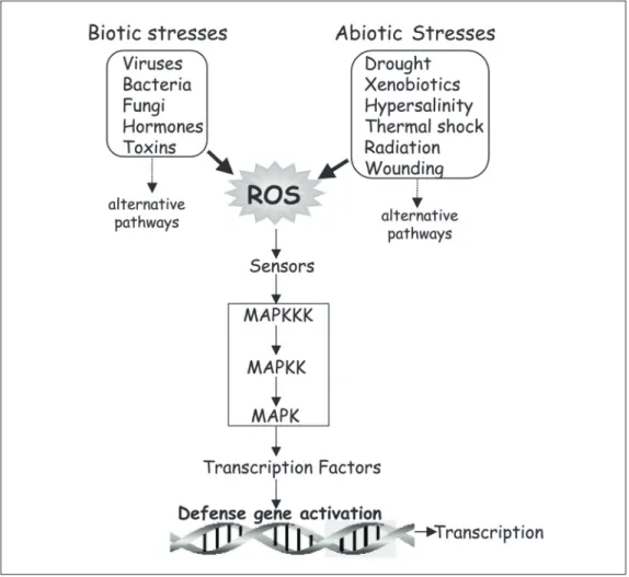
There is substantial evidence that a vari-ety of biotic and abiotic stresses induce ROS, which serve as a common factor in regulat-ing various signalregulat-ing pathways (Figure 9) (9,42). Similar stresses also activate MAPKs with kinetics that either precede or parallel H2O2 production, indicating that MAPKs
may be one of several converging points in the defense-signaling network (43). In addi-tion, exogenous application of several plant hormones and toxins has been shown to induce O2•− and H2O2 synthesis, leading to
differential induction of some antioxidant genes and isozymes (Figure 10) (9,37,41,44). Thus, the identification of all genes and proteins regulated by H2O2 is an important
Figure 8. Schematic representation of the promoter region of each of the three catalase (Cat) genes of maize indicating the locations of the NF-κB, ARE, AP-1, and ABRE motifs relative to the transcription start site (+1) of each gene. NF-κB = nuclear factor κB; ARE = antioxidant-responsive element: ABRE = abscisic acid-responsive element; AP-1 = activator protein-1.
tolerance to multiple, but interrelated, stresses. In addition to induction/repression of antioxidant defense genes, ROS are known to similarly affect expression of a variety of other genes involved in different signaling pathways in microbes (28), yeast (45), plants (46), and animals (19).
Genomic scale ROS-responsive gene expression
The advent of microarray expression anal-ysis makes possible the assessment of gene expression on a genomic scale, rendering tens of thousands of genes assayable in a single experiment (47). Thus, identification of ROS-responsive genes on a global scale is now tenable. DNA microarrays can compre-hensively examine gene expression networks during oxidative stress. There is now signifi-cant progress being made in surveying gene expression in response to H2O2 in
Esche-richia coli (48), yeast (4,45), animals (19), and higher plants (46).
A genome-wide transcription profile of E. coli cells exposed to H2O2 was examined
with a DNA microarray composed of 4169 E. coli open reading frames (48). Gene
ex-pression was measured in isogenic wild-type and oxyR deletion mutants (∆oxyR) to confirm that the H2O2-response regulator
OxyR activates most of the H2O2-inducible
genes. A very rapid and strong induction was observed of a set of OxyR-regulated genes in the wild type but not in the ∆oxyR, providing internal validation of the experi-ment and confirmation for the induction of the oxidative stress genes identified earlier by other means (38). Several new H2O2
-inducible genes were also identified: some were members of the OxyR regulon and some induced by an OxyR-independent mechanism suggestive of other H2O2
sen-sors and regulators in E. coli (38). Several genes repressed by OxyR were highly ex-pressed in the ∆oxyR mutant. Overall, the mRNA of 140 genes in the wild type, and 167 genes in the ∆oxyR were significantly induced after H2O2 treatment. It was also
found that soxS was induced by H2O2,
indi-cating an overlap with other regulatory path-ways. Two genes, Fpr and sodA, known to be members of the SoxRS regulon were also highly induced by H2O2 in both wild type
and ∆oxyR. The microarray data also showed an overlap between oxidative stress, heat
shock and SOS responses (38). The results from the E. coli microarrays clearly indicate that the activities of transcription factors in addition to OxyR and SoxRS are likely modu-lated by oxidative stress.
Whole-genome expression patterns in Saccharomyces cerevisiae cells exposed to H2O2, in addition to other stresses, indicated
that ~2/3 of the genome is involved in the response to environmental changes, and the global set of genes induced/repressed by each environmental signal were identified (4,45). The response to oxidative stress in-volves ~1/3 of the yeast genome and the maximal effects on gene expression occur slightly later relative to other stresses exam-ined during similar time-courses, with most of the transcriptome returning to prestress levels within 2 h following exposure to H2O2
(4). Genes that are repressed for ~60 min after exposure to H2O2 are only transiently
repressed in other stress time courses. Thus, genes encoding the translation apparatus and its regulators are remarkably coordinated for each environmental change, although the dynamics of each response are different. The expression programs following H2O2 or
O2•− treatment were essentially identical
despite the fact that different ROS are in-volved. There was strong induction of genes known to be involved in detoxification of both H2O2 and O2
•−
, such as CAT, SOD, and glutathione peroxidase, as well as genes in-volved in oxidative and reductive reactions (e.g., thioredoxin, glutathione reductase, glutaredoxin). The genes most strongly in-duced in response to H2O2 and O2•− were
dependent on the transcription factor Yap1p for their induction. Genes moderately in-duced by ROS and other signals are regu-lated by different transcription factors, de-pending on the conditions, and different up-stream signaling pathways may govern their response. It has also been demonstrated that in Schizosaccharomyces pombe H2O2
acti-vates the Sty1 (stress-activated MAPK) path-way in a dose-dependent manner via two
sensing mechanisms (49). At low H2O2
lev-els, a two-component signaling pathway, which feeds into either of the two (Wak1 or Win1) stress-activated MAPK kinase ki-nases, regulates Sty1. At high H2O2 levels,
however, Sty1 activation is controlled mainly by an independent two-component mechan-ism, which requires the function of both Wak1 and Win1. In addition, the individual bZip transcription factors, Pap1 and Atf1, were found to function within a limited range of [H2O2]: Pap1 activates target genes at low
[H2O2], whereas Atf1 controls transcriptional
responses to high [H2O2], with some minor
overlap. Some apparent cross talk among Sty1, Atf1, and Pap1 has been detected (50). Thus, S. pombe deploys a combination of stress-responsive regulatory proteins to gauge and trigger the appropriate transcrip-tional response to increasing H2O2
concen-trations (49). This organism mounts two sepa-rate responses to oxidative stress: an adap-tive response to low-level H2O2 exposure
that protects it from subsequent exposures to higher [H2O2], and an acute response that
allows the cell to survive a sudden, poten-tially lethal dose of H2O2.
Large-scale cDNA microarray analysis of the Arabidopsis transcriptome during oxi-dative stress identified 175 non-redundant expressed sequence tags from a sample of 11,000, which are regulated by H2O2. Of
these, 62 are repressed and 113 are induced. In addition, RNA blots showed that some of the H2O2-regulated genes are also
modu-lated by other signals known to involve oxi-dative stress (51). Furthermore, a substantial portion of these genes have predicted func-tions in defense response, cell rescue and signaling, and transcription, underscoring the pleiotropic effects of H2O2 in the
comparable to the situation in yeast (4). Of the 175 genes identified as H2O2-responsive,
most have no obvious direct role in oxida-tive stress but may be linked to oxidaoxida-tive stress indirectly, as a consequence of other biotic and abiotic stresses, explaining their sensitivity to H2O2. Among the genes
in-duced by H2O2 were genes encoding
trans-cription factors, suggesting that they may mediate downstream H2O2 responses. As in
other organisms, expression of the MAPKs in Arabidopsis is induced by oxidative stress, which in turn can mediate the induction of oxidative stress-responsive genes (52).
Toward an integrated view of oxy-stress responses
The traditional view of ROS as mere indiscriminate reactive byproducts of cellu-lar metabolism has recently undergone a
metamorphosis. This view came about by the discovery that ROS may act as signal-transducing molecules and that activation of intracellular transcription factors such as OxyR, SoxRS, NF-κB, and AP-1 occur via interaction with ROS, leading to gene trans-cription (Figure 11).
Genome sequencing and expression pro-filing using DNA or oligonucleotide mi-croarrays, and related technologies, have been used effectively in the study of global gene expression patterns in response to dif-ferent growth and environmental conditions to which organisms are exposed. Subsequent hierarchical clustering methods allow for the allocation of genes, coregulated tempo-rally or in response to a given signal, into specific expression groups or regulons (48, 53). The numbers of genes that can be de-tected by these methods in response to any given environmental or developmental
nal far exceed the limited number that could have been detected only a few years ago. With the current methods the expression of tens of thousands of genes can be detected in a single experiment in response to ROS, or to any given signal. Since transcription of genes into mRNA is governed by transcrip-tion factors which bind to cis-regulatory re-gions of the DNA in the vicinity of the target gene, the question arises as to whether large co-regulated groups of genes share cis-regu-latory elements that bind to common trans-cription factors. The data available with re-spect to oxidative stress seem to suggest this. Cis-acting elements within the promoters of ROS-activated genes are being defined as well as their cognate trans-acting factors. A comparative analysis of promoter sequences of genes with similar expression profiles should provide a basis for unraveling com-mon regulatory sequences and overlapping gene expression networks modulating ROS-responsive genes. The antioxidant enzymes CAT and SOD play key roles in modulating the levels of endogenous H2O2 and O2•−,
which in turn, at specific concentrations, act to modulate the expression of other ROS-responsive genes.
The use of microarrays and future deri-vations thereof, to examine global gene re-sponses to ROS is assured. It has become clear that there are far more genes and gene clusters responding to ROS than previously thought, and that ROS likely play far more key roles in cellular activities than antici-pated. As global gene responses to ROS are examined temporally and spatially along with different time-courses, and varied oxidant concentrations, even more complex regulons and regulatory cascades will emerge. It will also be instructive to examine gene expres-sion profiles in specific mutants to help de-lineate the roles of specific regulators and of ROS. The identification of alternate path-ways of ROS-dependent gene expression and characterization of the redox sensing mechanisms involved should point to new
insights and directions. Some genes have been identified whose transcription is re-sponsive to a variety of stresses in addition to oxystress, while others appear to be re-sponsive only to ROS or other specific sig-nals. Although some regulatory systems have been implicated in modulating such re-sponses, the complete network of regulators of ROS responses that activate such genes remains unclear.
Microarray results from different organ-isms clearly underscore the fluidity of ge-nomes to reorganize and respond to changes in the cellular and extracellular environment. However, characterization of global gene expression programs at the transcript level is only the first step toward defining the role or function of each ROS-responsive gene. It is not unlikely that small changes in gene ex-pression could lead to large alterations in protein levels. Consequently, proteomic analyses are essential to correlate mRNA changes with protein levels. There are in fact ample data demonstrating that ROS can alter the activity of cellular proteins.
being obtained on gene expression patterns with structural, topological, and functional parameters and interactions of the various proteins encoded by ROS-responsive regu-lons, and to view the cell in which they function holistically.
The paradox
The oxygen paradox is indeed the para-dox of evolution itself. Evolutionary pres-sures have made the best of a bad situation by generating mechanisms to curtail the un-desirable toxic effects of ROS, which are an unavoidable consequence of the aerobic lifestyle, and to put them to constructive uses. Indeed evolution has co-opted ROS to serve necessary and useful purposes in the maintenance of cellular homeostasis and in the communication of cells with the external environment. The focus must now be placed on a more thorough understanding of how ROS-mediated signals are perceived, trans-duced, and interpreted by the cell’s genetic machinery. Perhaps the most noteworthy observation to date concerning oxidative stress and the negative and positive roles of ROS is their universality among aerobic or-ganisms and the similarities emerging in the regulatory mechanisms underlying their roles in all species.
The classical view of ROS as villains that indiscriminately destroy biomolecules has undergone a shift, in which positive biologi-cal roles are considered as well. It is now accepted that ROS, particularly H2O2 and
O2
•−
, are carefully regulated metabolites ca-pable of signaling and communicating criti-cal information to the cell’s genetic machin-ery. Redox regulation of gene expression by oxidants and antioxidants is emerging as a vital mechanism in the health of all eukary-otes, including man.
It would indeed be interesting and chal-lenging to identify all the changes in gene expression regulated by oxidative stress, and to determine their commonality among
di-verse species. Such a global analysis of the effects of H2O2 and O2•− on the transcriptome
of eukaryotes has not yet been attained, but with the emergence of post-genomic tech-nologies it will likely be not far off.
Conclusions
The survival of organisms on earth de-pends upon the interactions of their genomes with the environments in which they exist. In the course of evolution, organisms evolved a complex array of mechanisms for adapting to both minor and major fluctuations in the environment. The emergence of oxygenic photosynthesis presented early life forms with the greatest environmental challenge and an opportunity. The challenge was to develop antioxidant defenses in order to sur-vive; the opportunity was to exploit the reac-tivity of oxygen for energy yielding and biosynthetic reactions. The opportunity led to the highly diversified life forms that evolved sufficient defenses and managed to exploit the aerobic lifestyle. Oxygen toxic-ity likely led to massive extinctions of those organisms unable to cope with it, unless they took refuge in isolated anaerobic niches. Thus, oxygen is a “double-edged sword” in that it makes life on earth possible, but in its reduced forms (ROS), it is highly toxic and lethal. Oxidative stress can arise from an imbalance between generation and elimina-tion of ROS leading to excess ROS levels inflicting indiscriminate damage to virtually all biomolecules, leading in turn to various diseases and cell death. The notion that ROS are merely toxic byproducts of O2
generated, an increase in intracellular oxi-dants results in two critical effects: damage to various cell components and activation of specific signaling pathways, influencing vari-ous cellular processes leading to proper cell functions or to cell death. Genomic tools are accelerating the discovery of ROS-respon-sive genes on a global scale and are expand-ing our understandexpand-ing of the oxidative stress response and the pleiotropic roles of ROS in signaling and gene expression.
Acknowledgments
I gratefully acknowledge the generous, and continuous, support of my research over many years by the U.S. National Institutes of Health, Department of Energy, Environmen-tal Protection Agency, National Science Foundation, and Department of Agriculture.
References
1. Scandalios JG (2002). The rise of ROS. Trends in Biochemical Sciences, 27: 483-486.
2. McClintock B (1984). The significance of responses of the genome to challenge. Science,226: 792-801.
3. Scandalios JG (2002). Oxidative stress responses: what have ge-nome-scale studies taught us? Genome Biology, 3: 1019.1-1019.6. 4. Gasch A, Spellman P, Kao C, Harel O, Eisen M, Storz G, Botstein D & Brown P (2000). Genomic expression programs in the response of yeast cells to environmental changes. Molecular Biology of the Cell, 11: 4241-4257.
5. Scandalios JG & Guan LM (2001). Transcription factors regulating antioxidant gene expression in response to biotic and abiotic sig-nals. In: Gene Families: Studies of DNA, RNA, Enzymes and Pro-teins. World Scientific, Singapore, 287-301.
6. Laloi C, Apel K & Danon A (2004). Reactive oxygen signaling: the latest news. Current Opinion in Plant Biology, 7: 323-328. 7. Thorpe GW, Fong CS, Alic N, Higgins VJ & Dawes IW (2004). Cells
have distinct mechanisms to maintain protection against different reactive oxygen species: Oxidative stress-response genes. Pro-ceedings of the National Academy of Sciences, USA, 101: 6564-6569.
8. Hancock JT, Desikan R & Neill SJ (2001). Role of reactive oxygen species in cell signaling pathways. Biochemical Society Transac-tions, 29: 345-350.
9. Scandalios JG (1997). Oxidative Stress and the Molecular Biology of Antioxidant Defenses. Cold Spring Harbor Laboratory Press, Plainview, NY, USA.
10. Elstner EF (1987). Metabolism of activated oxygen species. In: Davies DD (Editor), The Biochemistry of Plants. Vol. 11. Academic Press, New York, 253-315.
11. Dismukes GC, Klimov VV, Baranov SV, Kozlov YN, DasGupta J & Tyryshkin A (2001). The origin of atmospheric oxygen on Earth: The innovation of oxygenic photosynthesis. Proceedings of the National Academy Sciences, USA, 98: 2170-2175.
12. Fridovich I (1995). Superoxide radical and superoxide dismutases. Annual Review of Biochemistry, 64: 87-112.
13. Halliwell B (1996). Free radicals in biochemistry and medicine. In: Meyers RA (Editor), Encyclopedia of Molecular Biology and Molecu-lar Medicine. Vol. 2. Wiley-VCH, Weinheim, Germany, 330-337. 14. Scandalios JG (1997). Molecular genetics of superoxide dismutases.
In: Scandalios JG (Editor), Oxidative Stress and the Molecular Biol-ogy of Antioxidant Defenses. Cold Spring Harbor Laboratory Press, Plainview, NY, USA, 527-568.
15. Fink RC & Scandalios JG (2002). Molecular evolution and structure function relationships of the superoxide dismutase gene families in angiosperms, and their relationship to other eukaryotic and prokary-otic superoxide dismutases. Archives of Biochemistry and Biophys-ics, 399: 19-35.
16. Scandalios JG, Guan L & Polidoros AN (1997). Catalases in plants: Gene structure, properties, regulation, and expression. In: Scanda-lios JG (Editor), Oxidative Stress and the Molecular Biology of Antioxidant Defenses. Cold Spring Harbor Laboratory Press, Plainview, NY, USA, 343-406.
17. Guan L & Scandalios JG (1996). Molecular evolution of maize catalases and their relationship to other eukaryotic and prokaryotic catalases. Journal of Molecular Evolution, 42: 570-579.
18. Beckman KB & Ames BN (1998). The free radical theory of aging matures. Physiological Reviews, 78: 547-581.
19. Finkel T & Holbrook NJ (2000). Oxidants, oxidative stress and the biology of aging. Nature, 408: 239-247.
20. Beckman KB & Ames BN (1997). Oxidants, antioxidants, and aging. In: Scandalios JG (Editor), Oxidative Stress and the Molecular Biol-ogy of Antioxidant Defenses. Cold Spring Harbor Laboratory Press, Plainview, NY, USA, 201-246.
21. Helfand SL & Rogina B (2003). Molecular genetics of aging in the fly: is this the end of the beginning? BioEssays, 25: 134-141. 22. Chan SRWL & Blackburn EH (2004). Telomeres and telomerases.
Philosophical Transactions of the Royal Society of London, Series B. Biological Sciences, 359: 109-121.
23. McNight TD & Shippen DE (2004). Plant telomere biology. Plant Cell, 16: 794-803.
24. Von Zglincki T (2002). Oxidative stress shortens telomeres. Trends in Biochemical Sciences, 27: 339-344.
25. Sapolsky RM (2004). Organismal stress and telomeric aging: An unexpected connection. Proceedings of the National Academy of Sciences,USA, 101: 17323-17324.
26. Dalton TP, Shertzer HG & Puga A (1999). Regulation of gene expression by reactive oxygen. Annual Review of Pharmacology and Toxicology, 39: 67-101.
Meyers RA (Editor), Encyclopedia of Molecular Cell Biology and Molecular Medicine. Vol. 5. 2nd edn. Wiley-VCH, Weinheim, Ger-many, 489-512.
28. Storz G & Imlay JA (1999). Oxidative stress. Current Opinion in Microbiology, 2: 188-194.
29. Kiley PJ & Storz G (2004). Exploiting thiol modifications. PLoS Biology, 2: 1714-1717.
30. Ruis H & Schuller C (1995). Stress signaling in yeast. BioEssays, 17: 959-965.
31. Moradas-Ferreira P & Costa V (2000). Adaptive response of the yeast Saccharomyces cerevisiae to reactive oxygen species: de-fenses, damage and death. Redox Report,5: 277-285.
32. Delaunay A, Isnard A & Toledano MB (2000). H2O2 sensing through
oxidation of the Yap1 transcription factor. EMBO Journal, 19: 5157-5166.
33. Gabbita SP, Robinson K, Stewart C, Floyd R & Hensley K (2000). Redox regulatory mechanisms of cellular signal transduction. Ar-chives of Biochemistry and Biophysics, 376: 1-13.
34. Paulovich AG, Toczysky DP & Hartwell LH (1997). When check-points fail. Cell, 88: 315-321.
35. Babior BM (1984). The respiratory burst of phagocytes. Journal of Clinical Investigation, 73: 599-601.
36. Doke N (1997). The oxidative burst: roles in signal transduction and plant stress. In: ScandaliosJG (Editor), Oxidative Stress and the Molecular Biology of Antioxidant Defenses. Cold Spring Harbor Laboratory Press, Plainview, NY, USA, 785-813.
37. Guan LM, Zhao J & Scandalios JG (2000). Cis-elements and trans -acting factors that regulate expression of the maize Cat1 antioxidant gene in response to ABA and osmotic stress: H2O2 is the likely
intermediary signaling molecule for the response. Plant Journal,22: 87-95.
38. Christman MF, Morgan RW, Jacobson FS & Ames BN (1985). Positive control of a regulon for defenses against oxidative stress and some heat-shock proteins in Salmonella typhimurium. Cell, 41: 753-762.
39. Storz G & Zheng M (2000). Oxidative stress. In: Storz G & Henge-Aronis R (Editors), Bacterial Stress Responses. ASM Press, Wash-ington, DC, USA, 47-59.
40. Wu J & Weiss B (1991). Two divergently transcribed genes, soxR and SoxS, control a superoxide response regulon of Escherichia coli. Journal of Bacteriology, 173: 2864-2871.
41. Scandalios JG (2001). Molecular responses to oxidative stress. In: Hawkesford MJ & Buchner P (Editors), Molecular Analysis of Plant
Adaptation to the Environment. Kluwer Academic Publishers, Dor-drecht, The Netherlands, 181-208.
42. Somssich IE & Halbrock K (1998). Pathogen defence in plants - a paradigm of biological complexity. Trends in Plant Sciences, 3: 86-90.
43. Zhang S & Klessig D (2001). MAPK cascades in plant defense signaling. Trends in Plant Sciences, 6: 520-527.
44. Williamson J & Scandalios JG (1992). Differential response of maize catalases and superoxide dismutases to the photoactivated fungal toxin cercosporin. Plant Journal, 2: 351-358.
45. Causton H, Bing R, Koh S, Harbison C, Kanin E, Jennings E, Lee T, True H, Lander E & Young R (2001). Remodeling of yeast genome expression in response to environmental changes. Molecular Biol-ogy of the Cell, 12: 323-337.
46. Desikan R, Neill SJ & Hancock JT (2000). Hydrogen peroxide-induced gene expression in Arabidopsis thaliana. Free Radical Biol-ogy and Medicine, 28: 773-778.
47. Schena M, Shalon D, Davis R & Brown P (1995). Quantitative monitoring of gene expression patterns with complementary DNA microarray. Science, 270: 467-470.
48. Zheng M, Wang X, Templeton L, Smulski D, LaRossa R & Storz G (2001). DNA microarray-mediated transcriptional profiling of the Escherichia coli response to hydrogen peroxide. Journal of Bacteri-ology, 183: 4562-4570.
49. Quin J, Findlay VJ, Dawson K, Millar JB, Jones N, Morgan BA & Toone WM (2002). Distinct regulatory proteins control the graded transcriptional response to increasing H2O2 levels in fission yeast
Schizosaccharomyces pombe. Molecular Biology of the Cell, 13: 805-816.
50. Nguyen AN, Lee A, Place W & Shiozaki K (2000). Multistep phos-phorelay proteins transmit oxidative stress signals to the fission yeast stress-activated protein kinase. Molecular Biology of the Cell, 14: 1169-1181.
51. Desikan R, MacKerness A, Hancock JT & Neill SJ (2001). Regula-tion of the Arabidopsis transcriptome by oxidative stress. Plant Physiology, 127: 159-172.
52. Kovtun Y, Chiu WL, Tena G & Sheen J (2000). Functional analysis of oxidative stress-activated mitogen-activated protein kinases cas-cade in plants. Proceedings of the National Academy of Sciences, USA, 97: 2940-2945.